Abstract
Objective: To evaluate simultaneously the functional state of CD4+ and CD8+ T lymphocytes from Venezuelan HIV-1-infected pediatric patients.
Methods: Children were assigned to subgroups of rapid progressors (RPs) and slow progressors (SPs), based on clinical features. To determine the degree of CD4+ and CD8+ T-lymphocyte functionality, low cytometry techniques were used, and diverse parameters of the functionality of these cells were characterized by ex vivo tests, such as expression of CD95/Fas and CD127, and frequency of apoptosis. In addition, we determined, in cultured peripheral blood mononuclear cells, HIV-speciic proliferation and the production of interleukin-10 (IL-10), tumor necrosis factor alpha (TNF-α) and interferon gamma (IFN-γ), besides measuring intracellular IFN-γ in CD4+ T cells.
Results: Our results indicate that several molecular and cellular mechanisms of CD4+ and CD8+ T lymphocytes are deteriorated in RPs in comparison with SPs and controls. Indeed, both types of T lymphocytes from RPs exhibited an increased expression of CD95/Fas (p < 0.01), a signiicantly reduced expression of CD127 (p < 0.01), and an augmented frequency of apoptosis (p < 0.01). Furthermore, T cells from these patients displayed a diminished capacity of mitogen proliferation (p < 0.05), a reduced percentage of IFN-γ producing CD4+ T lymphocytes (p < 0.05), and a smaller capacity of IL-10, TNF-α and IFN-γ production (p< 0.01) in comparison with SP and control patients.
Conclusion: Our indings indicate that the decline of the normal T lymphocyte molecular and cellular responses is related to a rapid progression and a decreased resistance to HIV-1 infection in children.
J Pediatr (Rio J). 2012;88(2):161-8: HIV, progression, pediatric patients, immune function.
O
riginala
rticle Copyright © by Sociedade Brasileira de Pediatria161
Introduction
HIV infection causes high morbidity and mortality in children, accounting for more than 20% of the deaths related to HIV-1 infection in the world.1 A quarter of vertically infected children born in developed countries progress to serious
disease or death in the irst year of life without effective antiretroviral therapy, a proportion that rises to a half by 5 years of age.2 A bimodal clinical behavior has been identiied
in children infected by vertical transmission. Thus, 10 to 30%
Functional state of CD4+ and CD8+ T lymphocytes
and their role in the slow progression
of HIV infection in pediatric patients
Miguel Antonio Alfonzo,1 Alexandra Diaz,2 Luigina Siciliano,3
Maria Graciela López,3 Alida Hung,1 Juan Felix Garcia3
1. PhD. Universidad Central de Venezuela, Ciencias Fisiológicas, Caracas, Distrito Capital, Venezuela.
2. Graduate Student. Universidad Central de Venezuela, Ciencias Fisiológicas, Caracas, Distrito Capital, Venezuela. 3. MD. Hospital de Niños J.M. de los Ríos, Servicio de Infectologia, Caracas, Distrito Capital, Venezuela.
No conflicts of interest declared concerning the publication of this article.
Financial support:Consejo de Desarrollo Científico y Humanístico de la Universidad Central de Venezuela, CDCH-UCV (grantno. 09-00-5810-2005).
Suggested citation: Alfonzo MA, Diaz A, Siciliano L, López MG, Hung A, Garcia JF. Functional state of CD4+ and CD8+ T lymphocytes and their role in the slow progression of HIV infection in pediatric patients. J Pediatr (Rio J). 2012;88(2):161-8.
Manuscript submitted Oct 12 2011, accepted for publication Jan 18 2012.
of patients, which are named rapid progressors (RPs), start presenting AIDS symptoms in the irst months of life (within 1 year), whereas a minority group of patients, denominated slow progressors (SPs), remains asymptomatic for several years and can even arrive to the scholar age.3-5 Although some immunological and virological factors6,7 associated
with progression to disease have been identiied, such as clinical symptoms (hepatomegaly, splenomegaly, and lymphadenopathy manifested early in life),6,8 the cellular and molecular mechanisms underlying non-progression in HIV-1-infected children are not well understood.
The rate of progression to disease in HIV-1 infection varies considerably among individuals, due to a complex interplay between host’s genetic and immunological factors and the pathogenic potential of the virus.9,10 In children, we must consider several factors: relative immunological immaturity, HIV-1-mediated thymic destruction at a time of active thymopoiesis, and human leukocyte antigen class I sharing between mother and infant.11-13 Few studies have investigated the functional state of CD4+ and CD8+ T lymphocytesin RP and SP patients11 in order
to further establish some links between clinical outcome and immunological features. The relative contribution of host’s immunologic factors to delay progression to disease in Venezuelan children has not been examined precisely. Trying to explore these mechanisms, we decided to study a cohort of HIV-1-infected Venezuelan children, classiied by clinical features into RP and SP groups, to characterize diverse parameters of CD4+ and CD8+ T cellsby ex vivo tests such as expression of CD95/Fas (death receptor) and CD127 (receptor of surviving signals), and frequency of apoptosis. In addition, we determined, in cultured peripheral blood mononuclear cells (PBMCs) of these patients, HIV-speciic proliferation and the production of interleukin-10 (IL-10), tumor necrosis factor alpha (TNF-
α
) and interferon gamma (IFN-γ
), besides measuring intracellular IFN-γ
in CD4+ T cells. All these T-cell biological activities were compared against those observed in healthy children from the same hospital. These analyses might yield important information about HIV-1 pathogenesis and the role of protective immunity in HIV-1 infection in infants.Materials and methods
Patients and study design
We enrolled 15 HIV-1-infected Venezuelan children from the Hospital J.M. de Los Ríos (Caracas, Venezuela), who have been infected in uterus, during labor or by breastfeeding from seropositive mothers. All patients were naive to highly active antiretroviral therapy at the beginning of the study. We used 5 mL of whole blood samples to perform all assays. This pediatric group was divided into six SP children and nine RP children, which were monitored in the health center. The SP group had a median of 25.5 months of age
(19-36 months) and included patients with or without mild HIV-1 associated signs or symptoms (classiied in stages N2 or A2 according to the classiication of the Centers for Disease Control and Prevention, CDC). The RP group had a median of 12 months of age (3-26 months) and an onset of severe clinical manifestations (CDC category C) and/or signiicant immune suppression (CDC category 3) within the irst year of life. Eleven HIV-negative infants from the Healthy Children Service of the same hospital participated as controls. The control group had a median of 19 months of age (8-36 months). The hospital’s bioethical board approved this study and all children’s parents gave a written informed consent.
Viral load measurement
Plasma HIV-1 RNA levels were measured with a commercial quantitative reverse transcriptase polymerase chain reaction kit (Amplicor HIV Monitor Test, Roche Molecular Systems, USA), with a mean detection limit of 2.3 log10 copies/mL.
Cell immunophenotyping
Circulating T cells and their subsets were determined in whole blood samples by standard luorescence staining, followed by low cytometry analysis, using luorescent beads as an internal standard (Beckman Coulter, USA) and commercial monoclonal antibodies (MoAbs) from BD Biosciences, USA. The following MoAbs were used: anti-CD3-PerCP, anti-CD4-APC, anti-CD8-APC, anti-CD45RA-FITC, and anti-CD45RO-PE. Briely, 100 µL of blood samples were incubated with conjugated MoAbs for 30 min on ice, in the dark, and washed. Red blood cells were lysed with FACS Lysing Solution (BD Biosciences, USA), and samples were immediately applied to a BD FACSCalibur low cytometer(BD Biosciences, USA). Events were gated on a live lymphocyte region based on forward and side scatter parameters and analyzed using BD CellQuest software(BD Biosciences, USA).
Analysis of expression of Fas (CD95) and IL-7R
α
(CD127)irrelevant mouse antibodies were used as negative controls to determine background luorescence. Antigen expression was quantiied by low cytometry as the percentage of positive cells in a given cell population.
Detection of apoptosis
Spontaneous apoptosis was measured in fresh whole blood samples. Cells were stained with FITC-conjugated annexin V and propidium iodide using the Annexin V Apoptosis Detection kit I (BD Pharmingen, USA) as recommended by the manufacturer. The extent of apoptosis was quantiied by low cytometry as the percentage of annexin V-positive cells in a given cell population. Both live and apoptotic cells were included in the analysis and cell debris was excluded.
In vitro lymphocyte proliferation assays
PBMCs were isolated from ethylenediaminetetraacetic acid-treated blood samples by density gradient centrifugation (Histopaque, Sigma, USA). These PBMCs were then grown in 96-well culture plates at a density of 1x10⁵ cells/well
in 0.2 mL of RPMI-1640 (Gibco, Life Technology, USA), supplemented with 10% of heat-inactivated fetal bovine serum, 2 mM L-glutamine, 100 U/mL penicillin, and 100 mg/mL streptomycinsulfate (all from GIBCO, USA) in the presence of one of the following stimuli: 5 µg/mL of phytohaemagglutinin (PHA, Sigma, USA) and pooled HIV-1 envelope (HIV-1 Env) overlapping 20-mer peptides (each peptide at 1 µg/mL) (BD Biosciences, USA). Non-stimulated cells were used as controls. Lymphocyte proliferation was determined after 72 h of culture by the incorporation of bromodeoxyuridine(BrdU) (Sigma, USA), which was added in the last 4-5 h of culture. Cells were washed, ixed, permeabilized, and stained with FITC-conjugated anti-BrdU, anti-CD3-PerCP and anti-CD4-APC (BD Biosciences, USA) for 30 min. An additional wash step was performed, and cells were resuspended in 1% formaldehyde, applied to a BD FACSCalibur low cytometer(BD Biosciences, USA), and analyzed using BD CellQuest software (BD Biosciences, USA).
Measurement of cytokine production
Cytokine production was measured in supernatants of PBMCs cultured for 72 h in the presence of PHA or pooled HIV-1 Env overlapping 20-mer peptides, as above described. Supernatants were collected and stored at -80 ºC until analysis. A Cytometric Bead Array kit (BD Biosciences, USA) was used, according to the manufacturer’s instructions. Its methodology combines the principles of the “sandwich” immunoassays with the ability of low cytometry to measure the characteristics of multiple particles, so we were able to measure the level of three human cytokines (IL-10, TNF-
α
, and IFN-γ
) simultaneously in a single supernatant sample.Intracellular cytokine staining
Production of IFN-
γ
as an HIV-1-speciic CD4+ T cell response was quantiied by intracellular cytokine staining. Briely, 5 x 10⁵ PBMCs were stimulated with either PHA(5 µg/mL) or pooled HIV-1 Env overlapping 20-mer peptides (each peptide at 1 µg/mL) (BD Biosciences, USA). After 72 h, 10 µg/mL brefeldin A (Sigma, USA) was added to prevent export of intracellular cytokines. Following cell wash, cells were ixed, permeabilized, and stained with FITC-conjugated anti-IFN-
γ
, anti-CD3-PerCP, and anti-CD4-APC (all from BD Biosciences, USA). Cells were further washed and resuspended in 1% formaldehyde. Samples were applied to a BD FACSCalibur low cytometer (BD Biosciences, USA) and CD4+ T cells were gated for analysis using BD CellQuest software (BD Biosciences, USA).Statistical analysis
Experimental data are given as mean ± standard deviation. Differences (p values) were evaluated using the two-tailed, nonparametric Mann-Whitney U test and the two-tailed Student’s t test for normally distributed data. Differences were considered signiicant for p< 0.05.
Results
T cell subsets and viral load in HIV-infected children
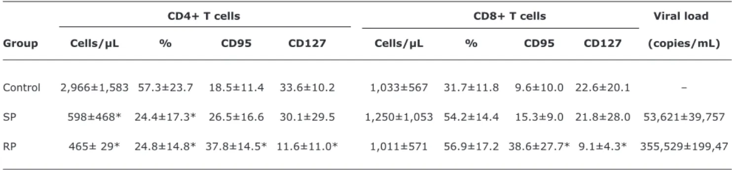
T lymphocyte subpopulations and viral loads of the studied groups are summarized in Table 1. A signiicant decrease in absolute and percentage values of blood CD4+ T cell counts was shown in both groups of HIV-infected patients in comparison to healthy infants (p < 0.01). Although SP patients showed a tendency to have higher absolute values of CD4+ T lymphocytes compared with RP patients, the difference was not statistically signiicant. On the other hand, the percentage of CD8+ T cells of both groups of HIV patients presented higher values than those of the control group, but there were not statistically signiicant differences. Similarly, it was observed that there was not statistically signiicant difference in the absolute number of CD8+ T lymphocytes between SP and RP patients. In relation to the viral load, there was not signiicant difference between both groups of HIV patients, although values were higher in RP patients compared with SP patients (Table 1).
IL-7R
α
(CD127) and Fas (CD95) expression in T cell subsetsTable 1 - Viral load, lymphocyte subpopulations, and expression of CD95 and CD127 in HIV-1-infected and healthy infants
CD4+ T cells CD8+ T cells Viral load
Group Cells/µL % CD95 CD127 Cells/µL % CD95 CD127 (copies/mL)
Control 2,966±1,583 57.3±23.7 18.5±11.4 33.6±10.2 1,033±567 31.7±11.8 9.6±10.0 22.6±20.1 –
SP 598±468* 24.4±17.3* 26.5±16.6 30.1±29.5 1,250±1,053 54.2±14.4 15.3±9.0 21.8±28.0 53,621±39,757
RP 465± 29* 24.8±14.8* 37.8±14.5* 11.6±11.0* 1,011±571 56.9±17.2 38.6±27.7* 9.1±4.3* 355,529±199,47
RP = rapid progressor; SP = slow progressor. * p < 0.01 vs. control.
The absolute, percentage and viral load values are expressed as mean ± standard deviation. All experimental assays were performed once. Differences (p values) were evaluated using the two-tailed, nonparametric Mann-Whitney Utest.
*p < 0.05 and **p < 0.01 vs. control.
RPs = rapid progressors; SPs = slow progressors.
Figure 1 - Spontaneous ex vivo apoptosis inCD4+ and CD8+ T cells. Data show the percentage of annexin V+/ PI- (early apoptotic) cells within gated CD4+ and CD8+ lymphocyte populations. Each value is the mean ± standard deviation. All experimental assays were performed twice in each sample
We evaluated T cell activities such as ex vivo expression percentages of IL-7R
α
subunit (CD127) in CD4+ and CD8+ T lymphocytes. It can be seen in Table 1 that receptor expression was signiicantly diminished in RP patients in relation to the control group, while SP patients presented similar values in comparison to the control group.Spontaneous apoptosis in CD4+ and CD8+ T lymphocytes from HIV-infected and control patients
We measured the frequency of cell death associated with spontaneous apoptosis. Both groups of infected patients presented signiicantly higher values of spontaneous apoptosis of CD4+ and CD8+ T cells compared with the group of healthy children. Noticeably, RP patients presented the highest values of apoptotic cells, even with a higher population of CD8+ T cells compared to SP patients (Figure 1).
Mitogen and viral-speciic lymphocyte proliferative
responses
Although both groups of patients presented a signiicant increment in the proliferation of their CD4+ and CD8+ T lymphocytes in the presence of PHA compared to the non-stimulated condition, proliferation responses never reached values of the control group, which were signiicantly higher (Figure 2). Conversely, when PBMCs were cultured in the presence of viral-speciic antigens, no relevant increment in the proliferation of neither CD4+ nor CD8+ T lymphocytes was observed in HIV-infected patients, presenting similar proliferation values than controls (Figures 2A and 2B).
Production of cytokines in PBMCs from HIV patients
PBMCs from the two different HIV- infected groups (SPs and RPs) were cultured for 72 h in different conditions of
Figure 3 - Production of cytokines (IL-10, TNF-α, and IFN-γ) (pg/mL) from PBMCs isolated from HIV-infected and controls infants. The quantiication of cytokines was performed in supernatants of cultured PBMCs for 72 h in the presence of PHA, viral antigens (Env), or medium alone (non-stimulated cells). Each value is the mean ± standard deviation. All cytokine tests were performed by triplicate
*p < 0.01 vs. non-stimulated cells.
IFN-γ = interferon gamma; IL-10 = interleukin-10; PBMCs = peripheral blood mononuclear cells; PHA = phytohaemagglutinin; RPs = rapid progressors; SPs = slow progressors; TNF-α = tumor necrosis factor alpha.
* p < 0.05 vs.non-stimulated cells (basal activity).
BrdU = bromodeoxyuridine; PBMCs = peripheral blood mononuclear cells; PHA = phytohaemagglutinin; RPs = rapid progressors; SPs = slow progressors.
Figure 2 - Lymphocyte proliferation. PBMCs were cultured for 72 h in the presence of PHA, viral antigens (Env) or medium alone (non-stimulated cells). Proliferation of CD4+ (A) and CD8+ (B)T lymphocytes was measured as the percentage of BrdU incorporation. Each value is the mean ± standard deviation. All proliferation assays were carried out by triplicate
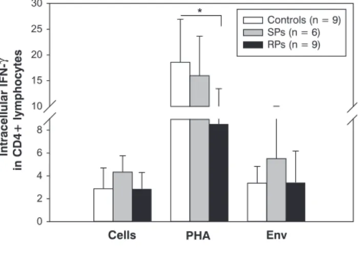
IFN-
γ
production in CD4+ T from control and HIV-infected childrenCD4+ T lymphocytes are the main source of production of IFN-
γ
,which is one of the most important antiviral cytokines in HIV-1 infection. We evaluated in vitro capacity of production of this cytokine by CD4+ T cells in pediatric patients, measured at the intracellular compartment. Results are shown in Figure 4. It was observed that, in cells cultured in the presence of PHA, the production of this cytokine was signiicantly increased in all studied groups. However, percentages of IFN-γ
-producing CD4+ T cells in both groups of HIV-infected patients were lower than those of the control group, being signiicant different in RPs (p < 0.01). The presence of the viral antigen (Env) did not modify the production of this cytokine in all studiedgroups, showing similar values to those in the absence of antigenic stimuli. However, SP patients displayed a tendency to higher values in comparison with controls.
Discussion
Our research hypothesis is that T cell functionality is relevant to the progression of vertically HIV-infected children, which may explain the distinctive clinical features described between RP and SP patients. Trying to address this hypothesis, we performed a study in which some of the CD4+ and CD8+ lymphocyte biological responses were evaluated simultaneously by means of ex vivo and in vitro analysis using PHA and viral antigens (Env) to assess the functionality of Tcells from Venezuelan pediatric HIV-infected patients with different clinical status, grouped as RPs and SPs, and to compare these biological activities with those from the T cells of Venezuelan healthy children.
Figure 4 - Intracellular cytokine staining of CD4+ T lymphocytes. The intracellular IFN-γ was quantiied in CD4+ T lymphocytes following stimulation for 72 h with PHA, viral antigens (Env), or medium alone (non-stimulated cells). Each value is the mean ± standard deviation. All cytokine staining was executed by triplicate * p < 0.05 vs. non-stimulated cells.
IFN-γ = interferon gamma; PHA = phytohaemagglutinin; RPs = rapid progressors; SPs = slow progressors.
as described.17 Interestingly, RP patients displayed a greater proportion of apoptotic CD4+ and CD8+ T cells in comparison to the group of SP patients.
To evaluate T cell biological activities, the expression of Fas/CD95 is a relevant apoptosis parameter. Thus, the ex vivo expression of Fas/CD95 observed in CD4+ and CD8+ T lymphocytes from our RP patients was higher than that from SP patients. Interestingly, the viral load in both groups of HIVpatients was different, being higher in RP compared to SP patients. It is possible that the lower expression of CD95 in both types of T lymphocytes in SP patients was due to their lower viral load, and consequently displays minor frequency of apoptosis in these cells, which has been described in HIV-infected adults.18
IL-7 is an important cytokine for an eficient development and maintenance of T lymphocyte subpopulations. This cytokine binds to the alpha subunit of its receptor (IL-7R
α
), located at the surface of T and B lymphocytes and natural killer cells.19,20 We demonstrated that IL-7Rα
expression in both CD4+ and CD8+ T lymphocytes is signiicantly reduced in the RP group, whereas the SP group presented similar values than the control group. This experimental inding may suggest that the T cells from SP patients have a greater probability to survive, and so these patients are able to maintain their cellular immunity more eficiently than RP patients. To support this assumption, higher levels of CD4+ and CD8+ T lymphocytes and lower frequency of apoptosis in these lymphocytes were found in SP patients. In this sense, our results are consistent with those of previous studies, which show that expression of IL-7Rα
isinversely correlated with immune activation and apoptosis, and positively correlated with CD4+ T-cell counts in HIV-infected patients.21 The loss of IL-7R
α
expression induced by immune activation may diminish Bcl-2 and Bcl-xl production, rendering cells more susceptible to apoptosis.22 Our datasupport the assumption that a lower expression of IL-7R
α
may be related with a higher frequency of apoptosis in these T cells.Going deeply into more molecular mechanisms, we studied several effector mechanisms of CD4+ and CD8+ T lymphocytes, such as the production of IL-10, TNF-
α
and IFN-γ
by PBMCs, and the proliferative responses of these T lymphocytes to mitogens or viral peptides. According to our analysis, RP patients exhibited a greater deterioration of the functional capacity of CD4+ and CD8+ T cells compared to SP patients. Indeed, we found in RP patients a signiicant partial loss of PHA proliferative response of CD4+ and CD8+ T lymphocytes, a lower production of IL-10, TNF-α
and IFN-γ
by PBMCs, and a diminished percentage of IFN-γ
producing CD4+ T cells, measured by intracellular cytokine staining. The greater loss of capacity of T cells to proliferate in the presence of PHA in RP patients coincide with results obtained previously showing that non-progressors adults have a greater capacity of T cell proliferation compared to RPs.23 Interestingly, all these effector responses were completely lost in both groups of patients when their PBMCs were cultured in the presence of the viral antigen (Env). This lack of response could be explained by the inhibitor effect of viral proteins and/or by the loss of HIV- speciic clones during the progression of infection,24,25 or by the fact that the viral infection affected T cell lines at thymus and destroyed the ability to produce anti-HIV clones as described.11-13 This experimental inding requires further investigation to identify the molecular mechanisms responsible for this lack of responses of HIV-infected children to viral antigen (Env).Classically, it has been established that, during HIV infection, a change in the Th1/Th2 immunological responses occurs. Th1 response (generally an antiviral response) diminishes as the disease progresses, and Th2 response (associated with a smaller antiviral protection) increases, compromising protection in the HIV-infected patients.26
References
1. UNAIDS. UNAIDS annual report 2008: towards universal access. Geneva: UNAIDS; 2009. http://data.unaids.org/pub/Report/2009/ jc1736_2008_annual_report_en.pdf. Access: 01/06/2009.
2. Gray L, Newell ML, Thorne C, Peckham C, Levy J; European Collaborative Study. Fluctuations in symptoms in human
immunodeiciency virus-infected children: the irst 10 years of
life. Pediatrics. 2001;108:116-22.
3. Dickover RE, Dillon M, Gillette SG, Deveikis A, Keller M, Plaeger-Marshall S, et al. Rapid increases in load of human
immunodeiciency virus correlate with early disease progression
and loss of CD4 cells in vertically infected infants. J Infect Dis. 1994;170:1279-84.
4. De Rossi A, Masiero S, Giaquinto C, Ruga E, Comar M, Giacca M, et al. Dynamics of viral replication in infants with vertically
acquired human immunodeiciency virus type 1 infection. J Clin Invest. 1996;97:323-30.
5. Salvatori F, Masiero S, Giaquinto C, Wade CM, Brown AJ, Chieco-Bianchi L, et al. Evolution of human immunodeiciency virus type
1 in perinatally infected infants with rapid and slow progression to disease. J Virol. 1997;71:4694-706.
6. Kalish LA, McIntosh K, Read JS, Diaz C, Landesman SH, Pitt J, et al. Evaluation of humanmmunodeiciency virus (HIV) type 1 load,
CD4 T cell level, and clinical class as time-ixed and time-varying markers of disease progression in HIV-1-infected children. J Infect Dis. 1999;180:1514-20.
7. Rich KC, Fowler MG, Mofenson LM, Abboud R, Pitt J, Diaz C, et al. Maternal and infant factors predicting disease progression in
human immunodeiciency virus type 1-infected infants. Women and Infants Transmission Study Group. Pediatrics. 2000;105:e8.
8. Children born to women with HIV-1 infection: natural history and risk of transmission. European Collaborative Study. Lancet. 1991;337:253-60.
9. Deacon NJ, Tsykin A, Solomon A, Smith K, Ludford-Menting M, Hooker DJ, et al. Genomic structure of an attenuated quasi species
of HIV-1 from a blood transfusion donor and recipients. Science. 1995;270:988-91.
10. Rhodes DI, Ashton L, Solomon A, Carr A, Cooper D, Kaldor J, et al.
Characterization of three nef-defective human immunodeiciency
virus type 1 strains associated with long-term nonprogression.
Australian Long-Term Nonprogressor Study Group. J Virol. 2000;74:10581-8.
11. Goulder PJ, Jeena P, Tudor-Williams G, Burchett S. Paediatric HIV infection: correlates of protective immunity and global perspectives in prevention and management. Br Med Bull. 2001;58:89-108.
12. Ometto L, Bertorelle R, Mainardi M, Zanchetta M, Tognazzo S, Rampon O, et al. Polymorphisms in the CCR 5 promoter
region inluence disease progression in perinatally human immunodeiciency virus type 1-infected children. J Infect Dis. 2001;183:814-8.
13. Kuhn L, Abrams EJ, Palumbo P, Bulterys M, Aga R, Louie L, et al. Maternal versus paternal inheritance of HLA class I alleles
among HIV-infected children: consequences for clinical disease
progression. AIDS. 2004;18:1281-9.
14. Grossman Z, Meier-Schellersheim M, Sousa AE, Victorino RM, Paul WE. CD4+ T-cell depletion in HIV infection: are we closer to understanding the cause? Nat Med. 2002;8:319-23.
15. Roederer M, Dubs JG, Anderson MT, Raju PA, Herzenberg LA, Herzenberg LA. CD8 naive T cell counts decrease progressively in HIV-infected adults. J Clin Invest. 1995;95:2061-6.
16. Casella CR, Rapaport EL, Finkel TH. Vpu increases susceptibility
of human immunodeiciency virus type 1-infected cells to fas killing. J Virol. 1999;73:92-100.
17. Samuelsson A, Broström C, van Dijk N, Sönnerborg A, Chiodi F. Apoptosis of CD4+ and CD19+ cells during human
immunodeiciency virus type 1 infection - correlation with clinical
progression, viral load, and loss of humoral immunity. Virology. 1997;238:180-8.
18. Siegel RM, Chan FK, Chun HJ, Lenardo MJ. The multifaceted role of Fas signaling in immune cell homeostasis and autoimmunity.
Nat Immunol. 2000;1:469-74.
19. Schluns KS, Kieper WC, Jameson SC, Lefrançois L. Interleukin-7
mediates the homeostasis of naïve and memory CD8 T cells in vivo. Nat Immunol. 2000;1:426-32.
20. Goldrath AW, Sivakumar PV, Glaccum M, Kennedy MK, Bevan MJ, Benoist C, et al. Cytokine requirements for acute and Basal
homeostatic proliferation of naive and memory CD8+ T cells. J Exp Med. 2002;195:1515-22.
21. Koesters SA, Alimonti JB, Wachihi C, Matu L, Anzala O, Kimani J, et al. IL-7Ralpha expression on CD4+ T lymphocytes decreases
with HIV disease progression and inversely correlates with immune activation. Eur J Immunol. 2006;36:336-44.
22. Colle JH, Moreau JL, Fontanet A, Lambotte O, Delfraissy JF, Thèze J. The correlation between levels of IL-7 Ralpha expression and
responsiveness to IL-7 is lost in CD4 lymphocytes from
HIV-infected patients. AIDS. 2007;21:101-3.
23. Rosenberg ES, Billingsley JM, Caliendo AM, Boswell SL, Sax PE, Kalams SA, et al. Vigorous HIV-1-speciic CD4+ T
cell responses associated with control of viremia. Science. 1997;278:1447-50.
24. Jowett JB, Planelles V, Poon B, Shah NP, Chen ML, Chen IS.
The human immunodeiciency virus type 1 vpr gene arrests infected T cells in the G2 + M phase of the cell cycle. J Virol. 1995;69:6304-13.
25. Finkel TH, Tudor-Williams G, Banda NK, Cotton MF, Curiel T, Monks C, et al. Apoptosis occurs predominantly in bystander cells and not in productively infected cells of HIV- and SIV-infected lymph nodes. Nat Med. 1995;1:129-34.
26. Clerici M, Shearer GM. The Th1-Th2 hypothesis of HIV infection: new insights. Immunol Today. 1994;15:575-81.
27. Groux H, Bigler M, de Vries JE, Roncarolo MG. Inhibitory and
stimulatory effects of IL-10 on human CD8+ T cells. J Immunol. 1998;160:3188-93.
in HIV-speciic CD8+ T cells, were observed in adults progressing to AIDS,29 and recent studies30 indicate that HIV-1 patients having a better management of the disease (known as long-term non-progressors) present signiicantly increased CD8+ Tcell populations compared to RPs.
In summary, we have found that the functional status of CD4+ and CD8+ T cells are distinct among pediatric infected Venezuelan patients. Thus, HIV-infected children with rapid progression to disease (RPs) presented more compromised functional immunity than infants whose disease progresses slowly (SPs). The identiication of molecular and cellular mechanisms that contribute to the maintenance of protective immune responses is an important hallmark for therapeutic and vaccine development.
Acknowledgements
Correspondence: Miguel Antonio Alfonzo
Laboratorio de Inmunoisiologia Celular, Universidad Central de Venezuela Escuela de Medicina Jose M. Vargas, piso 5 Esquina San Jose, Plaza San Lorenzo
San Jose, Caracas, Distrito Capital 1010 – Venezuela Tel.: +58 212 5628610
Fax: +58 212 5628610
E-mail: miguelacho1998@hotmail.com 28. Whitmire JK, Tan JT, Whitton JL. Interferon-gamma acts directly
on CD8+ T cells to increase their abundance during virus infection.
J Exp Med. 2005;201:1053-9.
29. Migueles SA, Laborico AC, Shupert WL, Sabbaghian MS, Rabin R, Hallahan CW, et al. HIV-speciic CD8+ T cell proliferation is coupled
to perforin expression and is maintained in nonprogressors. Nat Immunol. 2002;3:1061-8.
30. Betts MR, Nason MC, West SM, De Rosa SC, Migueles SA, Abraham J, et al. HIV nonprogressors preferentially maintain highly functional