p53 Mirrors Its Role in Tissue Differentiation
Oliver Couture, Eric Lombardi, Kendra Davis, Emily Hays, Nalini Chandar
*Department of Biochemistry, Chicago College of Osteopathic Medicine, Midwestern University, Downers Grove, Illinois, United States of America
Abstract
The tumor suppressor gene p5γ is involved in a variety of cellular activities such as cellular stress responses, cell cycle regulation and differentiation. In our previous studies we have shown p5γ’s transcription activating role to be important in osteoblast differentiation. There is still a debate in the literature as to whether p5γ inhibits or promotes differentiation. We have found p5γ heterozygous mice to show a p5γ dependency on some bone marker gene expression that is absent in knockout mice. Mice heterozygous for p5γ also show a higher incidence of osteosarcomas than p5γ knockout mice. This suggests that p5γ is able to modify the environment within osteoblasts. In this study we compare changes in gene expression resulting after either a transient or stable reduction in p5γ. Accordingly we reduced p5γ levels transiently and stably in CβC1β cells, which are capable of both myoblast and osteoblast differentiation, and compared the changes in gene expression of candidate genes regulated by the p5γ pathway. Using a PCR array to assay for p5γ target genes, we have found different expression profiles when comparing stable versus transient knockdown of p5γ. As expected, several genes with profound changes after transient p5γ loss were related to apoptosis and cell cycle regulation. In contrast, stable p5γ loss produced a greater change in MyoD and other transcription factors with tissue specific roles, suggesting that long term loss of p5γ affects tissue homeostasis to a greater degree than changes resulting from acute loss of p5γ. These differences in gene expression were validated by measuring promoter activity of different pathway specific genes involved in differentiation. These studies suggest that an important role for p5γ is context dependent, with a stable reduction in p5γ expression affecting normal tissue physiology more than acute loss of p5γ.
Citation: Couture O, Lombardi E, Davis K, Hays E, Chandar N (β01γ) Gene Expression Profiles Resulting from Stable Loss of p5γ Mirrors Its Role in Tissue Differentiation. PLoS ONE 8(11): e8β494. doi:10.1γ71/journal.pone.008β494
Editor: Simone Di Giovanni, Hertie Institute for Clinical Brain Research, University of Tuebingen., Germany Received May β4, β01γ; Accepted October β5, β01γ; Published November β8, β01γ
Copyright: © β01γ Couture et al. This is an open-access article distributed under the terms of the Creative Commons Attribution License, which permits unrestricted use, distribution, and reproduction in any medium, provided the original author and source are credited.
Funding: This work is supported by grants from the National Institutes of Health grant R15 AR055γ6β to Nalini Chandar and funds from Midwestern University. The funders had no role in study design, data collection and analysis, decision to publish, or preparation of the manuscript.
Competing interests: The authors have declared that no competing interests exist. * E-mail: [email protected]
Introduction
The tumor suppressor gene, p53, has been widely investigated as a transcription factor involved in multiple cellular processes including DNA damage, hypoxia, and cell cycle regulation [1-5]. In addition to its more traditional roles in stress responses and cell cycle regulation, p5γ also plays a role during development and cellular differentiation [6,7]. In our laboratory, we have found the osteocalcin gene, which codes for a protein associated with terminal osteoblast differentiation, to be regulated by p5γ in committed osteoblast cells [8]. Other studies have found several more tissue specific genes to be directly regulated by wild type p5γ, such as MyoD, a late signal of skeletal muscle differentiation [9,10]. While there are several studies indicating a role for p5γ in cell differentiation, not all of them are consistent for a role promoting differentiation. In neuronal cells, localization of p5γ to mitochondria is important in determining if cells undergo differentiation or apoptosis [γ]. In
mesenchymal stem cells, p5γ appears to keep progenitor cells from entering their cell type committed pathways by negatively regulating key differentiation transcription factors, such as Ppar for adipogenic differentiation, Myocd for myofibroblast differentiation, and Runxβ for osteogenic differentiation [11]. In embryonic stem cells, however, the role of p5γ is more complex. p5γ represses both Nanog and 4-Oct, both of which repress differentiation in embryonic stem cells. p5γ, however, can also activate the Wnt family of genes, resulting in inhibition of differentiation of stem cells [1β]. So while p5γ appears to inhibit early differentiation and keep cells in a more stem like state, late differentiation of certain cell fates are in part reliant upon the activity of p5γ.
defective spermatogenesis and skeletal abnormalities in a small percentage of the animals [15]. A high proportion of the tumors in the p5γ null mice are lymphomas followed by osteosarcoma and soft tissue tumors [1γ]. In p5γ heterozygous mice, the tumor spectrum is altered with a higher incidence of osteosarcomas and soft tissue tumors than lymphomas . This could mean that there are functionally redundant pathways that make up for loss of p5γ during development. However, its presence albeit in small amounts, is able to influence osteoblast function underscoring the important of p5γ in maintaining normal bone remodeling and preventing neoplastic transformation. Clinically osteosarcoma show heterogeneous tumor types thought to arise from the disruption of osteoblast differentiation at different times [16]. Since p5γ appears to normally inhibit entry into osteoblast differentiation, but is needed for terminal differentiation, it is not surprising that a partial disruption of p5γ promotes osteosarcoma development.
Since p5γ responds to multiple stimuli and can affect multiple, different cellular processes, changes in p5γ expression will affect cells differently depending on whether the change is transient or stable. In this study we test this by using shRNA interference to reduce p5γ levels in a bipotential cell line, CβC1β, transiently and compared the changes in gene expression of genes involved in p5γ pathways with a stable reduction of p5γ. We found greater changes in expression of MyoD and of other transcription factors with tissue specific roles in the stable p5γ knockdown cells, which suggests that long term partial loss of p5γ expression affects tissue homeostasis to a greater degree than changes resulting from transient loss of p5γ. Additionally, we show differences in the promoter activities of genes involved in differentiation between stable and transient loss of p5γ. Together this points toward the role of p5γ as being context dependent, with a stable loss affecting cellular differentiation more than a transient loss.
Materials and Methods
Cell line and growth conditions
In order to study the role of p5γ in the differentiation of mesenchymal stem cells, the bi-potential cell line CβC1β (ATCC, cat no. CRL-177β) was selected because it can differentiate into myotubes and can be induced to osteoblasts, depending on the culture media [17,18]. These cells were maintained in DMEM/F-1β media with 10% fetal bovine serum and incubated in T-75 flasks at γ7°C in 5% COβ/95% air.
Knockdown of p53 in C2C12 cells by shRNA
The p5γ HuSH-β9 shRNA plasmids (Origene®, cat. no. TF50000β), were used to create transient and stable knockdown lines of p5γ within the CβC1β cells. We used three different shRNA sequences to knockdown p5γ. Control cells received in a similar sized non effective scrambled sequence (TRβ000γ). SuperFect (QIAGEN, cat. no. γ01γ05) was used for both transient and stable transfections according to manufacturer’s protocol. Additionally, for obtaining stable clones, cells were selected post transfection using puromycin. Single cell clones were expanded, passaged and characterized before use.
RNA Extraction, Purification, and Quantification
To determine the changes at the transcriptional level, RNA was isolated either using TRI Reagent (Sigma, cat. no. T94β4) or the QIAGEN RNeasy Mini Kit (cat. no. 74104) prep system depending on the assay used. Genomic contamination of the RNA was removed using the Ambion DNA Free kit (Ambion, cat. no. AM1906). DNA free RNA was quantified using the Nanodropβ000 (Thermo Scientific, cat. no. ND-β000).
RT-PCR
Semiquantitative RT-PCR was used to measure the relative expression of p5γ after shRNA introduction as well as, the various bone and muscle markers.. RNA was reverse-transcribed using the QIAGEN OneStep RT-PCR kit (cat. no. β10β10) using equivalent amounts of RNA. The following primers were ordered from QIAGEN, and used according to their protocol: Gapdh (cat. no. QT0165869β) and p5γ (cat. no. QT00101906). To measure the intensity of the resulting PCR, the samples were run on a 1% agarose gel containing ethidium bromide and the image captured on a Kodak system. The image was then quantified using the UnscanIt™ software and the relative quantification was calculated using Gapdh to normalize the expression.
PCR Array
In addition to looking at the various differentiation markers, a more in depth analysis of pathway perturbations was also assayed using the QIAGEN/SABiosciences p5γ Signaling Pathway PCR Array (SABiosciences, cat. no. PAMM-0β7A). Genes included on the array are direct targets of p5γ, regulators of p5γ, or genes in a p5γ pathway that are downstream of p5γ, and cover many of the different pathways p5γ is found to affect, such as apoptosis, differentiation, and cell cycle regulation, as well as control genes for normalization, genomic DNA contaminating and RT efficiency. The RNA obtained for a stable line of shRNA plasmid γ4 (Sγ4), γ6 (Sγ6), as well as, a transient transfection created from plasmid γ6 (Tγ6) and a control line of CβC1β cells was made using QIAGEN RNeasy Mini Kit (cat. no. 74104) prep was first quantified using the Nanodrop β000, then 1.5 µg of total RNA was reverse-transcribed using the RTβ First Strand Synthesis
Kit (SABiosciences, cat. no. γγ0401) according to the standard protocol. A master mix using the RTβ SYBR Green/ROX kit
(SABioscineces, cat. no. γγ05β0) was made for each sample and then the array was then loaded and ran on an ABI Prism 7γ00 Real Time PCR System, using the settings as defined by SABiosciences for their arrays. Once run, the data was normalized using the included genes on the array, then fold changes were calculated by using the standard β-ΔΔCt method,
using the array of the control cells as the second Δ.
Quantitative RT-PCR
follows. p5γ (QIAGEN, cat. no. QT00101906), MyoD (Integrated DNA Technologies (IDT), Coralville, IA, forward 5’-GTTCTTCACGCCCAAAAGATG-γ’, reverse 5’-GGACAGTTGGGAAGAGTGTCATT-γ’), Pten (QIAGEN, cat. no.QT00141568), pγ00 (QIAGEN, cat. no.QT00β91900), Rb1 (IDT, forward 5’-TCGATACCAGTACCAAGGTTGA-γ’, reverse ACACGTCCGTTCTAATTTGCTG-γ’), Bag1 (IDT, forward 5’-GAGATGGTCCAGACGGAGGA-γ’, reverse ACCTTGCTGTGGGGTAACAA-γ’), Caspβ (IDT, forward 5’-TGGTGTAGATGGCAAACTGCT-γ’, reverse CCACGACATGCTTGGATGAAG-γ’), Casp9 (IDT, forward 5’-TGGCTCCTGGTACATCGAGA-γ’, reverse TTCGCAGAAACAGCATTGGC-γ’), Mcl1, (IDT, forward 5’-TAAGGACGAAACGGGACTGG-γ’, reverse TTCTGATGCCGCCTTCTAGG-γ’), Numb (IDT, forward 5’-GTACCTCGGCCACGTAGAAG-γ’, reverse 5’-TCCCGTTTTTCCAAAGAAGCC-γ’), and Prkca (IDT, forward 5’-GGCAACATGGAACTCAGGCA-γ’, reverse 5’-TCTGTCCAGGTTGTTGGATGG-γ’), with 18S (QIAGEN, cat. no. QT0β448075) used as a normalizer. These primers were used in an assay using SYBR Green QPCR on duplicate test samples. Samples of RNA free of DNA contamination were reverse transcribed using the Ambion High Capacity cDNA kit (Ambion, cat. no. 4γ68814). A master mix containing the Applied Biosystems PowerSYBR® Green βx Master Mix (Applied Biosystems, cat. no. 4γ67659), an equal amount of the cDNA reaction, and water to make β5 µl volume samples were created for each gene. The mix was then aliquoted into a 96 well plate, primer added, and ran on an Agilent 7γ00 Real Time PCR System. Fold changes were calculated using the β -ΔΔCt method using 18S as the first Δ with the control samples as
the second Δ.
Protein Extraction and Quantification
CβC1β clones with the different p5γ shRNAs were grown to confluency for stable lines or two days post transfection for transient transfected lines. Total protein was isolated from each plate using M-PER mammalian protein extraction reagent (Pierce, cat. no. 7850γ) with supplemented Roche Complete ULTRA Tablets protease inhibitor (Roche, cat. no. 5089β791001). Protein concentration was measured using Bradford assay (Thermo Scientific, cat. no. 1856β09). Following isolation and quantification, the proteins were subjected to western blot analyses. A β5 or 50 μg fraction of each protein extract was separated on a SDS PAGE gel, then electro transferred onto presoaked PVDF paper (Bio-Rad, cat. no. 16β-0177). The membranes were then blocked with 5% milk protein in PBS/T, and the primary antibody for either p5γ (Cell Signaling, cat. no. β5β4S) or -actin (Sigma, cat. no. Aβ066) was added and allowed to incubate overnight at 4°C. The membrane was then washed with PBS/T three times, and HRP conjugated secondary antibodies to mouse or rabbit IgGs (Thermo Scientific, ImmunoPure Antibody) was added, washed again, and exposed using the ECL Western blotting reagents (AmershamPharmicia Biotech, cat. no. RPNββ09). The image was then captured on a Kodak system and UnscanIt™ was used to obtain quantification.
Reporter Assays
Several gene specific reporter assays were carried out to examine the effect of transient and stable p5γ knockdown. Our reporter constructs consisted of mainly luciferase reporters but some of them were Chloramphenicol Acetyl Transferase (CAT) based reporters. Specific details for both assays are provided below.
When stable p5γ knockdown lines were used, cells received just the reporter construct. When the assay was carried out in cells with transient knockdown of p5γ, we either used a specific shRNA construct or a mixture of three p5γ shRNA constructs. Specific details are provided in figure legends. Control cells received a scrambled sequence of a similar length in the same plasmid backbone (for knockdown of p5γ) and/or the empty vector (reporter assays).
Luciferase Assays
Forty-eight hours after transfection, they were trypsinized, pelleted, and then resuspended in serum free media. An aliquot was used to count cells for normalization, the rest was assayed using Promega’s Bright GloTM Luciferase Assay Kit (Promega, cat. no. Eβ610). Equal volumes of the resuspended cells and Bright-GloTM were mixed and incubated for β minutes. A Turner Designs TDβ0/β0 Luminometer was used to record the luciferase activity for each sample. Samples were read for 10 seconds, each after a two second delay. Luciferase readings for each of the samples were then normalized according to the number of cells/ml. All measurements were carried out on triplicate samples and experiments were repeated at least thrice.
Measurement of p53 functional activity
In order to evaluate the functional activity of p5γ we transfected cells with, pG1γ-luc [19], a plasmid containing 1γ canonical p5γ binding site driving a luciferase reporter Control cells received a scrambled sequence (MG-15-luc) [19].
Chloramphenicol Acetyl Transferase Assays
Forty-eight hours after transfection the cells were lysed and equal amounts of protein were used to measure chloramphenicol acetyltransferase (CAT) activity using n-Butyryl CoA and 14C Chloramphenicol. The product was
extracted with xylene and radioactivity measured using a liquid scintillation counter. All measurements were carried out on triplicate samples and experiments were repeated at least thrice.
Developmental Pathways tested
promoters of a bone and muscle specific gene, osteocalcin and muscle creatine kinase, respectively, were analyzed using the CAT based pOSCATγ [ββ] and pγγ00MCKCAT [βγ] plasmids, respectively. All assays were carried out in triplicate and repeated at least thrice.
Statistics and bioinformatics
All statistics were done in GraphPad Prism 5 (Microsoft), using either unpaired t-tests or one-way ANOVA with Tuckey correction for multiple testing and post hoc tests, and requiring a p < 0.05 for significance. To visualized the difference between how Tγ6 and Sγ6 differ when compared to control cells, clustering was done using Clusterγ [β4] by using the fold change values calculated as above for the corresponding arrays, then adjusting the data by using the negative inverse for any value lower than one. These values were then mean centered before clustering. Hierarchical clustering was run using Euclidean distance as the similarity metric, and using centroid linkage as the method. The resulting clustering was viewed in TreeView [β5], which was also used to create a heat map.
Results
Generation of cells with stable and transient reduction of wild type p53
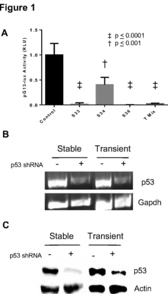
In order to create a stable knockdown of p5γ, we isolated puromycin resistant single cell clones after transfecting various p5γ shRNA constructs in a plasmid conferring puromycin resistance into CβC1β cells. Individual clones were expanded and characterized. The same constructs were also used for transient reduction of p5γ, but no selection was performed and cells were harvested after 48 hours for RNA and protein. Our intention was to generate cells with a similar knockdown in p5γ using the same shRNA construct. In order to rule out possible dosage dependent effects between and among the different constructs and type of tranfections, several methods were used to assess the knockdown Therefore in addition to determining extent of p5γ knockdown at the transcriptional and translational level, we tested the level of p5γ functional activity using a construct containing 1γ copies of a p5γ binding sequence [19]. This construct was transiently transfected into stable p5γ knockdown lines we had generated and p5γ functional activity was measured 48 hours later. In the case involving transient transfections both constructs (reporter and shRNA) were transfected together. As seen in Figure 1A, different levels of knockdown of p5γ activity was achieved with the different p5γ shRNA constructs when compared to cells receiving a non-specific scrambled sequence. Among the stable lines two out of three lines showed greater knockdown of p5γ activity. A mixture of all three shRNAs also produced an effective knockdown. As clone γγ and clone γ6 demonstrated effective knockdown of p5γ function we further proceeded to use the same shRNA constructs to transiently reduce p5γ in CβC1β cells. A representative RT-PCR of a stable clone γγ line is shown compared to stable control created with a scrambled sequence. Similarly using the same constructs in transient transfections we show a similar knockdown of p5γ (Figure 1A
lanes γ and 4). In Figure 1C we show representative results from use of a different shRNA that was used to create clone γ6. In all these cases we obtained knockdown of 65-90% of endogenous p5γ levels after analyses of band intensities. More importantly we chose to further analyze samples where the extent of knockdown in stable and transient cultures was comparable using the same shRNA sequence.
Realtime PCR arrays of p53 target genes show differences between stable and transient loss of p53
In order to look for more global differences between acute loss, from the transient knockdown, and a long term loss of p5γ the SABioscience Q-PCR array was used to assay the changes in genes related to p5γ. We chose to compare RNA from stable and transient knockdown created with the shRNA (Sγ6 vs. Tγ6). RNA from both these cells was first compared to control cells receiving a scrambled shRNA sequence. As expected, many of the targets showed changes with loss of p5γ expression. Hierarchical clustering of the expression changes between the two lines are shown as compared to control cells. Two types of change were observed one where genes are up-regulated in the acute loss compared to the long term loss of p5γ, and a second cluster with genes up-regulated in long term p5γ loss when compared to the acute loss. (Figure βA). As expected, when compared to control, the genes that are anti-apoptotic are down-regulated with transient loss of p5γ (Caspβ, Casp9, Cradd, Fasl, Bag1, etc.) (Figure βB). In contrast, these genes are slightly up-regulated when p5γ is knocked down stably. Similarly, the genes classified as differentiation related (Btgβ, Jun, MyoD, Numb, Sfn, and Wt1), with the exception of MyoD, are all up-regulated when p5γ was knocked down transiently. In stable p5γ knockdown lines they show variable expression (Figure βB). This may relate to the fact that some of them are tissue specific with little relevance to osteoblasts or myocytes.
Dosage of p53 does not alter target gene expression in stable p53 knockdown cells
Some of our previous studies demonstrated that the amount of p5γ within osteoblasts might affect cell proliferation and differentiation variably [β6-β8]. Since one of our stable clones (clone γγ) showed a smaller knockdown of p5γ activity (~50%) we compared this line to our stable line γ6 which has a greater knockdown of p5γ activity (~ 80%) to see if the difference in p5γ level affected its target genes is a dissimilar manner in CβC1β cells. A duplicate gene array to the one described in Figure βA was done using a control and the two stable p5γ knockdown lines. Our results showed very little if any variation between the two stable lines with differing dosage (Figure βC). This suggests that irrespective of the dosage, stable loss of p5γ affects target genes differently from that produced by transient p5γ loss.
Validation of array results with realtime PCR
Transient knockdown of p5γ was also carried out in triplicate. RNA from these newly generated cell lines were compared to results obtained from the gene array. As shown in Figure γ, the quantitative PCR results agreed with the array results in the direction of the change in expression. As expected of
Figure 1. Confirmation of stable and transient knockdown of p53. (A) Transcriptional activity of p5γ is reduced with reduction in p5γ levels Mean + S.E.M. of relative luciferase units (RLU) reported for stable (S) knockdown of p5γ using shRNA plasmids and a transient knockdown using a mix of all three plasmids (T Mix) when compared to controls receiving scrambled shRNA sequences. (B) RT-PCR of p5γ from a stable knockdown (Sγγ), a transient knockdown (Tγγ) and control (scrambled shRNA). (C) Immunoblot of p5γ expression in a stable (Sγ6) line and after transient knockdown of p5γ (Tγ6). The same blot was stripped and reprobed with anti-actin antibody. Similar results were obtained in three independent experiments and representative data are shown.
doi: 10.1γ71/journal.pone.008β494.g001
independent samples there was a variation in the extent of change between samples, but most importantly the directionality of the change was maintained for all of the genes tested, thus validating the results of the array. The variation seen in the case of some of the genes may relate to differences in the level of p5γ knockdown as three independent clones/transfections were compared to the array. Additionally they may represent changes in steady state levels of expression dependent on the status of the cells at the time of RNA isolation.
Developmental pathway markers differ in their responses to acute and stable p53 loss
In addition to the expression data, we wanted to determine if specific pathways related to differentiation are activated differently dependent on whether p5γ loss was stable or transient. The notch pathway is known to be important for tissue differentiation and has been shown to be important for both myoblast and osteoblast differentiation [β9,γ0]. When the differences in the promoter activity of a direct target in notch signaling, Hey1 [γ1],between stable and transient p5γ knockdowns were compared to control cells, there was a dramatic reduction, regardless of the type of transfection (p < 0.001; Figure 4A). We also tested the change of activity of vitamin D receptor which is important for both myoblast and osteoblast differentiation [γβ,γγ]. In contrast to the Hey1 reporter, the vitamin D pathway showed a different response depending on the length of p5γ knockdown. A stable reduction in p5γ resulted in a non-significant increase seen (p > 0.05; Figure 4B), whereas, an acute loss of p5γ resulted in a significant decrease in activity of the vitamin D receptor promoter (p < 0.05; Figure 4B).
Since the CβC1β cells can differentiate into either myocytes or osteoblasts, we tested the promoter activity of the genes Muscle Creatine Kinase (MCK), a late marker of myogenic differentiation, and osteocalcin, a late marker of osteoblast differentiation. When p5γ is stably reduced the loss results in a significant decrease in activity of the osteocalcin promoter (p < 0.001; Figure 5A). With transient loss, osteocalcin promoter activity is increased (p < 0.001; Figure 5B). In contrast to osteocalcin, the MCK promoter did not change after stable loss of p5γ when compared to control. (p > 0.05; Figure 5C). However, like osteocalcin, the promoter activity of MCK was also elevated initially by the knockdown of p5γ (p < 0.01; Figure 5D). This is in agreement with our previous work demonstrating a requirement of p5γ function for expression of the osteocalcin gene [8].
Discussion
complete loss of p5γ results in normal bone development and increased osteoblast differentiation [β6]. We have previously shown that in the presence of reduced dosage of wild type p5γ,
osteoblast differentiation is p5γ-dependent [β6]. This suggests that redundant pathways may exist, but in the presence of at least partial amounts of p5γ, the dosage of p5γ is a critical
Figure 2. Gene expression o f p53 target genes after p53 loss. (A) Heat map generated by centroid linkage of the Euclidian distance of mean centered fold change values of transient transfection of shRNA plasmid γ6 (Tγ6) and stable line created with plasmid γ6 (Sγ6) using a control line (created with scrambled shRNA) as a common divisor, β-((Tγ6-Normalizers) – Control) compared to β -((Sγ6-Normalizers) – Control), (n = 1). The negative inverse used for fold changes between 0 and 1. Clusters were then divided into two categories,
those with higher expression in Tγ6 and those with higher expression in Sγ6. (B) Fold change values plotted for genes involved in apoptosis or differentiation with a fold change between treated and control cells greater than two (C) Comparison of stable clones of varying p5γ expression. Plot of the same gens as in (B) but between Sγ6 and a stable line created with clone γ4 (Sγ4) show stable p5γ knockdown irrespective of the level of knockdown, have a much more similar expression pattern than seen between transient and stable.
determinant of osteoblast function. However, targeted knockout of p5γ in bone has consistently shown this gene to have an inhibitory effect on osteoblast proliferation and differentiation [γ4]. In our studies we have used committed osteoblasts to demonstrate that osteocalcin, a bone matrix protein expressed in advanced osteoblast differentiation, is only produced when p5γ is present [β7,β8]. We have also shown the osteocalcin gene is a direct transcriptional target of p5γ [8]. Osteoblast differentiation progresses through stages of proliferation and expression of several matrix proteins followed by quiescence and expression of osteocalcin and generation of a mineralized matrix [γ5]. Osteoblasts arise from common mesenchymal progenitors and progress through commitment to the osteoblast lineage to preosteoblasts and osteoblasts. It is therefore possible that p5γ has distinct stage specific roles during development and differentiation. In the context of osteosarcomas, p5γ is generally absent due to a p5γ gene rearrangement [γ6] and replacement of wild type p5γ in these cells is associated with inhibition of tumor growth and the appearance of a calcified matrix [β8]. Osteosarcoma tumors also generally do not express osteocalcin [γ7]. Since bone remodels through life, it is possible that the presence of p5γ is important to maintain homeostasis in post natal bone. This suggests that p5γ may influence the environment within bone to allow orderly differentiation in addition to maintaining a balance of cell proliferation and cell death.
In an attempt to separate the different roles of p5γ we studied the short and long term effect of p5γ loss on its target genes. In this study we utilized CβC1β, a bipotential cell line capable of differentiation towards myoblast and osteoblast, to determine the effect of transient and stable knockdown of p5γ using RNA interference. An acute loss of p5γ lowered the mRNA expression for a number of genes related to apoptosis. This is expected, as one of the major roles of p5γ is in
regulating apoptosis, a function that represents an immediate response to prevent damaged DNA and or cells from survival. However, these same genes show a small increase in expression after long term loss of p5γ; perhaps as an adaptation to reduced p5γ levels. Specifically, members of the caspase family of genes, Caspβ and Casp9, as well as the Caspβ assembly protein Cradd, are all down-regulated with an acute loss of p5γ, whereas they appear to be up-regulated when p5γ is reduced for the long term. It is also possible that the change seen in caspases with stable p5γ loss may reflect other functions unrelated to apoptosis as has been seen for some cell types [γ8-40]. Similarly, the expression of Prkca was also greatly reduced in the transient lines and elevated in the stable lines. While Prkca function in apoptosis is well understood [41] and is represented as an inducer of apoptosis in this gene array (http://www.sabiosciences.com/ rt_pcr_product/HTML/PAMM-0β7A.html), several roles have been attributed to Prkca and its class of proteins in signaling and cell proliferation [41]. Therefore, the response seen after stable p5γ loss may be symbolic of change in growth control. Irrespective of the reasons, the differences in responses appear to be consistent with the type of p5γ loss.
The apoptotic pathway is not the only pathway that appears to differ according to the length of p5γ knockdown. Genes involved in p5γ related differentiation pathways varied in expression based on the type of p5γ loss. MyoD and Numb are well known differentiation and cell fate related genes [4β,4γ]. Validation of these genes with realtime PCR show that these changes are consistent with the type of p5γ loss. Our array data show Btgβ, Wt1, MyoD, Jun, and Numb all have a higher expression in an acute loss of p5γ. This may reflect a generalized stress response produced as a result of a sudden decrease in p5γ expression. Numb is a repressor of the Notch signaling pathway [44], and its elevation after transient loss of
Figure 3. Array Confirmation. Confirmation of targets from gene array by Quantitative PCR verifies array expression values. QPCR of selected genes that showed variation in expression in arrays were selected and tested with three independently isolated single cell clones using the same shRNA as the array(Sγ6), Transient transfections were carried out in triplicate with the same shRNA , and control cells received the scrambled control plasmid (mean + S.E.M., n = γ) for verification. For each gene, the change in expression matched that of the array in both types of transfections.
p5γ may relate to p5γ’s role as an enhancer of notch signaling [45]. However, under conditions of stable loss such an effect is absent and may reflect other complementary roles of Numb in regulating p5γ function [46] MyoD has the largest decrease after stable loss of p5γ, consistent with evidence that it is a p5γ regulated gene [47] and myogenesis represents the default pathway in CβC1β cells [17]. While the changes in the other genes are difficult to extrapolate to CβC1β differentiation, it is clear that these differentiation associated genes are affected markedly only after chronic loss of p5γ suggesting that the
Figure 4. Activities of Notch and Vitamin D receptor pathways after p53 loss. (A) Hey1-luc reporter shows a significant reduction in Notch signaling activity of Sγγ, Sγ4, and Sγ6 when compared to controls. Similar results are seen for T Mix when compared to control. (B) Vitamin D receptor reporter plasmid shows non-significant changes with long term p5γ reduction. However, a loss was observed in T Mix cells when compared to control. Three novel clones (n = γ) were measured in triplicate, and the mean of each triplicate was used to calculate the presented mean ± S.E.M. for each group.
doi: 10.1γ71/journal.pone.008β494.g004
deficiency of p5γ has brought about a change in the environment within these cells.
We studied lineage specific, as well as, lineage independent signaling pathways that might be affected by the knockdown of p5γ in CβC1β cells. We investigated activity of the Notch pathway and did not find differences between stable and transient knockdown as in both cases this pathway was depressed with loss of p5γ. In a genome wide study carried out to determine p5γ regulators, the notch pathway, especially Hey1, was found to be members of an evolutionarily conserved network governing p5γ function [48]. It was interesting to note that our studies show a mutual regulation with loss of p5γ affecting the Notch pathway. In the case of vitamin D receptor activity, while a significant effect was not seen with chronic loss of p5γ, a transient loss produced a reduction in the receptor’s
Figure 5. Bone and muscle differentiation markers show different patterns based on the type of transfection. (A) Osteocalcin gene promoter shows loss of activity between Sγ6 and control cells created with a scrambled shRNA sequence. (B) Conversely, in T Mix samples, an increase in the activity on the osteocalcin promoter when compared with control cells receiving scrambled shRNA. (C) Muscle creatine kinase (MCK) gene promoter activity, showed no difference in activity when Sγ6 is compared to control cells. (D) Promoter activity increased in T Mix when compared to control cells. Three novel clones (n = γ) were measured in triplicate each, using the mean to calculate the presented mean ± S.E.M. for each group.
function. Vitamin D and its receptor are known to have potent genomic as well as non-genomic action in myocytes and on calcium and phosphate metabolism [49]. Our results probably represent these complex pathways that are affected with p5γ loss. We also studied activities of two genes of advanced differentiation in CβC1β cells, one for each of the two common lineages. MCK is seen late during myocyte differentiation and osteocalcin is a late marker of bone differentiation. One of the immediate changes after loss of p5γ, as measured with the transient samples, was an up-regulation in activity of these genes. However, after a long term loss of p5γ only osteocalcin activity was decreased. This is consistent with other studies that have shown p5γ to be necessary for osteogenic reprogramming of skeletal muscle committed cells [11].
These studies indicate that while the role of p5γ is important to protect cells from damage by regulating key apoptotic genes, p5γ has an additional role in maintaining tissue homeostasis. This may be especially important in osteoblasts since bone remodels throughout life. The presence of p5γ may be critical to allow for orderly progression of the different steps of the differentiation process. p5γ is a direct regulator of matrix proteins osteopontin and osteocalcin [8,50]. Osteocalcin
expression coincides with a decrease in proliferation and matrix deposition and the initiation of the mineralization process. Loss of p5γ in tumors may therefore produce a constitutive proliferative state with the lack of osteocalcin, a feature commonly seen in osteosarcomas. This study shows a distinct difference in expression of differentiation markers with long term loss of p5γ may result from the loss of ability of p5γ to maintain the balance between regulatory factors, and or acutely sense and modify the environment for orderly differentiation.
Acknowledgements
We thank Drs. B. Vogelstein, M. Gessler, P. Hauschka and P. Chambon for their kind gift of reagents.
Author Contributions
Conceived and designed the experiments: NC. Performed the experiments: OC KD EL EH. Analyzed the data: OC NC. Wrote the manuscript: NC OC.
References
1. Bai L, Zhu W-G (β006) p5γ: Structure, Function and Therapeutic Applications. J Cancer Mol β: 1β.
β. Ryan KM, Phillips AC, Vousden KH (β001) Regulation and function of the p5γ tumor suppressor protein. Curr Opin Cell Biol 1γ: γγβ-γγ7. doi: 10.1016/S0955-0674(00)00β16-7. PubMed: 11γ4γ904.
γ. Solá S, Morgado AL, Rodrigues CM (β01γ) Death receptors and mitochondria: two prime triggers of neural apoptosis and differentiation. Biochim Biophys Acta 18γ0: β160-β166. doi:10.1016/j.bbagen. β01β.09.0β1. PubMed: βγ041071.
4. Freed-Pastor WA, Prives C (β01β) Mutant p5γ: one name, many proteins. Genes Dev β6: 1β68-1β86. doi:10.1101/gad.190678.11β. PubMed: ββ71γ868.
5. Smith ND, Rubenstein JN, Eggener SE, Kozlowski JM (β00γ) The p5γ tumor suppressor gene and nuclear protein: basic science review and relevance in the management of bladder cancer. J Urol 169: 1β19-1ββ8. doi:10.1097/01.ju.0000056085.58ββ1.80. PubMed: 1β6β9γγβ.
6. Molchadsky A, Rivlin N, Brosh R, Rotter V, Sarig R (β010) p5γ is balancing development, differentiation and de-differentiation to assure cancer prevention. Carcinogenesis γ1: 1501-1508. doi:10.109γ/carcin/ bgq101. PubMed: β0504879.
7. Menendez S, Camus S, Izpisua Belmonte JC (β010) p5γ: guardian of reprogramming. Cell Cycle 9: γ887-γ891. doi:10.4161/cc.9.19.1γγ01. PubMed: β0948β96.
8. Chen H, Hays E, Liboon J, Neely C, Kolman K et al. (β011) Osteocalcin gene expression is regulated by wild-type p5γ. Calcif Tissue Int 89: 411-418. doi:10.1007/s00ββγ-011-95γγ-x. PubMed: β19649γ0. 9. Tamir Y, Bengal E (1998) p5γ protein is activated during muscle
differentiation and participates with MyoD in the transcription of muscle creatine kinase gene. Oncogene 17: γ47-γ56. doi:10.10γ8/sj.onc. 1β019β9. PubMed: 9690516.
10. Cam H, Griesmann H, Beitzinger M, Hofmann L, Beinoraviciute-Kellner R et al. (β006) p5γ family members in myogenic differentiation and rhabdomyosarcoma development. Cancer Cell 10: β81-β9γ. doi: 10.1016/j.ccr.β006.08.0β4. PubMed: 17045β06.
11. Molchadsky A, Shats I, Goldfinger N, Pevsner-Fischer M, Olson M et al. (β008) p5γ plays a role in mesenchymal differentiation programs, in a cell fate dependent manner. PLOS ONE γ: eγ707. doi:10.1γ71/ journal.pone.000γ707. PubMed: 1900ββ60.
1β. Solozobova V, Blattner C (β011) p5γ in stem cells. World J Biol Chem β: β0β-β14. doi:10.4γγ1/wjbc.vβ.i9.β0β. PubMed: β1949570.
1γ. Donehower LA, Harvey M, Slagle BL, McArthur MJ, Montgomery CA et al. (199β) Mice deficient for p5γ are developmentally normal but susceptible to spontaneous tumours. Nature γ56: β15-ββ1. doi: 10.10γ8/γ56β15a0. PubMed: 155β940.
14. Donehower LA, Harvey M, Vogel H, McArthur MJ, Montgomery CA et al. (1995) Effects of genetic background on tumorigenesis in p5γ-deficient mice. Mol Carcinog 14: 16-ββ. doi:10.100β/mc.β940140105. PubMed: 7546β19.
15. Sah VP, Attardi LD, Mulligan GJ, Williams BO, Bronson RT et al. (1995) A subset of p5γ-deficient embryos exhibit exencephaly. Nat Genet 10: 175-180. doi:10.10γ8/ng0695-175. PubMed: 766γ51β. 16. Scott MC, Sarver AL, Gavin KJ, Thayanithy V, Getzy DM et al. (β011)
Molecular subtypes of osteosarcoma identified by reducing tumor heterogeneity through an interspecies comparative approach. Bone 49: γ56-γ67. doi:10.1016/j.bone.β011.05.008. PubMed: β16β1658. 17. Yaffe D, Saxel O (1977) Serial passaging and differentiation of
myogenic cells isolated from dystrophic mouse muscle. Nature β70: 7β5-7β7. doi:10.10γ8/β707β5a0. PubMed: 56γ5β4.
18. Katagiri T, Yamaguchi A, Komaki M, Abe E, Takahashi N et al. (1994) Bone morphogenetic protein-β converts the differentiation pathway of CβC1β myoblasts into the osteoblast lineage. J Cell Biol 1β7: 1755-1766. doi:10.108γ/jcb.1β7.6.1755. PubMed: 7798γβ4.
19. Kern SE, Kinzler KW, Bruskin A, Jarosz D, Friedman P et al. (1991) Identification of p5γ as a sequence-specific DNA-binding protein. Science β5β: 1708-1711. doi:10.11β6/science.β047879. PubMed: β047879.
β0. Maier MM, Gessler M (β000) Comparative analysis of the human and mouse Hey1 promoter: Hey genes are new Notch target genes. Biochem Biophys Res Commun β75: 65β-660. doi:10.1006/bbrc. β000.γγ54. PubMed: 10964718.
β1. Klein-Hitpass L, Schorpp M, Wagner U, Ryffel GU (1986) An estrogen-responsive element derived from the 5' flanking region of the Xenopus vitellogenin Aβ gene functions in transfected human cells. Cell 46: 105γ-1061. doi:10.1016/009β-8674(86)90705-1. PubMed: γ46γ4γγ. ββ. Morrison NA, Shine J, Fragonas JC, Verkest V, McMenemy ML et al.
(1989) 1,β5-dihydroxyvitamin D-responsive element and glucocorticoid repression in the osteocalcin gene. Science β46: 1158-1161. doi: 10.11β6/science.β588000. PubMed: β588000.
βγ. Jaynes JB, Johnson JE, Buskin JN, Gartside CL, Hauschka SD (1988) The muscle creatine kinase gene is regulated by multiple upstream elements, including a muscle-specific enhancer. Mol Cell Biol 8: 6β-70. PubMed: γγγ6γ66.
β4. de Hoon MJ, Imoto S, Nolan J, Miyano S (β004) Open source clustering software. Bioinformatics β0: 145γ-1454. doi:10.109γ/ bioinformatics/bth078. PubMed: 14871861.
β6. Chandar N, Donehower L, Lanciloti N (β000) Reduction in p5γ gene dosage diminishes differentiation capacity of osteoblasts. Anticancer Res β0: β55γ-β559. PubMed: 1095γγβ6.
β7. Chandar N, Campbell P, Novak J, Smith M (199γ) Dependence of induction of osteocalcin gene expression on the presence of wild-type p5γ in a murine osteosarcoma cell line. Mol Carcinog 8: β99-γ05. doi: 10.100β/mc.β94008041γ. PubMed: 8β80γ78.
β8. Chandar N, Swindle J, Szajkovics A, Kolman K (β005) Relationship of bone morphogenetic protein expression during osteoblast differentiation to wild type p5γ. J Orthop Res βγ: 1γ45-1γ5γ. doi: 10.1016/j.orthres.β005.04.010.1100βγ0616. PubMed: 15994055. β9. Engin F, Lee B (β010) NOTCHing the bone: insights into
multi-functionality. Bone 46: β74-β80. doi:10.1016/j.bone.β009.05.0β7. PubMed: 195β0195.
γ0. Mourikis P, Gopalakrishnan S, Sambasivan R, Tajbakhsh S (β01β) Cell-autonomous Notch activity maintains the temporal specification potential of skeletal muscle stem cells. Development 1γ9: 45γ6-4548. doi:10.1β4β/dev.084756. PubMed: βγ1γ6γ94.
γ1. Fischer A, Schumacher N, Maier M, Sendtner M, Gessler M (β004) The Notch target genes Hey1 and Heyβ are required for embryonic vascular development. Genes Dev 18: 901-911. doi:10.1101/gad.β91004. PubMed: 1510740γ.
γβ. Tanaka M, Kishimoto KN, Okuno H, Saito H, Itoi E (β01γ) Vitamin D receptor gene silencing effects on differentiation of myogenic cell lines. Muscle Nerve: ([MedlinePgn:]) PubMed: βγ87γγ55.
γγ. Kommagani R, Whitlatch A, Leonard MK, Kadakia MP (β010) p7γ is essential for vitamin D-mediated osteoblastic differentiation. Cell Death Differ 17: γ98-407. doi:10.10γ8/cdd.β009.1γ5. PubMed: 19779497. γ4. Wang X, Kua HY, Hu Y, Guo K, Zeng Q et al. (β006) p5γ functions as a
negative regulator of osteoblastogenesis, osteoblast-dependent osteoclastogenesis, and bone remodeling. J Cell Biol 17β: 115-1β5. doi:10.108γ/jcb.β00507106. PubMed: 16γ804γ7.
γ5. Lian JB, Stein GS (199γ) The developmental stages of osteoblast growth and differentiation exhibit selective responses of genes to growth factors (TGF beta 1) and hormones (vitamin D and glucocorticoids). J Oral Implantol 19: 105-1γ6; discussion:
γ6. Chandar N, Billig B, McMaster J, Novak J (199β) Inactivation of p5γ gene in human and murine osteosarcoma cells. Br J Cancer 65: β08-β14. doi:10.10γ8/bjc.199β.4γ. PubMed: 17γ9619.
γ7. Ambroszkiewicz J, Gajewska J, Klepacka T, Chełchowska M, Laskowska-Klita T et al. (β010) Clinical utility of biochemical bone
turnover markers in children and adolescents with osteosarcoma. Adv Med Sci 55: β66-β7β. PubMed: β1084β54.
γ8. Fernando P, Kelly JF, Balazsi K, Slack RS, Megeney LA (β00β) Caspase γ activity is required for skeletal muscle differentiation. Proc Natl Acad Sci U S A 99: 110β5-110γ0. doi:10.107γ/pnas.16β17β899. PubMed: 1β1774β0.
γ9. Miura M, Chen XD, Allen MR, Bi Y, Gronthos S et al. (β004) A crucial role of caspase-γ in osteogenic differentiation of bone marrow stromal stem cells. J Clin Invest 114: 1704-171γ. doi:10.117β/JCIβ004β04β7. PubMed: 15599γ95.
40. Lamkanfi M, Festjens N, Declercq W, Vanden Berghe T, Vandenabeele P (β007) Caspases in cell survival, proliferation and differentiation. Cell Death Differ 14: 44-55. doi:10.10γ8/sj.cdd.440β047. PubMed: 1705γ807.
41. Nakashima S (β00β) Protein kinase C alpha (PKC alpha): regulation and biological function. J Biochem 1γβ: 669-675. doi:10.109γ/ oxfordjournals.jbchem.a00γβ7β. PubMed: 1β417014.
4β. Yokoyama S, Asahara H (β011) The myogenic transcriptional network. Cell Mol Life Sci 68: 184γ-1849. doi:10.1007/s00018-011-06β9-β. PubMed: β1γ18β6γ.
4γ. Gulino A, Di Marcotullio L, Screpanti I (β010) The multiple functions of Numb. Exp Cell Res γ16: 900-906. doi:10.1016/j.yexcr.β009.11.017. PubMed: 19944684.
44. Katoh M (β006) NUMB is a break of WNT-Notch signaling cycle. Int J Mol Med 18: 517-5β1. PubMed: 16865βγ9.
45. Roemer K (β01β) Notch and the p5γ clan of transcription factors. Adv Exp Med Biol 7β7: ββγ-β40. doi:10.1007/978-1-4614-0899-4_17. PubMed: ββγ99γ51.
46. Colaluca IN, Tosoni D, Nuciforo P, Senic-Matuglia F, Galimberti V et al. (β008) NUMB controls p5γ tumour suppressor activity. Nature 451: 76-80. doi:10.10γ8/nature0641β. PubMed: 1817β499.
47. Puri PL, Bhakta K, Wood LD, Costanzo A, Zhu J et al. (β00β) A myogenic differentiation checkpoint activated by genotoxic stress. Nat Genet γβ: 585-59γ. doi:10.10γ8/ng10βγ. PubMed: 1β415β71. 48. Huang Q, Raya A, DeJesus P, Chao SH, Quon KC et al. (β004)
Identification of p5γ regulators by genome-wide functional analysis. Proc Natl Acad Sci U S A 101: γ456-γ461. doi:10.107γ/pnas. 0γ0856β100. PubMed: 14990790.
49. Ceglia L (β008) Vitamin D and skeletal muscle tissue and function. Mol Aspects Med β9: 407-414. doi:10.1016/j.mam.β008.07.00β. PubMed: 187β79γ6.