Expression of p53, p16 and Ki67 proteins in ductal
adenocarcinoma of the pancreatic head and their relation
with survival and cell differentiation
Expressão das proteínas p53, p16 e ki67 no adenocarcinoma da cabeça do pâncreas e sua
relação com a sobrevida e diferenciação celular
Mário Benjamin Goitia-Durán1, Marcelo Moura Linhares2, Ricardo Artigiani Neto3, Franz Robert Apodaca-Torrez4, Edson José Lobo5, Alberto Goldenberg6
ABSTRACT
Objective: To determine the expression of p53, p16 and Ki-67 and its relevance in survival and cell differentiation. Methods:
Fifteen duodenopancreatectomized patients were included. Immunohistochemical expression of p53, p16 and Ki-67 was determined in paraffin embedded tumor blocks. The relation of these expressions with different variables was studied. Results: Ninety-three per cent of tumors showed expression of p53 and p16. Ki-67 was expressed in 86.66% of tumors (labeling index – LI 11.91
± 9.47). The presence of combined alterations was not related to significant differences in tumor type, stage or survival; similar results were obtained analyzing isolated expressions. When groups of p16 and Ki-67 expressions where created, the median survival was not significant. However, there was a slightly better survival in patients with focal expression of p16 (median survival 20.75 versus 14.34),
when compared to patients with diffuse expression. Conclusion: The overexpression of p53, p16 and Ki-67 was not related to survival or tumor grade, when comparing isolated or combined expressions.
Keywords: Tumor suppressor proteins/analysis; Cell cycle proteins/ analysis; Ki-67 antigen/analysis; Tumor suppressor protein p53; Pancreatic neoplasms; Survivorship
RESUMO
Objetivo: Determinar a expressão de p53, p16 e Ki-67 e sua relevância na sobrevida e diferenciação celular. Métodos: Foram incluídos 15 pacientes submetidos a duodenopancreatectomia. A expressão imunohistoquímica
de p53, p16 e Ki-67 foi determinada em blocos tumorais embebidos em parafina. Foi estudada a relação dessas expressões com as variáveis.
Resultados: Noventa e três por cento dos tumores apresentaram expressão de p53 e p16. Ki-67 estava expresso em 86,66% dos tumores (índice proliferativo – IP 11,91 ± 9,47). A presença de alterações combinadas não estava relacionada a diferenças significativas no tipo tumoral, no estágio ou na sobrevida; resultados semelhantes foram obtidos com a análise de expressões isoladas. Quando foram criados os grupos de expressões de p16 e Ki-67, a sobrevida mediana não era significativa. Entretanto, havia uma sobrevida discretamente melhor nos pacientes com expressão focal do p16 (sobrevida mediana 20,75
versus 14,34) em comparação com pacientes com expressão difusa.
Conclusões: A superexpressão das proteínas p53, p16 e Ki-67 não estavam relacionadas à sobrevida ou ao grau tumoral quando se compararam as expressões isoladas ou combinadas.
Descritores: Proteínas supressoras de tumor/análise; Proteínas de ciclo celular/análise; Antígeno KI-67/análise; Proteína supressora de tumor p53; Neoplasias pancreáticas; Sobrevida
INTRODUCTION
Pancreatic cancer (PC) is one of the deadliest cancers. Its incidence and mortality are similar and it represents a Public Health problem in Western countries(1-3).
Most of the time late diagnosis contributes to poor prognosis and precludes adequate surgical treatment(4).
Understanding carcinogenesis and its relation with
Study carried out at the Department of Surgical Gastroenterolgy of the Universidade Federal de São Paulo – UNIFESP, São Paulo (SP), Brazil. 1 MD; Master in Sciences at the Universidade Federal de São Paulo – UNIFESP, São Paulo (SP), Brazil.
2 PhD in Sciences at the Universidade Federal de São Paulo – UNIFESP, São Paulo (SP), Brazil.
3 PhD in Sciences at the Universidade Federal de São Paulo – UNIFESP, São Paulo (SP), Brazil.
4 PhD in Sciences at the Universidade Federal de São Paulo – UNIFESP, São Paulo (SP), Brazil.
5 Professor; Head of the Biliary Tract and Pancreas Group at the Universidade Federal de São Paulo – UNIFESP, São Paulo (SP), Brazil.
6 PhD; Adjunct Professor of Surgical Gastroenterolgy at the Universidade Federal de São Paulo – UNIFESP, São Paulo (SP), Brazil.
Corresponding author:Mário Benjamin Goitia-Durán – E-mail: [email protected]
clinicopathological behavior and possible prognostic value is a challenge for many researchers(5-8).
An association between mutation of K-ras(9-11),
p53(12,13), and p16(14-16) (the genes most frequently mutated
in pancreatic cancer) and survival of patients was described. Tumor supressor genes, such as p53 and p16, play a role in regulating cell cycle and tumor progression. The first, located at 17p13, encodes a 53 KDa and 393 amino acid protein that accumulates in the cell when the gene is mutated, due to stabilization. This stable protein is easily detected by immunohistochemistry (IH)(17).
Similarly, p16 gene, located at 9p21, when inactivated by deletion or hypermethylation precludes cell cycle regulation by inhibition of cyclin-cdk complexes that negatively regulate protein Rb1(16). The p16 protein,
similarly to p53, is easily detected by IH(18,19). This
expression detection reflects gene mutation or alteration in approximately 85% of the samples (20).
Besides suppressor genes, some other markers were used to determine aggressiveness and differentiation of the tumor(21,22). A commonly used marker is Ki-67, which
was discovered at Kiev University, Ukraine. This 345-395 KDa protein is expressed in every proliferating tissue and in all cellular phases but G0, due to phosphorilation processes. Since its discovery in 1979, it has been widely used to determine tumor growth patterns and cell differentiation. This enabled researchers to use it as a prognostic factor instead of a predictive only(21,23).
OBJECTIVE
To determine the expression of p53, p16 and Ki-67 and its relevance in survival and cell differentiation and compare the results of combined and isolated immunostaining for each marker.
METHODS
Patients and samples
Fifteen cases of PC – all ductal adenocarcinomas of pancreatic head – were studied. All patients were seen and operated on at Surgical Gastroenterology Department of the Universidade Federal de São Paulo (UNIFESP-EPM), in São Paulo, Brazil, from 1993 to 2003. The sample consisted of 45 routinely neutral-formalin fixed and paraffin-embedded blocks strictly containing tumor tissue, and there were three for each patient. The subjects were 11 males (73.33%) and 4 females (26.66%) with a median age of 56 years (range of 45-66 years). According to the Union for International Cancer Control (UICC), 40% (6 patients) had IIB stage, 33.33% (5 patients) had IIA stage, and 13.33% (2 patients) had IB and III stages each. Tumors were graded and 6 were classified as well
differentiated while 9 as moderately differentiated. By the end of the study, 2 patients were still alive with 34 and 119 months of survival each.
Antibodies and immunohistochemical staining
The primary antibodies were anti-p53 DO-7 (DakoCytomation, Glostrup, Denmark) against human p53, anti-p16 clone Ab7 16PO7 (Neomarkers, Fremont, CA, USA) against p16, and anti-Ki67 clone MIB-1 (Immunotech, Marseille, France) against Ki-67 antigen. Immunohistochemistry was performed according to Hsu et al.(24). Briefly, 4-μm thick sections were deparaffined
and incubated with primary antibodies against human proteins and antigen was diluted to 1:20 for p53, 1:100 for p16, and 1:80 for Ki-67, at 4°C, overnight. A microwave irradiation procedure was applied for antigen retrieval. Immunolocalization was performed using the Streptavidin Biotin Complex IHRP; Duet, Mouse/Rabbit (DakoCytomation, Glostrup, Denmark) kit. The color of the reactions was developed using 3-3’diaminobenzidine (DAB) (SIGMA Chemical Co., St. Louis, MO, USA) and counterstaining was carried out with Harris hematoxylin. For positive controls sections of colon adenocarcinoma for p53, sections of cervical cancer for p16, and sections of olidendroglioma for Ki-67 were included. For negative controls sections of normal pancreatic tissue were included. Positive staining included nuclear and cytoplasmic staining for p53 of more than 10% of target cells; for p16, more than 5% and any immunostained nuclei, regardless of intensity, for Ki-67 and expressed for labeling index (LI).
Statistical analysis
The data were analyzed by Fisher’s exact test (χ2)
to compare proportions of isolated and combined staining. A cutoff was used in immunostaining of p16 and Ki-67 of 25 and 15% of the stained cells, respectively. Overall survival estimates were obtained according to the actuarial method of Kaplan-Meyer(25).
Survival curves below and above the chosen cutoff were plotted monthly and compared by log rank. Finally, to determine significance of the risk factor, Cox proportional univariate analysis(26) was used. Statistical
tests were paired and a p value > 0.05 was determined. Statistical software SAS version 8.02 (SAS Institute, Cary, NC, USA) was used.
RESULTS
According to TNM stage and cutoff point, expression data are shown in table 2.
25% of cells was 14.34 (95%CI: 5.39-18.46) compared to expression below 25% that was 20.75 (95%CI: 9.07-24.45), log rank p = 0.088 and Hazard Ratio (HR) 2.821 (95%CI: 0.817-9.740) (Table 3).
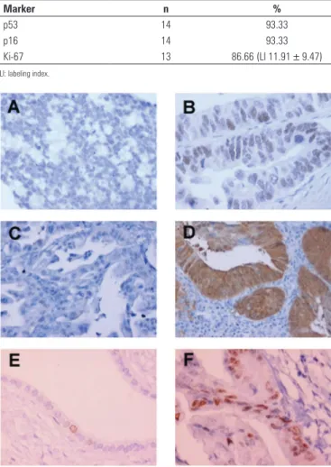
Marker n %
p53 14 93.33
p16 14 93.33
Ki-67 13 86.66 (LI 11.91 ± 9.47)
Table 1. Overall immunostaining
LI: labeling index.
Table 2. Immunostaining and TNM stage
Stage Total (%) p53 p16 Ki-67
< 25% > 25% < 15% > 15%
IB 2 (13.3) 2 1 1 2 0
IIA 5 (33.3) 5 1 4 2 3
IIB 6 (40.0) 5 2 3 2 2
III 2 (13.3) 2 2 0 1 1
Total 15 (100) 14 (93.3) 6 (42.9) 8 (57.1) 7 (53.8) 6 (46.2)
Survival (months) p-value
log rank Hazard Ratio (HR)
p53 15.7 (4-34) NS –
p16 > 25% 14.34 (95%CI: 5.39-18.46) p = 0.088 2.821 (95%CI: 0.817-9.740)
p16 < 25% 20.75 (95%CI: 9.07-24.45)
Ki-67 > 15% 17.46 (95%CI: 5.39-27.56) p = 0.1955 0.419 (95%CI: 0.108-1.618)
Ki-67 < 15% 17.71 (95%CI: 9.07-19.94)
Table 3. Immunostaining and survival using cutoff point
NS: non significant. Figure 1. Representative photomicrographs of immunostaining. (A)negative
staining for DO-7; (B) positive staining for DO-7, 400x; (C) negative staining for Ab7 16PO7; (D) positive staining for Ab7 16PO7, 200x; (E) negative staining for MIB-1; (F) positive staining for MIB-1, 400x
The association of p53 immunostaining with mortality did not show significance (p = 0.667) even considering staining with cell differentiation. Similar findings were obtained comparing p16 (p ≥ 0.05) and Ki-67 (p > 0.05). Median global survival was 17.71 months (95%CI: 9.07-20.47) (Figure 2). Survival of p53 positive patients was 15.7 months. Using the cutoff point, survival of patients with expression of p16 above
1.0
0.7
0.5
0.2
0.0
Survival Distribution F
unction
0 2 4 6 8 10 12
Time (months)
Figure 2. Overall survival.
Patients with staining for MIB-1 above 15% of cells showed survival of 17.46 months (95%CI: 5.39-27.56), compared to those with below 15% who survived 17.71 months (95%CI: 9.07-19.94), log rank p = 1,955 and HR 0.419 (95%CI: 0.108-1.618) (Table 3).
Finally, association of immunoexpressions was calculated and 53% of the patients shared p53 and p16 (> 25%); 33.3% of the patients presented p53 and Ki-67 (> 15%), 26.7% showed association between p16 (> 25%) and Ki-67 (> 15%), and those accumulating p53, p16 (> 25%), and Ki-67(>15%) expression represented 26.7%. None of the combined expressions appear to be related to survival (p ≥ 0.05).
DISCUSSION
technology involved in detection and identification of such alterations was expensive and not always available. PC carcinogenesis is a multiple step process and would certainly be better understood by the study of more than one gene or marker involved at a time. The possibility of immunohistochemical detection of protein expressions is an important tool in genetic research. This is specially considered in gene p53, since mutation or alteration in its function confers stability and increases half-life of the nuclear protein. Similarly, p16 expression would be easily determined by IH(19,27-29). Since its first description,
Ki-67 has been employed to accurately measure proliferation of cell fraction, which would be a marker of cell differentiation. This would create a great deal of possible uses as an excellent marker for prognosis and not only for prediction(23).
In this study, all samples showed positive reaction for DO-7 for more than 50% of cells, except for patient 14 who was negative, and was the only one who survived more than 5 years (119 months). Ninety-three percent of the samples were positive for Ab7 16PO7. These proportions were not significant and the results influenced the decision to establish groups using a cutoff point of positive expressions. Two publications showed interesting findings. One published that lack of expression of p16 was related to better survival(14),
and the other reported that mutation of p16 was related to clearly longer survival rates(16). LI was quite
heterogeneus (11.91 ± 9.47) and not significant. Some authors(16,30) published similar findings in PC and other
gastrointestinal tumors due to heterogeneous expression of Ki-67. On the other hand, this was not observed in some neurologic tumors, in which LI would be easily used as a predictor and a prognostic factor as well(23,31).
Correlation of TNM with protein expression was also described(32,33), but this association was not clear
enough and requires further studies. Our group could not find this association, neither a p value > 0.05. In this study, using the Kaplan-Meyer method overall survival estimates were carried out on samples with expressions below and above cutoff point. Although it was not possible to use a cutoff point of p53 expressions, interesting observations in p16 groups were found. Survival of patients with expressions focally positive for Ab7 16PO7 (< 25%) was better than diffuse positive (> 25%) ones. It was not possible to find statistically significant differences between these groups, and the Ki-67/LI groups as well; besides, it is possible that the findings reflected the small number of subjects studied.
There are several publications analyzing genetic mutations and altered expressions of various genes as responsible for differences in survival(10,21). However,
likewise in our study, the negative results could be due to the small sample(10,16). That bias could explain our
findings of association of expressions and the lack of differences in survival.
CONCLUSION
In this study, overexpression of p53, p16 and Ki-67 was not related to survival or tumor grade, when comparing isolated or combined expressions.
Taken together, the present results encourage us to create a cooperative group to study not only these mutations, but also a pool of them in an expressive series using other specific technologies, such as microarrays and gene sequencing.
REFERENCES
1. Ahlgren JD. Epidemiology and risk factors in pancreatic cancer. Semin Oncol. 1996;23(2):241-50.
2. Sarmiento JM, Nagomey DM, Sarr MG, Farnell MB. Periampullary cancers: are there differences? Surg Clin North Am. 2001;81(3):543-55.
3. Greenlee RT, Hill-Harmon MB, Murray T, Thun M. Cancer statistics, 2001. CA Cancer J Clin. 2001;51(1):15-36. Erratum in: CA Cancer J Clin. 2001;51(2):144. 4. Rivera JA, Fernández-del Castillo C, Warshaw AL. The preoperative staging of
pancreatic adenocarcinoma. Adv Surg. 1997;30:97-122.
5. Yokota J, Sugimura T. Multiple Steps in Carcinogenesis Involving Alterations of Multiple Tumor Supressor Genes. FASEB J. 1993;7(10):920-5.
6. Seymour AB, Hruban RH, Redston M, Caldas C, Powell SM, Kinzler KW, et al. Allelotype of pancreatic adenocarcinoma. Cancer Res. 1994;54(10):2761-4. 7. Ueki T, Toyota M, Sohn T, Yeo CJ, Issa JPJ, Hruban RH, et al. Hypermethylation
of multiple genes in pancreatic adenocarcinoma. Cancer Res. 2000; 60(7):1835-9.
8. Yeo TP, Hruban RH, Leach SD, Wilentz RE, Sohn TA, Kern SE, et al. Pancreatic cancer. Curr Probl Cancer. 2002;26(4):176-275.
9. Rall CJ, Yan YX, Graeme-Cook F, Beauchamp R, Yamdel DW, Povoski SP, et al. Ki-ras and p53 mutations in pancreatic ductal adenocarcinoma. Pancreas. 1996;12(1):10-7.
10. Dergham ST, Dugan MC, Kucway R, Du W, Kamarauskiene DS, Vaitkevicius VK, et al. Prevalence and clinical significance of combined K-ras mutation and p53 aberration in pancreatic adenocarcinoma. Int J Pancreatol. 1997;21(2):127-43. 11. Esteller M, González S, Risques RA, Marcuello E, Mangues R, Germà Ret al.
K-ras and p16 Aberrations Confer Poor Prognosis in Human Colorectal Cancer. J Clin Oncol. 2001;19(2):299-304.
12. Redston MS, Caldas C, Seymour AB, Hruban RH, da Costa L, Yeo CJ, et al. p53 Mutations in pancreatic carcinoma and evidence of common involvement of homocopolymer tracts in DNA microdeletions. Cancer Res. 1994;54(11):3025-33. 13. Havrilesky LJ, Alvarez AA, Whitaker RS, Marks JR, Berchuck A. Loss of
expression of the p16 tumor supressor gene is more frequent in advanced ovarian cancer lacking p53 mutations. Gynecol Oncol. 2001;83(3):491-500. 14. Hu YX, Watanabe H, Ohtsubo K, Yamaguchi Y, Ha A, Okai T, et al. Frequent
loss of p16 expression and its correlation with clinicopathological parameters in pancreatic carcinoma. Clin Cancer Res 1997;3(9):1473-7.
15. Biankin AV, Biankin SA, Kench JG, Morey AL, Lee CS, Head DR, et al. Aberrant p16(INK4A) and DPC4/Smad4 expression in intraductal papillary mucinous tumors of the pancreas is associated with invasive ductal adenocarcinoma. Gut. 2002;50(6):861-8.
17. Levine AJ. p53, the cellular gate-keeper for growth and division. Cell. 1997;88(3):323-31.
18. Gerdes J, Li L, Schlueter C, Duchrow M, Wohlenberg C, Gerlach C, et al. Immunobiochemical and molecular biologic characterization of the cell proliferation-associated nuclear antigen that is defined by monoclonal antibody Ki-67. Am J Pathol. 1991;138(4):867-73.
19. Lebe B, Sağol O, Ulukus C, Coker A, Karademir S, Astarcioglu H, et al. The
importance of cyclin D1 and Ki67 expression on the biological behavior of pancreatic adenocarcinomas. Pathol Res Pract. 2004;200(5):389-96. 20. Kaino M. Alterations in the tumor supressor genes p53, RB, p16/MTS1,
and p15/MTS2 in human pancreatic cancer and hepatoma cell lines. J Gastroenterol. 1997;32(1):40-6.
21. Balcom JH 4th, Keck T, Warshaw AL, Antoniu B, Graeme-Cook F, Fernández-del Castillo C. Telomerase activity in periampullary tumors correlates with aggressive malignancy. Ann Surg. 2001;234(3):344-51.
22. Hiyama E, Kodama T, Shinbara K, Iwao T, Itoh M, Hiyama K, et al. Telomerase activity is detected in pancreatic cancer but not in benign tumors. Cancer Res. 1997;57(2):326-31.
23. Brown DC, Gatter KC. Ki67 protein: the immaculate deception? Histopathology. 2002;40(1):2-11.
24. Hsu SM, Raine L, Fanger H. Use of avidin-biotin-peroxidase complex (ABC) in immunoperoxidase techniques: a comparison between ABC and unlabelled antibody (PAP) procedures. J Histochem Cytochem. 1981;29(4):577-80.
25. Kaplan EL, Meier P. Nonparametric estimation from incomplete observation. J Am Stat Assoc. 1958;53(282):457-81.
26. Cox DR. Regression models and life tables. J Roy Stat Soc. 1972;34:187-220. 27. Guan RJ, Fu Y, Holt PR, Pardee AB. Association of K-ras mutations with p16
methylation in human colon cancer. Gastroenterology. 1999;116(5):1063-71. 28. Savitskaia N, Lumma A, Eilert C, Naumann M, Schmiegel W. Genetic
alterations in cell-cycle inhibitors p16/MTS1 and p15/MTS2 and growth control in pancreatic tumors. Gastroenterology. 1996;110:A428.
29. Al-Aynati MM, Radulovich N, Ho J, Tsao MS. Overexpression of G1-S cyclins and cyclin-dependent kinases during multistage human pancreatic duct cell carcinogenesis. Clin Cancer Res. 2004;10(19):6598-605.
30. Forones NM, Carvalho AP, Gianotti-Filho O, Lourenço LG, Oshima CTF. Cell Proliferation and Apoptosis in Gastric Cancer and Intestinal Metaplasia. Arq Gastroenterol 2005;42(1):30-4.
31. Kyzer S, Gordon PH. Determination of proliferative activity in colorectal carcinoma using monoclonal antibody Ki67. Dis Colon Rectum. 1997;40(3):322-5. 32. Sagol O, Yavuzsen T, Oztop I, Ulukus C, Ylmaz U, Alakavuklar M, et al. The
effect of apoptotic activity, survivin, Ki-67, and P-glycoprotein expression on prognosis in pancreatic carcinoma. Pancreas. 2005;30(4):343-8.