UNIVERSIDADE FEDERAL DE UBERLÂNDIA
INSTITUTO DE GENÉTICA E BIOQUÍMICA
PÓS-GRADUAÇÃO EM GENÉTICA E BIOQUÍMICA
Marcadores Biológicos de Adaptação ao Treinamento Esportivo: Salivares e Sanguíneos
Aluno: Miguel Mauricio Díaz Gómez
Orientador: Prof. Dr. Foued Salmen Espindola
UNIVERSIDADE FEDERAL DE UBERLÂNDIA
INSTITUTO DE GENÉTICA E BIOQUÍMICA
PÓS-GRADUAÇÃO EM GENÉTICA E BIOQUÍMICA
Marcadores Biológicos de Adaptação ao Treinamento Esportivo: Salivares e Sanguíneos
Aluno: Miguel Mauricio Díaz Gómez
Orientador: Prof. Dr. Foued Salmen Espindola
Tese
apresentada
à
Universidade
Federal
de
Uberlândia como parte dos
requisitos para obtenção do
Título de Doutor em Genética e
Bioquímica. Área: Bioquímica.
iii
UNIVERSIDADE FEDERAL DE UBERLÂNDIA
INSTITUTO DE GENÉTICA E BIOQUÍMICA
PÓS-GRADUAÇÃO EM GENÉTICA E BIOQUÍMICA
Marcadores Biológicos de Adaptação ao Treinamento Esportivo: Salivares e Sanguíneos
ALUNO: Miguel Mauricio Díaz Gómez
COMISSÃO EXAMINADORA
Presidente: Prof. Dr. Foued Salmen Espindola (Orientador).
Examinadores:
Prof. Dr. Candido Celso Coimbra
Prof. Dr. Rinaldo Wellerson Pereira
Prof. Dr. Guilherme Morais Puga
Prof. Dr. Robinson Sabino
Data da Defesa: 11/02/2014
As sugestões da Comissão Examinadora e as Normas PGGB para o formato
da Tese foram contempladas.
Dedicatória
Agradecimentos
Aos cidadãos brasileiros que, com seus impostos, permitem o acceso a uma educação gratuita; À CAPES, pelo investimento desses recursos financeiros no
SUMÁRIO
Dedicatória iv
Agradecimentos v
Apresentação 8
Capítulo 1 10
Fundamentação Teórica 10
Referências 16
Capítulo 2 19
Differential Expression of Circulating miRNAs Following Resistance Exercise and
Carbohydrate/Protein Supplementation 19
Resumo 20
Abstract 21
Introduction 22
Materials and Methods 24
Subjects 24
Design 24
Diet and supplementation 25
RNA extraction 26
RT-qPCR 26
Statistical Analysis 27
Results 27
Discussion 28
Acknowledgements 33
References 34
Figure Legends 43
Figure 1 44
Figure 2 45
Figure 3 46
Capítulo 3 47
Salivating over Biological Markers; A Call for a Closer Collaboration between Coaches
and Sports Scientists 47
Resumo 48
Abstract 49
Introduction 50
Training load 52
Fatigue 55
Lactate 55
Hydration status 57
Performance 59
Other applications 60
Conclusions 61
References 63
Table 1 69
Salivary Surrogates of Plasma Nitrite and Catecholamines During a 21-week Training
Season in Swimmers 80
Resumo 81
ABSTRACT 82
INTRODUCTION 83
METHODS 85
Ethics Statement 85
Design 85
Sample collection 86
Catecholamines 86
Nitrite 87
Salivary proteins 87
Determination of sAA activity 88
STATISTICAL ANALYSIS 88
RESULTS 89
Markers of autonomic activity 89
Nitrite 89
Correlation between biochemical markers and training outcomes 89
DISCUSSION 90
REFERENCES 95
Table 1 101
Table 2 102
Table 3 103
Figure 1 104
Figure 2 105
Figure 4 107
Apresentação
Os estudos apresentados nesta tese foram delineados com o intuito de 1) examinar a o potencial de microRNAs (miRNAs) circulantes no sangue na identificação de processos moleculares associados ao exercício físico e 2) o potencial da saliva como ferramenta na valoração da adaptação ao treinamento esportivo em atletas profissionais. Por um lado, a recente identificação de miRNAs tornou possível um melhor entendimento de processos moleculares que controlam diversos estados fisiopatológicos. A pesquisa envolvendo miRNAs é de grande relevância clínica já que os miRNAs regulam quase dois terços do genoma de mamíferos. Os miRNAs podem ser secretados no sangue em microvesículas e da mesma forma que hormônios, podem exercem funções em células alvo. Infelizmente, embora o exercício físico seja uma das ferramentas mais eficazes para a manutenção da saúde, a pesquisa sobre miRNAs nesta área ainda é limitada. Assim, no capitulo 1 são resumidos os estudos sobre miRNAs circulantes e exercício físico enquanto que no capitulo 2 são descritos os resultados experimentais da mensuração de 12 miRNAs circulantes em sujeitos fisicamente ativos submetidos a uma sessão de exercício resistido seguido de suplementação nutricional.
microRNAs e Exercício Físico1
A recente identificação de microRNAs (miRNAs) tornou possível o melhor entendimento de processos moleculares que controlam diversos estados fisiopatológicos1. Os miRNAs são pequenas moléculas de RNA endógeno não codificadores de proteínas que possuem aproximadamente de 18 a 24 nucleotídeos em extensão. Estes miRNAs atuam na célula regulando a expressão gênica de RNAs mensageiros (mRNA) alvos ao nível pós transcricional2. A regulação gênica mediada por miRNAs ocorre através do
silenciamento do gene alvo ou através de sua degradação. Estes processos são direcionados devido a uma complementaridade entre as moléculas efetoras, miRNAs, e regiões específicas dos respectivos mRNA alvos, mais precisamente na região 3' UTRs (3' não traduzidas)3. Os miRNAs também podem atuar ao nível de cromatina levando a metilação do DNA em regiões promotoras e, portanto, podendo afetar a expressão de genes-alvo 3. Os miRNAs são considerados reguladores essenciais de processos intracelulares de expressão gênica inerentes à adaptação ao exercício tais como angiogênese4, inflamação5, metabolismo mitocondrial6 e regeneração muscular 7,8.
Neste contexto, foi comprovado por exemplo, que o miR-696 é altamente sensível à prática de exercício aeróbico em ratos 9. A expressão do miR-696
1 A fundamentação teórica aqui apresentada descreve de forma sucinta o
diminui após o exercício aeróbico enquanto que após imobilização unilateral dos membros inferiores esta expressão encontra-se aumentada. Interessantemente, o miR-696 controla a expressão do gene do receptor ativado por proliferadores de peroxissoma gama (PPARg), regulador central da biogênese mitocondrial 9. Devido à função dos miRNAs no silenciamento de genes alvo, a expressão do mRNA e da proteína PPARg foi maior quando os animais foram submetidos ao exercício físico. Estes resultados fornecem fortes evidências, ao nível molecular, da importância dos miRNAs no controle da expressão gênica em diferentes situações fisiológicas geradas pelo exercício físico e treinamento.
Recentemente, foi descoberto que os miRNAs são secretados na corrente sanguínea no repouso, em resposta à lesão muscular assim como a diferentes condições patológicas10–12. Estes miRNAs circulantes (c-miRNAs) encontram-se principalmente em microvesículas e, em consequência, são mais resistentes a degradação por nucleases 13. Além disso, os c- miRNAs podem ser transportados desde o corrente sanguíneo e regular funções no interior de células alvo13. Os exosomas podem ser formados por dobras da membrana celular ao interior da célula levando à formação de microvesículas que subsequentemente se fundem com a membrana plasmática liberando os exosomas na circulação14. Além do sangue, exosomas ricos em miRNA são encontrados em outros fluidos como a saliva, lagrimas e leite materno15.
Evidencia recente sugere que o empacotamento de miRNA em exosomas não é aleatório e os níveis de expressão de c-miRNAs são diferentes aos da célula de origem13. Quase o 30% dos miRNA secretados não reflete o perfil de expressão das células de origem, o que sugere que miRNAs específicos são selecionados para serem mantidos no meio interior ou secretados em exosomas16. O mecanismo detalhado da secreção de c-miRNAs ainda não foi
vesículas extracelulares como exosomas, corpos apoptóticos assim como as proteínas HDL, LDL e argonauta estão envolvidos no processo de secreção e transporte de c-miRNAs17. Assim, considera-se que os c-miRNAs, de forma similar aos hormônios, determinam varias interações não somente entre células mas também entre diferentes tecidos.
miRNAs específicos a um tecido são aqueles com um nível de expressão maior a 20 vezes sua expressão em outros tecidos18. Recentemente, foi reportado que vários miRNAs altamente expressos no músculo, mioMIRs, podem ser detectados no plasma e no soro e cujos níveis mudam em desordens musculares. Por exemplo, os níveis séricos de miR-1, miR-133a, e miR-206 são maiores na síndrome de distrofia muscular de Duchenne (DMD), no modelo de distrofia muscular deficiente de distrofina em camundongos (mdx), assim como na distrofia muscular canina ligada ao cromossomo X quando comparados com animais normais19. A maior expressão de 1, 133a, 133b, e miR-206 também tem sido demostrada em pacientes com DMD quando comparados a sujeitos controles da mesma idade20,21. Finalmente, o grupo de Karolina e colaboradores demonstrou no 2011 que os níveis circulantes de miR-144, são maiores em humanos e outros animais com diabetes tipo 2. Este aumento esta negativamente correlacionado com o substrato de receptor de insulina 1e em consequência, a elevação de c-miR-144 pode estar associada ao desenvolvimento da resistência à insulina no musculo esquelético22.
Ainda que vários c-miRNAs tenham sido propostos como biomarcadores de doenças, poucos estudos tem avaliado a dinâmica de c-miRNAs em função do exercício. O primeiro relato foi publicado no 2011 e neste os autores reportaram a expressão diferenciada dos c-miRNAs -20a, -21, -146a, -221 e 222 após três meses de treinamento23. Embora o origem destes miRNA ainda não
também foram relatadas23. Resultados similares foram reportados pelo grupo de Bye e colaboradores no 2013, onde c-miR-21, -210 e -222 apresentaram maiores níveis de expressão no grupo de sujeitos com baixo VO2max24.Embora
nenhuma correlação foi encontrada entre fatores de risco de doenças cardiovasculares com os níveis de miRNAs, os autores sugeriram que estes c-miRNAs podem ser considerados como marcadores de aptidão física e futuro risco de desenvolvimento de doença cardiovascular.
Recentemente, o grupo de Aoi e colaboradores investigaram o efeito de um programa de exercício sobre os níveis circulantes de miRNAs específicos ao músculo esquelético em sujeitos sedentários. Foi encontrado que um grande numero de mioMIRs (miR-1, -133a, -133b, -206, -208b, e -499) apresenta uma baixa expressão no soro. Além disso, os níveis de c-miR-486 diminuíram após uma sessão de exercício de 60 min ao 70% do VO2max e após 4 semanas de
treinamento25. Devido a que segundo os autores, 60 minutos de exercício não é tempo suficiente para provocar mudanças na expressão (diminuição) e subsequente secreção de mioMIRs no sangue, a redução em c-miR-486 pode ser explicada por uma maior captação do miRNA induzida pelo exercício.
Alguns estudos tem sugerido que a maior expressão de mioMIRs na circulação em resposta ao exercício é consequência de extravasamento do conteúdo celular após dano à membrana plasmática. Isto principalmente devido a que só o exercício excêntrico, mas não concêntrico, provocou a liberação de miR-1, -133a, -133b, e -208b26 e que o perfil de expressão no sangue é similar ao do músculo em resposta ao exercício27. Além disso, foi reportado que mioMIRs e outros miRNAs expressos no musculo cardíaco e no endotélio vascular aumentam em resposta a uma maratona em sujeitos fisicamente ativos28,27,29. No entanto, três horas de exercício em bicicleta (exercício
mioMIRs relacionados a miopatias como miR-9, -23a, -23b, e -3130. Isto pode estar associado a uma melhoria na função muscular subsequente ao exercício30.
Os estudos mencionados acima sugerem que a secreção de miRNAs pelas fibras musculares, endoteliais e/ou sanguíneas esta associada à sua habilidade de influenciar o ambiente para seu próprio beneficio. Após serem transcritos no núcleo e exportados no citoplasma nas células de origem, as moléculas de miRNA são empacotadas em microvesículas que quando ligadas à membrana celular, secretam exosomas na circulação. Estes exosomas, a sua vez, são captados por endocitose e doam os miRNAs na células alvo promovendo e/ou regulando um amplo leque de processos moleculares31.
Referências
1. Güller, I. & Russell, A. P. MicroRNAs in skeletal muscle: their role and regulation in development, disease and function. J. Physiol. 588, 4075–4087
(2010).
2. Reinhart, B. J. et al. The 21-nucleotide let-7 RNA regulates developmental
timing in Caenorhabditis elegans. Nature403, 901–906 (2000).
3. Ebert, M. S. & Sharp, P. A. Roles for microRNAs in conferring robustness to biological processes. Cell149, 515–524 (2012).
4. Zhang, C. MicroRNAs in vascular biology and vascular disease. J
Cardiovasc. Transl. Res.3, 235–240 (2010).
5. Davidson-Moncada, J., Papavasiliou, F. N. & Tam, W. MicroRNAs of the immune system: roles in inflammation and cancer. Ann. N. Y. Acad. Sci. 1183,
183–194 (2010).
6. Chan, S. Y. et al. MicroRNA-210 controls mitochondrial metabolism during
hypoxia by repressing the iron-sulfur cluster assembly proteins ISCU1/2. Cell
Metab.10, 273–284 (2009).
7. Williams, A. H. et al. MicroRNA-206 delays ALS progression and promotes
regeneration of neuromuscular synapses in mice. Science 326, 1549–1554
(2009).
8. Davidsen, P. K. et al. High responders to resistance exercise training
demonstrate differential regulation of skeletal muscle microRNA expression. J.
Appl. Physiol. Bethesda Md 1985110, 309–317 (2011).
9. Aoi, W. et al. The microRNA miR-696 regulates PGC-1{alpha} in mouse
skeletal muscle in response to physical activity. Am. J. Physiol. Endocrinol.
Metab.298, E799–806 (2010).
10. Mitchell, P. S. et al. Circulating microRNAs as stable blood-based markers
for cancer detection. Proc. Natl. Acad. Sci. U. S. A. 105, 10513–10518 (2008).
11. Laterza, O. F. et al. Plasma MicroRNAs as sensitive and specific
12. Heneghan, H. M., Miller, N. & Kerin, M. J. Circulating miRNA signatures: promising prognostic tools for cancer. J. Clin. Oncol. Off. J. Am. Soc. Clin. Oncol.
28, e573–574; author reply e575–576 (2010).
13. Valadi, H. et al. Exosome-mediated transfer of mRNAs and microRNAs is
a novel mechanism of genetic exchange between cells. Nat. Cell Biol. 9, 654–
659 (2007).
14. Théry, C., Zitvogel, L. & Amigorena, S. Exosomes: composition, biogenesis and function. Nat. Rev. Immunol.2, 569–579 (2002).
15. Michael, A. et al. Exosomes from human saliva as a source of microRNA
biomarkers. Oral Dis.16, 34–38 (2010).
16. Pigati, L. et al. Selective release of microRNA species from normal and
malignant mammary epithelial cells. PloS One5, e13515 (2010).
17. Turchinovich, A., Weiz, L., Langheinz, A. & Burwinkel, B. Characterization of extracellular circulating microRNA. Nucleic Acids Res.39, 7223–7233 (2011).
18. Lee, E. J. et al. Systematic evaluation of microRNA processing patterns in
tissues, cell lines, and tumors. RNA14, 35–42 (2008).
19. Mizuno, H. et al. Identification of Muscle-Specific MicroRNAs in Serum of
Muscular Dystrophy Animal Models: Promising Novel Blood-Based Markers for Muscular Dystrophy. PLoS ONE6, e18388 (2011).
20. Vignier, N. et al. Distinctive Serum miRNA Profile in Mouse Models of
Striated Muscular Pathologies. PLoS ONE8, e55281 (2013).
21. Cacchiarelli, D. et al. miRNAs as serum biomarkers for Duchenne
muscular dystrophy. EMBO Mol. Med.3, 258–265 (2011).
22. Karolina, D. S. et al. MicroRNA 144 Impairs Insulin Signaling by Inhibiting
the Expression of Insulin Receptor Substrate 1 in Type 2 Diabetes Mellitus. PLoS
ONE6, e22839 (2011).
23. Baggish, A. L. et al. Dynamic regulation of circulating microRNA during
acute exhaustive exercise and sustained aerobic exercise training. J. Physiol.
589, 3983–3994 (2011).
24. Bye, A. et al. Circulating MicroRNAs and Aerobic Fitness – The
25. Aoi, W. et al. Muscle-enriched microRNA miR-486 decreases in circulation
in response to exercise in young men. Front. Physiol. 4, 80 (2013).
26. Banzet, S. et al. Changes in circulating microRNAs levels with exercise
modality. J. Appl. Physiol. Bethesda Md 1985 (2013).
doi:10.1152/japplphysiol.00075.2013
27. Baggish, A. L. et al. Rapid upregulation and clearance of distinct
circulating microRNAs after prolonged aerobic exercise. J. Appl. Physiol.
Bethesda Md 1985116, 522–531 (2014).
28. Gomes, C. P. C. et al. Circulating miR-1, miR-133a, and miR-206 levels
are increased after a half-marathon run. Biomark. Biochem. Indic. Expo.
Response Susceptibility Chem. 1–5 (2014). doi:10.3109/1354750X.2014.952663
29. Mooren, F. C., Viereck, J., Krüger, K. & Thum, T. Circulating microRNAs as potential biomarkers of aerobic exercise capacity. Am. J. Physiol. Heart Circ.
Physiol.306, H557–563 (2014).
30. Russell, A. P. et al. Regulation of miRNAs in human skeletal muscle
following acute endurance exercise and short-term endurance training. J.
Physiol.591, 4637–4653 (2013).
31. Cortez, M. A. et al. MicroRNAs in body fluids—the mix of hormones and
Capítulo 2
Differential Expression of Circulating
miRNAs Following Resistance Exercise and
Carbohydrate/Protein Supplementation
Resumo
Neste estudo foram investigados os níveis de expressão de 12 microRNAs circulantes (c-miRNAs) envolvidos na proliferação e diferenciação celular, na angiogênese, na inflamação e no controle glicêmico, após uma sessão de exercício resistido (ER) e suplementação nutricional. Doze indivíduos foram submetidos a 10 séries de 10 repetições de exercício de extensão do joelho com o 80% da sua respetiva repetição máxima (RM), e seguido de suplementação com carboidrato ou carboidrato/proteína. Esta suplementação foi feita num delineamento simples cego e aleatório onde os indivíduos foram seus próprios controles. Amostras de sangue foram coletadas antes e depois (3h e 24h) do ER. A expressão relativa dos c-miRNAs foi analisada através da análise de variância com dois factores (ANOVA) com medidas repetidas. A resposta molecular do grupo que recebeu suplementação com proteína foi maior nos c-miRNAs envolvidos em miogênese, em particular o hsa-miR-133a e -503. Ambos os tratamentos produziram uma expressão diferenciada nos c-miRNAs miR-126 e 16, ambos reguladores de processos de angiogênese. O hsa-miR133a está associado à proliferação de células satélite e é parcialmente responsável pela hipertrofia muscular após o ER. Além disso, a maior e a menor expressão do hsa-miR-126 e do -16, respectivamente, sugerem processos de neovascularização.
Palavras Chave: Proliferação Celular; Angiogênese; Parácrino; Plasma.
Abstract
We investigated the levels of expression of 12 circulating miRNAs (c-miRNAs) involved in cell proliferation, differentiation, angiogenesis, inflammation and glycemic control following resistance exercise (RE) and dietary supplementation. Twelve subjects performed 10 sets of 10 repetitions with 80% of their respective one-repetition maximum (1RM) followed by either carbohydrate or carbohydrate/protein supplementation in a randomized single-blind counter-balanced design. Samples of blood were collected before RE, 03 and 24 hours afterwards. The relative expression data of all of the genes were analyzed using a two-way analysis of variance with repeated measures. The molecular response in the group that supplemented with protein was more pronounced for c-miRNAs involved in myogenesis, particularly hsa-miR-133a and -503. Both treatments revealed a differential expression of the c-miRNAs hsa-miR-126 and -16, which regulate angiogenesis. We argue that hsa-miR-133a is associated with satellite cell proliferation, and might be partially responsible for muscle hypertrophy following RE. Further, both the up- and down-regulation of hsa-miR-126 and -16, respectively, are likely to reflect neovascularization.
Introduction
In response to resistance exercise (RE) and dietary protein supplementation, there is an accretion of myofibrillar proteins, which normally results in hypertrophy and gains in strength (Yang et al. 2012; Moore et al. 2014; Witard et al. 2014).
MicroRNAs (miRNAs) are small non-coding molecules of RNA that regulate gene expression at the posttranscriptional level. MiRNAs bind to the 3’UTR sites of
target mRNA, which suppresses protein synthesis or triggers mRNA degradation. Consequently, miRNAs occupy critical positions in a broad range of physiological processes (Sita-Lumsden et al. 2013; Ambros 2004). Accumulating evidence suggests that muscle-specific miRNAs, referred to as myomirs, regulate muscle differentiation and growth. For instance, Dicer-knockout mice show substantially
reduced muscle mass and abnormal fiber morphology (O’Rourke et al. 2007).
Further, the expression of miR-208, -208b and -499 determines the type and content of myosin heavy chain and the type of the muscle fiber (Rooij and Quiat 2009). Following RE and dietary protein supplementation in young men, the expression of the myomir hsa-miR-1 is decreased (Drummond et al. 2008). This is attributed to an enhanced rate of muscle protein synthesis as hsa-miR-1 inhibits the insulin-like growth factor I (IGF-I)- protein-kinase B (AKT) pathway (Elia et al. 2009).
networks of cellular communication that probably involve not only cell-to-cell, but also tissue-to-tissue crosstalk.
To date, only a handful of studies have investigated changes on the levels of c-miRNAs in response to exercise. In a recent study, the expression levels of hsamir181b and 214 increased after uphill walking whereas hsamiR1, 133a, -133b and -208b increased following downhill walking. The number, but not the function, of c-miRNAs secreted in response to downhill exercise was attributed to a higher degree of muscle injury (Banzet et al. 2013). A similar pattern of expression was observed in response to exhaustive exercise before and after three months of training. While some c-miRNAs are differentially expressed following a single bout of exhaustive exercise in untrained subjects, others are expressed only in trained individuals (Baggish et al. 2011).
Materials and Methods
Subjects
The subjects were 12 recreationally active young males (aged 22.02 ± 2.49 years, height 177.10 ± 8.05 cm, BMI 23.60 ± 0.71 kg/m2). All of the subjects declared having participated in ~3 h/wk of structured physical activity for at least two months previous to this study. None of them smoked, had significant medical history or was taking regular or incidental medication during the study. All of the subjects gave their written informed consent before participating in the study. The experimental protocol was in accordance with the Declaration of Helsinki and approved by the respective Institutional Review Board (protocol 332.117).
Design
blood (± 4mL) from the antecubital vein were collected into EDTA-coated tubes from each subject under resting conditions before the RE session, 03 and 24 h afterwards. These points were chosen to examine both the recovery and adaptation to RE (Mahoney et al. 2008). The blood samples were obtained by a qualified phlebotomist using standardized venipuncture techniques. The samples were obtained each from a different puncture to avoid the influence of local inflammation on the levels of c-miRNAs.
Diet and supplementation
RNA extraction
The plasma was obtained by centrifugation at 1.900 x g for 10 min at 4°C shortly after blood collection. The supernatant (upper yellow phase) was transferred to conical microtubes for a second high-speed centrifugation step at 16.000 x g for 10 min at 4°C. All of the samples were processed on ice. Total RNA enriched with small RNAs including miRNAs was extracted from plasma using the miRNeasy Mini Kit (Qiagen, USA). Total RNA was extracted from 200 µL of plasma and mixed with 1mL of Qiazol. Chloroform of an equal volume of the starting sample was added and shaken vigorously for 30 s. The samples were then centrifuged at 12.000 x g for 15 min at 4°C. The upper aqueous phase was transferred to a new collection tube and mixed with 900 µL of 100% ethanol. Using RNeasy MinElute spin columns, the solution was centrifuged at 8.000 x g for 15 s and total RNA eluted in 12 µL of RNase-free water. The concentration of RNA in the samples was quantified by spectrophotometer (Nanodrop ND-100; Nanodrop Technologies, Inc. USA). The concentration of RNA extracted from plasma ranged from 13 to 62 ng/µL. Cel-miR-39 was spiked-in and used as a positive control for PCR. The samples were stored frozen at -80°C until analysis.
RT-qPCR
miScript Universal Primer, 1 mL RNAse-free water and 100µL cDNA template was prepared. For each well, 25 µL of the solution were used. Each plate was then incubated at 95 °C for 15 min, followed by 40 amplification cycles of 94 °C for 15 s, 55 °C for 30 s, and 70 °C for 30 s. Transcripts of Cel-miR-39 were quantified as the reference gene. The fluorescence data were analyzed with the software Step One Plus (Applied Biosystems, USA) with automated settings for the determination of baselines and quantifications cycles. All of the miR expression data are normalized to the reference gene Cel-miR-39. The miR expression data corresponding to 03 h and 24 h after the RE session are normalized to the levels of miR before each RE session, which were determined at a relative expression of 1.
Statistical Analysis
We tested the effect of treatment (carbohydrate vs. carbohydrate/protein) over time on the levels of expression of c-miRNAs. To accomplish this, the relative expression data of all of the genes were analyzed using a two-way analysis of variance (ANOVA) with repeated measures followed by the Bonferroni post hoc test for multiple comparisons of means when appropriate. For all of the analyses, significant results were defined at the level of p<0.05.
Results
first 03 h after RE with decreasing levels over the following 24 h. Comparison between the groups revealed a significant effect of time for hsa-miR-503 [F (2, 33) = 4.45, p = 0.019] and both time [F (2, 33) = 20.64, p > 0.0001] and treatment [F (1, 33) = 12.67, p = 0.001] for hsa-miR-133a. On the other hand, we found opposite profiles of expression for the miRNAs involved in angiogenesis. Carbohydrate supplementation triggered increases on the levels of hsa-miR-126 [F (1, 33) = 5.66, p = 0.023] shortly after RE. Supplementation with protein almost reached significance [F (2, 33) = 3.21, p = 0.053]. Contrary to this, protein ingestion led to decreased levels of hsa-miR-16 [F (2, 33) = 6.31, p = 0.004] 24 h after RE with a tendency for significance between treatments [F (1, 33) = 3.89, p = 0.057]. As expected, these findings suggest that dietary protein supplementation induces a greater molecular response following RE than exercise alone (Figures 2 and 3). Lastly, we found no significant effects in other c-miRNAs related to angiogenesis (hsa-miR-20a, -21, -210, -221 and -222), inflammation/response to hypoxia (hsa-miR-146), glycemic control (hsa-miR-223) or cancer (hsa-miR-34a).
Discussion
During the immediate hours after exercise, the skeletal muscle recovers by resynthesizing glycogen and repairing mechanical and free radical induced-damage. On the other hand, adaption occurs over the subsequent days as a result of mitochondrial biogenesis and myofibrillar protein synthesis (Mahoney et al. 2005; Mahoney et al. 2008). Resistance exercise alone or accompanied by dietary protein supplementation has repeatedly been shown to increase the rate of myofibrillar protein synthesis up to 48 h after exercise (Reitelseder et al. 2014; Drummond et al. 2008). Convincing evidence now supports the notion that down-regulation of some of the inhibitors of the AKT-mTOR pathway, such as protein regulated in development and DNA damage responses (REDD1), tuberous sclerosis 1 and 2 (TSC1 and 2) and myostatin, coupled with a higher expression of positive regulators, such as Ras homolog enriched in brain (Rheb) and MyoD are amongst the mechanisms responsible for fiber hypertrophy (Fujita et al. 2007; Drummond et. al. 2009). These observations are in strong agreement with recent findings showing that validated targets of muscle-specific miRNAs influence cell growth and satellite cell proliferation (McCarthy and Esser 2007; Simon et al. 2008).
cocktail of TNF-like weak inducer of apoptosis (TWEAK), a cytokine known to produce muscle atrophy, significantly reduced miR-133a/b (Panguluri et al. 2010). In humans, the levels of expression within the muscle of hsa-miR-133a have been reported to increase after 1 h of cycling (Nielsen et al., 2010), and to remain unaltered following a single bout or 12 weeks of RE (Drummond et al. 2008; Davidsen et al. 2011). However, in agreement with our findings others have found differentially up-regulated levels of circulating hsa-miR-133a/b after RE and eccentric exercise in young males (Banzet et al. 2013; Uhlemann et al. 2012).
Based on these data, it is difficult to interpret the exact role of hsa-miR-133a during skeletal muscle recovery or adaptation. Hsa-miR-133a might have a role in satellite cell proliferation during muscle hypertrophy. Recent evidence indicates that the myomirs 1 and 206 as well as miR 27b regulate the expression of paired-box transcription factor Pax3 and Pax7, proteins required for the survival and migration of satellite cells during myogenesis (Chen et al. 2010; Crist et al. 2009). Particularly, these miRNAs allow the transition from the proliferation to the differentiation stage by down-regulating Pax3 and Pax7. Consequently, in a similar fashion, higher levels of hsa-mir-133a might be associated with satellite cell proliferation, and be partially responsible for muscle hypertrophy following RE.
repressors of vascular endothelial growth factor (VEGF), miR-126 enhances VEGF signaling, angiogenesis and vascular integrity in vivo (Fish et al. 2008). Fascinatingly, in response to tissue injury, apoptotic bodies enriched in miR-126 serve as a paracrine signal by inducing the production of CXC motif chemokine 12 (CXCL12), and the subsequent recruitment of endothelial progenitor cells (Zernecke et al. 2009). This mechanism has been extended into humans with type 2 diabetes mellitus and endothelial dysfunction in whom the loss of circulating hsa-miR-126 was inversely proportional to the level of blood glucose (Zampetaki et al. 2010). Hence, the increase in hsa-miR-126 in our study is most likely associated with angiogenesis and overall vascular adaptation in response to exercise.
modulate several targets, interpretations of the cell differentiation and angiogenic properties of hsa-miR-503 and -16, respectively, should be made with caution.
Some limitations to this study should be acknowledged. First, at this point no statements can be made in regard to the source and release of c-miRNAs. Previous attempts to correlate the expression of c-miRNAs to known markers of muscle injury or circulating growth factors such as creatine kinase or IGF-1, respectively, have failed or shown only weak correlations (Sawada et al. 2013). More importantly, even if strong correlations were found, this would not necessarily demonstrate that c-miRNAs were passively leaked out, rather than being actively released into the circulation. Especially, considering that concentric endurance exercise, which does not induce muscle injury, has been known to elicit a distinctive profile of c-miRNAs (Baggish et al. 2011; Uhlemann et al. 2012). Of further interest will be to investigate if membrane proteins are involved in the release of exosomes enriched in miRNAs following mechanical or nutritional stimuli. Secondly, our subjects had had previous experience with RE and constituted a somewhat modest sample. By including untrained subjects, the c-miRNA response to RE could have probably been more comprehensive. On the other hand, our sample was very homogeneous and the subjects served as their own control. Thus, we believe that the controlled diet and the random counter-balanced design allowed us to observe important differences in the profile of c-miRNAs in response to RE when only dietary supplementation was modified.
Acknowledgements
We are grateful to participants for their involvement and understanding towards the instructions giving throughout the study. This study was supported by from a grant from the National Council for Scientific and Technological Development (CNPQ). M.M.D.G. and O.L.B.J. received graduate fellowships from the Coordination for the Improvement of Higher Education- Personnel (CAPES) and the Brazilian Program for Post Graduate Students (PECPG - CAPES), respectively. F.S.E. received the fellowship Research Productivity from CNPQ. The funders had no role in study design, data collection and analysis, decision to publish, or preparation of the manuscript.
Conflict of Interest: None reported.
References
Ambros, V. 2004. The functions of animal microRNAs. Nature. 16;431(7006):350-5.
Arroyo, J.D.;, Chevillet, J.R.; Kroh, E.M.; Ruf, I.K.; Pritchard, C.C.; Gibson, D.F.; Mitchell, P.S.; Bennett, C.F.; Pogosova-Agadjanyan, E.L.; Stirewalt, D.L.; Tait, J.F.; Tewari, M. 2011. Argonaute2 complexes carry a population of circulating microRNAs independent of vesicles in human plasma. Proc Natl Acad Sci U S A. ;108(12):5003-8. doi: 10.1073/pnas.1019055108.
Baggish, A.L.; Hale, A.; Weiner, R.B.; Lewis, G.D.; Systrom, D.; Wang, F.; Wang, T.J.; Chan, S.Y. 2011. Dynamic regulation of circulating microRNA during acute exhaustive exercise and sustained aerobic exercise training. J Physiol.; 589(Pt 16):3983-94. doi: 10.1113/jphysiol.2011.213363.
Banzet, S.1.; Chennaoui, M.; Girard, O.; Racinais, S.; Drogou, C.; Chalabi, H.; Koulmann, N. 2013. Changes in circulating microRNAs levels with exercise modality. J Appl Physiol (1985).; 1;115(9):1237-44. doi: 10.1152/japplphysiol.00075.2013.
Chamorro-Jorganes, A.; Araldi, E.; Penalva, L.O.; Sandhu, D.; Fernández-Hernando, C.; Suárez, Y. 2011. MicroRNA-16 and microRNA-424 regulate cell-autonomous angiogenic functions in endothelial cells via targeting vascular endothelial growth factor receptor-2 and fibroblast growth factor receptor-1. Arterioscler Thromb Vasc Biol.; 31(11):2595-606. doi: 10.1161/ATVBAHA.111.236521.
Chen, J.F.; Callis, T.E.; Wang, D.Z. 2009. microRNAs and muscle disorders. J Cell Sci.; 122(Pt 1):13-20. doi: 10.1242/jcs.041723.
Chen, J.F.; Mandel, E.M.; Thomson, J.M.; Wu, Q.; Callis, T.E.; Hammond, S.M.; Conlon, F.L.; Wang, D.Z. 2006. The role of microRNA-1 and microRNA-133 in skeletal muscle proliferation and differentiation. Nat Genet.; 38(2):228-33. doi: 10.1038/ng1725.
Chen, J.F.; Tao, Y.; Li, J.; Deng, Z.; Yan, Z.; Xiao, X.; Wang, D.Z. 2010. microRNA-1 and microRNA-206 regulate skeletal muscle satellite cell proliferation and differentiation by repressing Pax7. J Cell Biol.; 190(5):867-79. doi: 10.1083/jcb.200911036.
Dalbo, V.J.; Roberts, M.D.; Hassell, S.; Kerksick, C.M. 2013. Effects of pre-exercise feeding on serum hormone concentrations and biomarkers of myostatin and ubiquitin proteasome pathway activity. Eur J Nutr.; 52(2):477-87. doi: 10.1007/s00394-012-0349-x.
Davidsen, P.K.1.; Gallagher, I.J.; Hartman, J.W.; Tarnopolsky, M.A.; Dela, F.; Helge, J.W.; Timmons, J.A.; Phillips, S.M. 2011. High responders to resistance exercise training demonstrate differential regulation of skeletal muscle microRNA expression. J Appl Physiol (1985).; 110(2):309-17. doi: 10.1152/japplphysiol.00901.2010.
Dreyer, H.C.; Drummond, M.J.; Pennings, B.; Fujita, S.; Glynn, E.L.; Chinkes, D.L.; Dhanani, S.; Volpi, E.; Rasmussen, B.B. 2008. Leucine-enriched essential amino acid and carbohydrate ingestion following resistance exercise enhances mTOR signaling and protein synthesis in human muscle. Am J Physiol Endocrinol Metab.; 294(2):E392-400. doi: 10.1152/ajpendo.00582.2007.
Drummond, M.J.; Dreyer, H.C.; Pennings, B.; Fry, C.S.; Dhanani, S.; Dillon, E.L.; Sheffield-Moore, M.; Volpi, E.; Rasmussen, B.B. 2008. Skeletal muscle protein anabolic response to resistance exercise and essential amino acids is delayed with aging. J Appl Physiol (1985).; 104(5):1452-61. doi: 10.1152/japplphysiol.00021.2008.
Drummond, M.J.; McCarthy, J.J.; Fry, C.S.; Esser, K.A.; Rasmussen, B.B. 2008. Aging differentially affects human skeletal muscle microRNA expression at rest and after an anabolic stimulus of resistance exercise and essential amino acids. Am J Physiol Endocrinol Metab; 295(6):E1333-40. doi: 10.1152/ajpendo.90562.2008.
Drummond, M.J.1.; Miyazaki, M.; Dreyer, H.C.; Pennings, B.; Dhanani, S.; Volpi, E.; Esser, K.A.; Rasmussen, B. B. 2009. Expression of growth-related genes in young and older human skeletal muscle following an acute stimulation of protein synthesis. J Appl Physiol (1985).; 106(4):1403-11. doi: 10.1152/japplphysiol.90842.2008.
Elia, L.; Contu, R.; Quintavalle, M.; Varrone, F.; Chimenti, C.; Russo, M.A.; Cimino, V.; De Marinis, L.; Frustaci, A.; Catalucci, D.; Condorelli, G. 2009. Reciprocal regulation of microRNA-1 and insulin-like growth factor-1 signal transduction cascade in cardiac and skeletal muscle in physiological and pathological conditions. Circulation.;120(23):2377-85. doi: 10.1161/CIRCULATIONAHA.109.879429.
Fish, J.E.; Santoro, M.M.; Morton, S.U.; Yu, S.; Yeh, R.F.; Wythe, J.D.; Ivey, K.N.; Bruneau, B.G.; Stainier, D.Y.; Srivastava, D. 2008. miR-126 regulates angiogenic signaling and vascular integrity. Dev Cell.; 15(2):272-84. doi: 10.1016/j.devcel.2008.07.008.
regulation of human muscle protein synthesis. J Physiol.; 582(Pt 2):813-23. doi: 10.1113/jphysiol.2007.134593
Hunter, M.P.; Ismail, N.; Zhang, X.; Aguda, B.D.; Lee, E.J.; Yu, L.; Xiao, T.; Schafer, J.; Lee, M.L.; Schmittgen, T.D.; Nana-Sinkam, S.P; Jarjoura, D.; Marsh, C.B. 2008. Detection of microRNA expression in human peripheral blood microvesicles. PLoS One.; 3(11):e3694. doi: 10.1371/journal.pone.0003694.
Mahoney, D.J.; Parise, G.; Melov, S.; Safdar, A.; Tarnopolsky, M.A. 2005. Analysis of global mRNA expression in human skeletal muscle during recovery from endurance exercise. FASEB J.; 19(11):1498-500. doi: 10.1096/fj.04-3149fje.
Mahoney, D.J.; Safdar, A.; Parise, G.; Melov, S.; Fu, M.; MacNeil, L.; Kaczor, J.; Payne, E.T.; Tarnopolsky, M.A. 2008. Gene expression profiling in human skeletal muscle during recovery from eccentric exercise. Am J Physiol Regul Integr Comp Physiol.; 294(6):R1901-10. doi: 10.1152/ajpregu.00847.2007.
McCarthy, J.J.; Esser, K.A. 2007. MicroRNA-1 and microRNA-133a expression are decreased during skeletal muscle hypertrophy. J Appl Physiol (1985).; 102(1):306-13. doi: 10.1152/japplphysiol.00932.2006
Moore, D.R.; Camera, D.M.; Areta, J.L.; Hawley, J.A. 2014. Beyond muscle hypertrophy: why dietary protein is important for endurance athletes. Appl Physiol Nutr Metab. 7:1-11. doi: 10.1139/apnm-2013-0591.
exercise in human skeletal muscle. J Physiol.; 588(Pt 20):4029-37. doi: 10.1113/jphysiol.2010.189860.
O'Rourke, J.R.; Georges, S.A.; Seay, H.R.; Tapscott, S.J.; McManus, M.T.; Goldhamer D.J.; Swanson, M.S.; Harfe, B.D. 2007. Essential role for Dicer during skeletal muscle development. Dev Biol ;311(2):359-68.
Panguluri, S.K.; Bhatnagar, S.; Kumar, A.; McCarthy, J.J.; Srivastava, A.K.; Cooper, N.G.; Lundy, R.F.; Kumar, A. 2010. Genomic profiling of messenger RNAs and microRNAs reveals potential mechanisms of TWEAK-induced skeletal muscle wasting in mice. PLoS One.; 5(1):e8760. doi: 10.1371/journal.pone.0008760.
Reitelseder, S.; Agergaard, J.; Doessing, S.; Helmark, I.C.; Schjerling, P.; van Hall, G.; Kjaer, M.; Holm, L. 2014. Positive muscle protein net balance and differential regulation of atrogene expression after resistance exercise and milk protein supplementation. Eur J Nutr.;53(1):321-33. doi: 10.1007/s00394-013-0530-x.
Rosenberg, M.I.; Georges, S.A.; Asawachaicharn, A.; Analau, E.; Tapscott, S.J. 2006. MyoD inhibits Fstl1 and Utrn expression by inducing transcription of miR-206. J Cell Biol.; 175(1):77-85. doi: 10.1083/jcb.200603039
down-regulation of Cdc25A. Mol Biol Cell.; 21(13):2138-49. doi: 10.1091/mbc.E10-01-0062.
Sawada, S.; Kon, M.; Wada, S.; Ushida, T.; Suzuki, K.; Akimoto, T. 2013. Profiling of circulating microRNAs after a bout of acute resistance exercise in humans. PLoS One.; 8(7):e70823. doi: 10.1371/journal.pone.0070823.
Simon, D.J.; Madison, J.M.; Conery, A.L.; Thompson-Peer, K.L.; Soskis, M.; Ruvkun, G.B.; Kaplan, J.M.; Kim, J.K. 2008. The microRNA miR-1 regulates a MEF-2-dependent retrograde signal at neuromuscular junctions. Cell.; 133(5):903-15. doi: 10.1016/j.cell.2008.04.035.
Sita-Lumsden, A.; Dart, D.A.; Waxman, J.; Bevan, C.L. 2013. Circulating microRNAs as potential new biomarkers for prostate cancer. Br J Cancer. 28; 108(10):1925-30. doi: 10.1038/bjc.2013.192
Uhlemann, M.; Möbius-Winkler, S.; Fikenzer, S.; Adam, J.; Redlich, M.; Möhlenkamp, S.; Hilberg, T.; Schuler, G.C.; Adams, V. 2014. Circulating microRNA-126 increases after different forms of endurance exercise in healthy adults. Eur J Prev Cardiol.; 21(4):484-91. doi: 10.1177/2047487312467902.
Verdijk, L.B1.; Jonkers, R.A.; Gleeson, B.G.; Beelen, M.; Meijer, K.; Savelberg, H.H.; Wodzig, W.K.; Dendale, P.; van Loon, L.J. 2009. Protein supplementation before and after exercise does not further augment skeletal muscle hypertrophy after resistance training in elderly men. Am J Clin Nutr.; 89(2):608-16. doi: 10.3945/ajcn.2008.26626.
Vickers, K.C.; Palmisano, B.T.; Shoucri, B.M.; Shamburek, R.D.; Remaley, A.T. 2011. MicroRNAs are transported in plasma and delivered to recipient cells by high-density lipoproteins. Nat Cell Biol.; 13(4):423-33. doi: 10.1038/ncb2210.
Zampetaki, A.; Kiechl, S.; Drozdov, I.; Willeit, P.; Mayr, U.; Prokopi, M.; Mayr, A.; Weger, S.; Oberhollenzer, F.; Bonora, E.; Shah, A.; Willeit, J.; Mayr, M. 2010. Plasma microRNA profiling reveals loss of endothelial miR-126 and other microRNAs in type 2 diabetes. Circ Res.; 107(6):810-7. doi: 10.1161/CIRCRESAHA.110.226357.
Witard, O.C.; Cocke, T.L.; Ferrando, A.A.; Wolfe, R.R.; Tipton, K.D. 2014. Increased net muscle protein balance in response to simultaneous and separate ingestion of carbohydrate and essential amino acids following resistance exercise. Appl Physiol Nutr Metab. 39(3):329-39. doi: 10.1139/apnm-2013-0264.
Figure Legends
Figure 1. Timeline of the RE sessions and collection of samples. The subjects attended the RE sessions in a randomized single-blind counter-balanced design. The time at which the subjects had dinner during the days of the RE sessions is not shown.
Figure 2. Changes in c-miRNAs following RE and carbohydrate or carbohydrate/ protein supplementation. Values shown (n = 12 subjects per group; means ± SD) are the fold regulation relative to baseline, which was determined at a relative expression of 1.0. Significant results were defined at p < 0.05. α = Significant effects over time. β = Significant effects between treatments.
44
Figure 1 T e n d a y s1-RM strength tests
Standardized meals given to the subjects Breakfast
Blood collection
1stRE session + dietary supplementation
Blood collection Blood collection Tw o w e e k s 2 4 h o u rs > 1 .5 h o u rs 3 h o u rs ~ 2 0 h o u rs Lunch
Standardized meals given to the subjects Breakfast
Blood collection
Figure 2 + 3h + 24 h
0 20 40 60 80
1
0
0
ab
hsa-miR-133a
fold regulation relative to baseline
+ 3h + 2 4h -2 0 0 -1 0 0 0 1 0 0 2 0 0 a b hsa-miR-503
fold regulation relative to baseline
+ 3 h + 2 4 h -1 0
0 -50 0 50
1
0
0
b
hsa-miR-16
fold regulation relative to baseline
+ 3h + 24 h -4
0 -20 0 20 40
b
hsa-miR-126
fold regulation relative to baseline
46
Figure
3
-5
0 0 50
1 0 0 1 5 0 hsa-miR-133a
fold regulation relative to baseline
-6 0 0 -4 0 0 -2 0 0 0 2 0 0 hsa-miR-503
fold regulation relative to baseline
-1
0
0 -50 0 50
1
0
0
hsa-miR-126
fold regulation relative to baseline
-2 0 0 -1 0 0 0 1 0 0 hsa-miR-16
fold regulation relative to baseline
Capítulo 3
Salivating over Biological Markers; A Call for
a Closer Collaboration between Coaches and
Sports Scientists
Resumo
Durante a última década, a medicina esportiva tem dado um maior interesse ao uso da saliva como matriz substituta do sangue. A saliva oferece claras vantagens sobre o sangue, no que diz respeito não só à sua coleta, mas também ao seu manuseamento e armazenamento. Ainda assim, embora muitos sejam já os métodos analíticos aplicados para estudar a resposta de uma vasta gama de moléculas na saliva à prática esportiva, na nossa opinião, a informação daí retirada é ainda pouco útil. Parece haver um desacordo entre a abordagem da procura de biomarcadores e da informação relevante para o treinador e, por isso, é crítico discriminar entre os efeitos que o exercício promove nos parâmetros salivares e a informação que o marcador, utilizado para avaliar a resposta fisiológica ao treinamento, deve oferecer. Nenhuma molécula por si só deve ser considerada um marcador biológico devido à diferença estatística na sua concentração em resposta ao exercício. Por outras palavras, na medicina esportiva, os marcadores biológicos devem estar associados à variação da carga de treinamento; devem identificar um estado de recuperação ou inadaptação; ou devem ter o potencial de prever o sucesso atlético. Por outro lado, alguns estudos têm de fato revelado um enorme potencial dos marcadores salivares no monitoramento do treinamento esportivo. No presente trabalho, são discutidos os estudos que identificaram as moléculas salivares que, quando usadas juntamente com os parâmetros clássicos de desempenho na fisiologia do exercício, podem oferecer uma compreensão mais aprofundada de como os atletas lidam com o treinamento e, portanto, podem auxiliar no planeamento e prescrição do mesmo. Esta revisão é uma tentativa de, em primeiro lugar, enfatizar a forma como os marcadores biológicos salivares devem ser utilizados e interpretados e, em segundo lugar, demonstrar o potencial dos marcadores salivares na medicina desportiva.
Abstract
During the last decade considerable interest has been directed towards using saliva as a surrogate matrix for blood in sports medicine. Saliva offers clear advantages over blood in regards to collection, handling and storage. Event though many analytical methods have been applied to study the response of a broad range of salivary analytes to sports and exercise, in our opinion little useful information has been accumulated thus far. There seems to be a disagreement in our approach to look for biomarkers and the information that is relevant for coaches. Thus, it is critical that we be able to discriminate between the effects that exercise evokes in salivary parameters and the information that a marker used to evaluate the physiological response to training should offer. Any molecule should not be considered a biological marker solely because of a statistical difference in their concentration in response to exercise. Phrased differently, biological markers in sports medicine must be associated with the variation of training load, identify a state of recovery or maladaptation or bear the potential to predict athletic success. On the other hand, some studies have in fact revealed great potential of salivary markers to monitor sports training. Here, we discuss the studies that identified salivary molecules that when used together with classical parameters of performance in exercise physiology might offer a deeper understanding on how athletes cope with training and hence, assist in the planning and prescription of training. In order to produce data that can be translated into the field, both scientists and coaches should participate in the aim and design of the studies as well as the interpretation and application of the findings. This review is an attempt to 1) emphasize how salivary biological markers should be employed and interpreted and 2) demonstrate the translational potential of salivary markers in sports medicine.
Introduction
During the last decade considerable interest has been directed towards using saliva as a surrogate matrix for blood in sports medicine. Saliva offers clear advantages over blood in regards to collection, handling and storage. Furthermore, considering some of the limitations imposed by the International Olympic Committee to obtain blood samples from professional athletes, monitoring the physiological adaptation to training by means salivary markers would be ideal for both coaches and athletes.
To date, many analytical methods have been applied to study the response of a broad range of salivary analytes to sports and exercise. However, in our opinion little useful information has been accumulated thus far because a large number of these findings are not strictly relevant to sports medicine. To be useful, any marker should offer information on the physiological status of the athlete. As such, markers are used to diagnose how efficient training is and the impact it has on the athletes. For instance, the use of power output and heart rate is critical to determine the training load (TL) in cycling. The combination of the two allows coaches to estimate the physiological cost of training at precise speeds. Similarly, maximal oxygen consumption (VO2max) and the percentage at which
sports training markers allow coaches to identify adaptation to training (improved performance) or inappropriate recovery.
In general, most studies on salivary markers have focused on either proteins, such as salivary alpha-amylase (sAA) or salivary immunoglobulin A (sIgA), hormones such as cortisol (sC) and testosterone (sT), or metabolites such as lactate (sLac) and nitrite (sNO2). Whereas most proteins are actively secreted
into saliva from the salivary glands after sympathetic innervation, other substances such as steroid hormones and sLac diffuse passively from blood. The pioneer work of Chatterton et al, 2,3 on the reactivity of the sympathetic
nervous system reported increases in sAA after exercise and parachute jumping. They reasoned that because salivary glands received sympathetic stimulation, proteins released into saliva in response to certain stimuli, such as exercise, might be used as a proxy for catecholamines. Because catecholamines, to a certain extent, mediate the fight-or-flight response, they suggested sAA as a marker of stress. Since then, several studies in sports science adopted a similar
experimental design and have considered as a marker of “physical stress” any
salivary protein or molecule that increases in response to exercise4–6. However, in this context increases in heart rate, respiration rate, skin conductance, rate of perceive exertion or even transpiration rate could be considered as markers of
“physical stress”. More importantly, “physical stress” as used in this context is an
Thus, there seems to be a disagreement in our approach to look for biomarkers and the information that is relevant for coaches. It is critical that we be able to discriminate between the effects that exercise evokes in salivary parameters and the information that a marker used to evaluate the physiological response to training should offer. Any molecule should not be considered a biological marker solely because of a statistical difference in their concentration in response to exercise. Phrased differently, biological markers in sports medicine must be associated with the variation of TL, identify a state of recovery or maladaptation or bear the potential to predict athletic success.
On the other hand, some studies have in fact revealed great potential of salivary markers to monitor sports training. Here, we discuss the studies that identified salivary molecules in a way that, when used together with classical parameters of performance in exercise physiology might offer a deeper understanding on how athletes cope with training and hence, assist in the planning and prescription of TL. A summary of these data is shown in Table 1. A closer collaboration between us, as sports scientists, and coaches is essential in the search for (salivary) biological markers. In order to produce data that can be translated into the field, both scientists and coaches should participate in the aim and design of the studies as well as the interpretation and application of the findings8. This review is an attempt to 1) emphasize how salivary biological markers should be employed and interpreted and 2) demonstrate the translational potential of salivary markers in sports medicine.
Training load
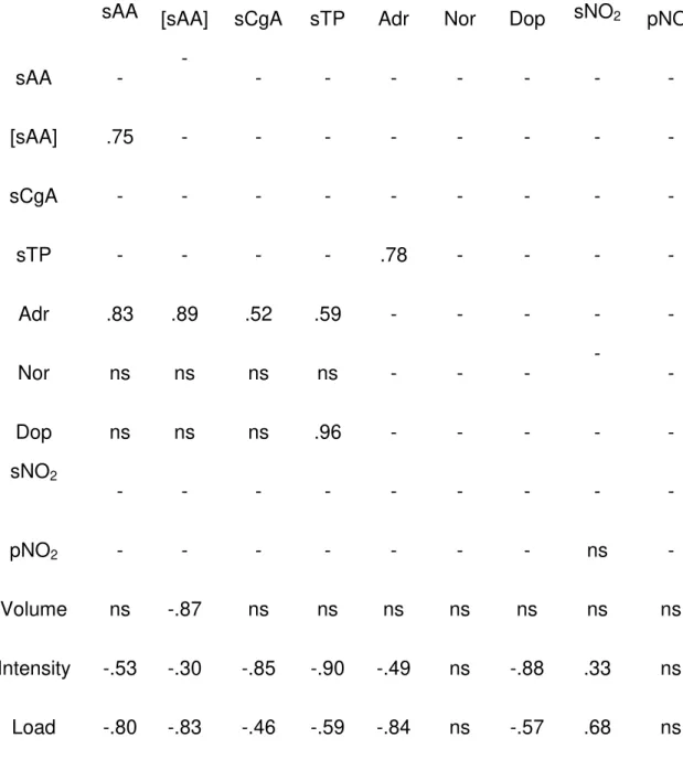
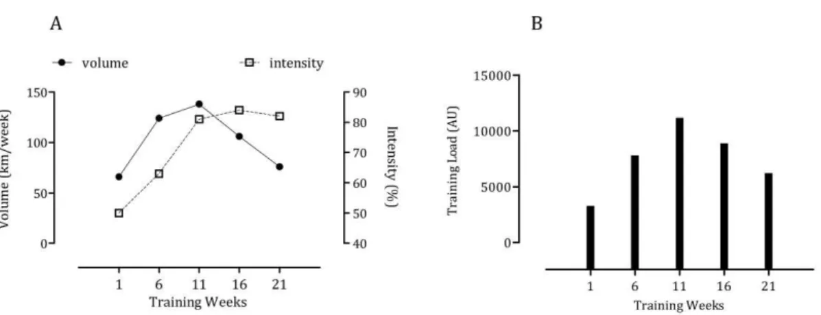
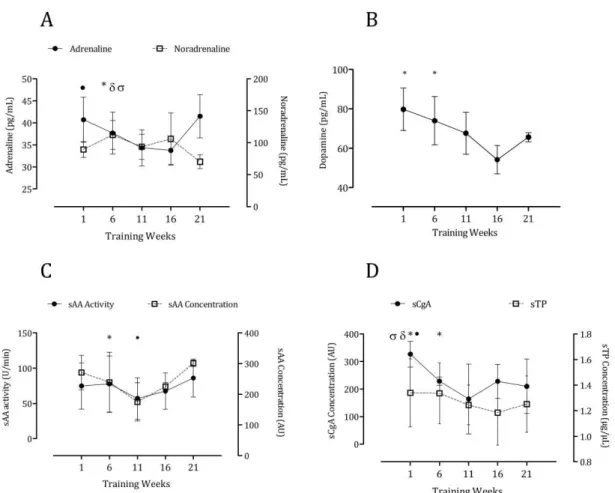
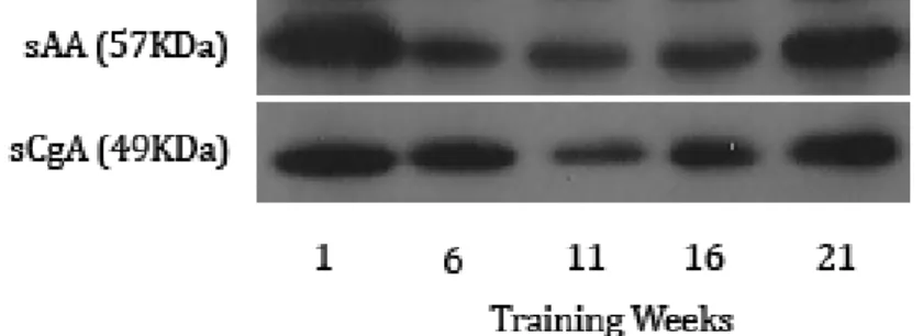
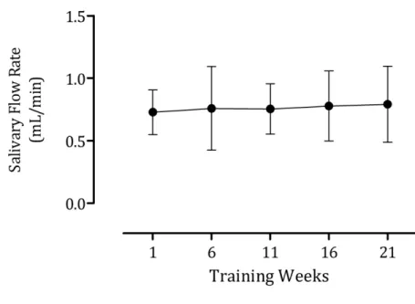
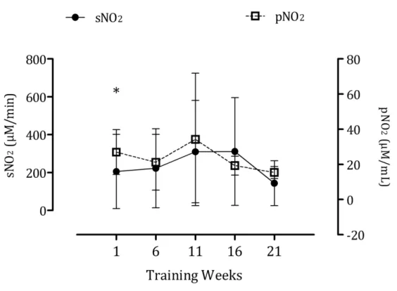
these markers might relate to inappropriate recovery and adaptation. Catecholamines occupy critical positions in the regulation of physiological processes during exercise10. On the other hand, it has been shown that circulating nitrite predicts exercise capacity in trained subjects11. With this mind, we investigated the response of salivary proteins and nitrite to training in elite swimmers and compared it against plasma catecholamines and nitrite, respectively9,10. We found strong correlations between sPT (r=0.59), sAA (r=0.89) and sCgA (r=0.52) and plasma adrenaline and between all of the above against the intensity and load of training. Event though no significant correlations were found between salivary and plasma nitrite, the former also predicted training intensity (r=0.33) and load (r=0.68) as has been previously reported9.
Other salivary proteins such as immunoglobulin A (sIgA) and lysozyme (sLys) have been found to decrease in response to elite training12. In this study, 31 professional rugby players were monitored for eleven months. Even though no correlation analysis was performed between salivary parameters and training load, it was found that the athletes were more disposed to infections of the upper respiratory tract (URTI) when training intensity was higher and this was associated with a decreased in salivary proteins and elevations in salivary cortisol (sC)12.
In addition to salivary proteins, the concentration of hormones in saliva, mainly testosterone (sT) (or its metabolic intermediates) and sC have been used to track the physiological impact of elite training as markers of anabolic and catabolic status, respectively. In this regard, 36 female professional handball players participated in a 16-week training program that consisted of 8 weeks of strength training at ~80% of the one-repetition maximum (1RM) and endurance training at the LT and 8 weeks of interval and high intensity training. The training program lead to an increase in the concentration of dehydroepiandrosterone (sDHEA)13.
Interestingly, the sDHEA/sC ratio showed a negative linear relationship (r = -0.73) with TL at the end of the 16 weeks13. Similar findings were observed in
endurance, power and agility and one subsequent week of tapering14. Whereas sC correlated positively with TL (r=0.64), the sT/sC ratio correlated negatively (r=-0.77). The response of salivary hormones to training was also strongly associated with negative mood states14. In 18 Naval Special Warfare operators submitted to four months of physical training aimed to promote power and strength endurance sT, sC, sDHEA as well as the sDHEA/sC ratio increased during the months with higher volume and intensity and decreased concomitantly with TL. However, no changes were observed for the sT/sC ratio15.
The cortisol awakening response (CAR) is a steep increase in the circulating levels of cortisol approximately 30 minutes after awakening. The CAR is thought to reflect the reactivity of the hypothalamic-pituitary-adrenal (HPA) axis to lasting and strenuous physiological and psychological demands16. After 16 weeks of endurance, strength and power training in 12 professional female tennis players, the diurnal rhythm and the levels upon awakening and 30 min afterwards of sC decreased when compared to pre-training levels. This was accompanied by concomitant increases in sAA upon awakening and as such, it was suggested that progressive increases in training load induce asymmetry in the activation of the HPA axis and the sympathetic nervous system, respectively17. Blunted rhythms of cortisol in response to excessive training have been suggested to represent a subclinical form of hypocortisolism17. However, the clinical meaning of such variation during elite training remains inconclusive.
Fatigue
Only a couple of studies have been able to diagnose fatigue by means of salivary markers in trained subjects. Ideally, the appropriate monitoring of TL would allow the necessary strategies for recovery to avoid overtraining. Yet, in ultra-endurance sports or especially in military training where sleep deprivation is common, the exceptionally high volume of exercise might lead to fatigue. The ratio between two peptides found in acidic and basic proline-rich salivary proteins was shown to increase in line with the rate of perceived exertion when nine amateur cyclists performed sets of 19-minute walking and 20-minute cycling at 70% of their maximum ventilator threshold with 10-minute rest between sets for ten consecutive hours19. These findings were later replicated by the same group in a larger cohort of subjects in an experimental model of mental fatigue20. Cognitive performance and mood states were assessed in 19 subjects (+16 controls) during 48 hours of sleep deprivation. The Fatigue Biomarker Index, as coined by the authors, could identify poor performance and mental fatigue with both selectivity and specificty20. On the other hand, sC showed a similar pattern
than the work/rest cycles in 11 subjects taking part in the United States Air Force Special Tactics Officer selection. This process consists of five days of exercise such as running, swimming and ruck marching, several-hour missions that include swimming, land navigation, and skill tests as well as leadership reaction courses21. However, no correlation analyses were performed to confirm the
association between salivary markers and fatigue in these studies.
Lactate
The LT and the maximum lactate steady state (MLSS) are indices of aerobic endurance commonly used to adjust the intensity of training in swimmers and rowers22,23. Lactate diffuses passively into saliva from blood and its accumulation
have studied the accumulation of lactate in blood for decades and significant understanding in regard to its dynamics and application to sports medicine have been gathered. Unfortunately, very few studies have been directed towards investigating the suitability of surrogates of blood lactate (bLac) to predict training intensity in elite athletes. Based on the principle that, in general, sympathetic stimulation to the salivary glands leads to the secretion of electrolytes and proteins Chicharro and colleagues had already demonstrated twenty years ago that the salivary concentration of Na+ and Cl- show a similar behavior than bLac (r = 0.82) and catecholamines (r=0.75) in healthy males submitted to incremental exercise24. Similar results by their group were reported in ten-year-old children a
year later25. Other studies have followed this rationale and have identified the
viability of salivary proteins to predict the salivary LT in active subjects and elite athletes during sub-maximal or maximal exercise. Strong correlations have been reported between bLac and both the activity (r=0.95)26 and the concentration of
sAA (r=0.84 and r=0.81)27,28, sPT (r=0.90; r=0.78 and r=0.93)27–29 and sCgA (r=0.82)28. Others studies instead tackled the issue directly and determined the potential of salivary lactate (sLac) to predict bLac accumulation. Clearly, directly quantifying salivary lactate (sLac) would provide more useful information. In this regard, it has been demonstrated that sLac is a viable and reliable index to predict MLSS in amateur cyclists with strong correlations between bLac and sLac when the intensity of exercise was expressed in terms of VO2 (r=0.89) and power
(r=0.92)30. Using a graded cycle ergometer test with 25-W increments every three minutes until volitional exhaustion in amateur athletes, Segura and colleagues reported a high degree of concordance (r=0.81) between bLac and sLac, with the latter being highly accurate to predict the LT31. Similar associations in the pattern of accumulation between bLac and sLac (r=0.71) have been described in experienced marathon runners after a 30-km race32. Due to the
passive diffusion of lactate from blood into saliva, it was suggested that more rapid increases in workload might complicate the determination of LT31. However,
concentration of sLac is between 15 and 50% that the one found in blood during exercise28,31,32. Stronger correlations have been found in athletes (r=0.511) than in non-athletes (r=0.385) after maximal exercise with the latter group showing higher concentration of sLac at the workloads than the athletes33. Finally, equivalent to bLac measurements, sLac is higher in sprinters than long distance runners after a 400-m sprint and a 3000-m run, respectively34. Many of the studies mentioned above used common electro enzymatic methods to quantify lactate. Hence, from a methodological standpoint, portable lactate analyzers with lower detection limits could be employed to measure sLac in the field. On the other hand, in the laboratory, sLac has been proven to remain stable for 40 days when stored at 4C31. The absence of red blood cells in saliva eliminates the need of pre-treating the samples with sodium fluoride to preserve glucose and avoid further production of lactate28.
Hydration status
Euhydration is critical to maintain muscle strength and endurance35,36. However,
beyond the clear importance of maintaining an appropriate hydration status for athletic performance, monitoring water loss, for instance, is pivotal in fighting and equestrian sports in which fast weight loss practices in the form of dehydration are employed prior to a competition in order to meet a required weight limit. Interesting findings have been accumulated regarding the potential of salivary parameters to track acute dehydration. Some studies have reported strong correlations between saliva osmolality (sOSM; r=0.94), salivary total protein (sPT; r=0.97) and salivary flow rate (r=-0.88) with the percentage of body mass loss (BML) up to 2.9% when subjects exercised at 60% of their VO2max for