Dinosaurs
Michael D. D’Emic1*., John A. Whitlock2., Kathlyn M. Smith3
, Daniel C. Fisher4, Jeffrey A. Wilson4
1Anatomical Sciences Department, Stony Brook University, Health Sciences Center, School of Medicine, Stony Brook, New York, United States of America,2Science and Mathematics Department, Mount Aloysius College, Cresson, Pennsylvania, United States of America,3Department of Geology & Geography and Georgia Southern University Museum, Georgia Southern University, Statesboro, Georgia, United States of America,4Museum of Paleontology and Department of Earth & Environmental Sciences, University of Michigan, Ann Arbor, Michigan, United States of America
Abstract
Background:Tooth replacement rate can be calculated in extinct animals by counting incremental lines of deposition in tooth dentin. Calculating this rate in several taxa allows for the study of the evolution of tooth replacement rate. Sauropod dinosaurs, the largest terrestrial animals that ever evolved, exhibited a diversity of tooth sizes and shapes, but little is known about their tooth replacement rates.
Methodology/Principal Findings: We present tooth replacement rate, formation time, crown volume, total dentition volume, and enamel thickness for two coexisting but distantly related and morphologically disparate sauropod dinosaurs Camarasaurus and Diplodocus. Individual tooth formation time was determined by counting daily incremental lines in dentin. Tooth replacement rate is calculated as the difference between the number of days recorded in successive replacement teeth. Each tooth family inCamarasaurushas a maximum of three replacement teeth, whereas eachDiplodocus tooth family has up to five. Tooth formation times are about 1.7 times longer inCamarasaurusthan inDiplodocus(315 vs. 185 days). Average tooth replacement rate inCamarasaurusis about one tooth every 62 days versus about one tooth every 35 days inDiplodocus. Despite slower tooth replacement rates inCamarasaurus, the volumetric rate ofCamarasaurustooth replacement is 10 times faster than inDiplodocusbecause of its substantially greater tooth volumes. A novel method to estimate replacement rate was developed and applied to several other sauropodomorphs that we were not able to thin section.
Conclusions/Significance:Differences in tooth replacement rate among sauropodomorphs likely reflect disparate feeding strategies and/or food choices, which would have facilitated the coexistence of these gigantic herbivores in one ecosystem. Early neosauropods are characterized by high tooth replacement rates (despite their large tooth size), and derived titanosaurs and diplodocoids independently evolved the highest known tooth replacement rates among archosaurs.
Citation:D’Emic MD, Whitlock JA, Smith KM, Fisher DC, Wilson JA (2013) Evolution of High Tooth Replacement Rates in Sauropod Dinosaurs. PLoS ONE 8(7): e69235. doi:10.1371/journal.pone.0069235
Editor:Alistair Robert Evans, Monash University, Australia
ReceivedNovember 9, 2012;AcceptedJune 6, 2013;PublishedJuly 17, 2013
Copyright:ß2013 D’Emic et al. This is an open-access article distributed under the terms of the Creative Commons Attribution License, which permits unrestricted use, distribution, and reproduction in any medium, provided the original author and source are credited.
Funding:Funding was provided by the Scott D. Turner Award (University of Michigan) to MDD. The funders had no role in study design, data collection and analysis, decision to publish, or preparation of the manuscript.
Competing Interests:The authors have declared that no competing interests exist.
* E-mail: michael.demic@stonybrook.edu
.These authors contributed equally to this work.
Introduction
Large or complex dentitions generally experience attrition through abrasion against food, substrates, or other teeth. In mammals, food intake and tooth use tend to increase with body size, so larger animals tend to exhibit increased tooth wear [1]. During their nearly 300-million-year evolutionary history [2], vertebrate herbivores evolved numerous mechanisms to cope with increased tooth wear, including changes in the mechanical properties of tooth tissues [3,4], increases in the number of teeth that are functional at one time [5,6], continuous tooth growth and eruption throughout the life of the animal [7], increases in the number of tooth-bearing bones, changes in crown volume and/or shape [8–11], and increased tooth replacement rate [12,13].
Sauropod dinosaurs achieved the largest adult body sizes of any terrestrial herbivore, and so would have required a large food
Here, we measure tooth formation time, replacement rate, crown volume, and enamel thickness in sectioned teeth of
CamarasaurusandDiplodocus, two neosauropod dinosaurs from the Late Jurassic Morrison Formation of North America. The largest exemplars of these two genera are similar in body mass (e.g., femur length ca. 1.8 m, sum of femoral and humeral circumference ca. 1.3 m; MDD unpublished data), but they belong to distantly related neosauropod clades that differ substantially in skull morphology, body proportions, and inferred feeding ecology [17–20]. The rarity of sauropod craniodental materials that can be sacrificed for histological sampling limits the taxonomic scope across which we can measure these features. We explore the distribution of these features more broadly within Sauropodomor-pha by developing a method to estimate tooth replacement rates for several taxa that have craniodental material but cannot be sampled histologically.
Materials and Methods
Permission was received to access the relevant specimens from museum collections managers. Specimens were loaned from the Yale Peabody Museum, Utah Museum of Natural History, Staatliches Museum fu¨r Naturkunde, and Iziko South African Museum. Computed tomography (CT) images were acquired at the Canton Health Center, University of Michigan, using a General Electric Lightspeed Pro 8 CT scanner, GE Medical systems, Milwaukee, Wisconsin. CT slices were taken using 140 Kv and 325 mA, with 1.250 mm thick slices and 0.625 mm overlap. Incremental lines were counted in thin section. Each tooth was mechanically removed from the jaws. Specimens were embedded in epoxy resin, cut longitudinally on a Buehler Isomet saw with a diamond wafering blade, mounted on a glass slide, cut to a thick section, and hand-sanded and polished until incremental lines were visible. Thin sections were photographed using a Spot CCD camera (Spot Insight 11.2 Color Mosaic, Diagnostic Instruments) mounted on a Nikon SMZ 1500 microscope. Increments were counted in ImageJ [21,22] using the IncMeas v1.11 plug-in [23].
Tooth formation time and replacement rate inDiplodocusand
Camarasauruswere measured by counting incremental lines of von Ebner (Fig. 1), which have been shown to represent daily fronts of dentin deposition in several groups of extant amniotes [12,13,24– 26]. We define tooth replacement rate to be the time required to replace one tooth in a given alveolus. This rate is sometimes expressed in days, with the unit numerator implicit. Replacement rate was calculated by subtracting tooth formation times for successive teeth within one family, following Erickson [12,13]. Recently, Scheyer and Moser [27] questioned the identification of incremental lines of von Ebner in sauropods, suggesting that they could represent longer-period increments (e.g., Andresen lines). We examined our thin sections and did not find smaller increments between the lines spaced ca. 15 microns apart in areas where preservation seems excellent, so we interpret these lines as daily fronts of deposition. The ca. 15-micron spacing of
incremental lines of von Ebner observed in Diplodocus and
Camarasaurus is close to the mean value observed in labelling studies of adultAlligator[13].
Enamel thickness was measured in ImageJ on photographs of thin sections. Thickness was measured perpendicular to the enamel-dentin junction at seven locations around the tooth crown (three labial, three lingual, and one apical). Fewer measurements were made on teeth for which enamel was chipped or missing in certain locations. Labial and lingual measurements were taken at roughly evenly spaced locations along the apicobasal axis (one
near the tooth tip or apex, one near the mid-length of the crown, and one near the crown-root junction). Enamel thickness varies around the tooth crown, so comparison of labial and lingual thicknesses for each tooth was based on the average of three labial measurements and the average of three lingual measurements. An overall average of all measurements taken on a single tooth was also calculated. Raw enamel measurements are presented in Supporting Information (Raw Data S1).
Volumes of both the entire tooth and the crown (i.e., the part of the tooth covered in enamel, including the pulp cavity) were measured by water displacement via suspension three times and averaged (see Raw Data S1) [28]. Total erupted tooth volume (the sum of the volumes of all ‘fresh’ [functional but unworn] teeth in the jaw) and crown volume (the sum of the crown volumes of all fresh teeth in the jaw) were estimated for Camarasaurus and
Diplodocus. Tooth crowns are similar in volume for adjacent teeth throughout and among jaw elements for all tooth positions except for the last few in these species. For each species, the antepenultimate and penultimate tooth crown volumes were estimated as 75% of the average measured tooth crown volume, and the last tooth position was estimated as 50% of the average measured tooth crown volume. In contrast to total crown volumes, total functional tooth volumes were more complicated to estimate because the alternating pattern of tooth replacement in these species yields tooth roots of substantially different size in adjacent teeth. The average of total functional tooth volumes for two large teeth was used as the functional individual tooth volume. As with crowns, total functional tooth volumes for the antepenultimate, penultimate, and ultimate tooth positions were estimated as 75%, 75%, and 50% of the volume of the largest teeth, respectively. Finally, in Diplodocus, dentary teeth are about 10% smaller in volume than premaxillary or maxillary teeth [29], so estimates of the volumes of dentary teeth and crowns were adjusted accordingly.
In many cases, destructive sampling of a specimen was not possible. For these taxa, we developed a non-invasive approach for estimating replacement rate, based on use of Camarasaurus as a model for taxa with broad-crowned teeth (Patagosaurus, Mamench-isaurus) and Diplodocusas a model for taxa with narrow-crowned teeth (Nigersaurus, Rı´o Negro titanosaur). Both models were used for estimation of replacement rate inMassospondylus, which has an intermediate crown breadth. Tooth length was measured for teeth ofCamarasaurusandDiplodocusfor which ages were already known via counts of incremental lines of von Ebner. For each genus, regression of tooth formation time on tooth length generated an equation that was used to estimate tooth formation time in teeth that were not sampled histologically.
We estimated volumetric tooth replacement rate (the time required to replace the total dentition-in-use) by dividing total erupted tooth volume by average tooth replacement rate. We made a similar estimate using only tooth crown volumes (volumetric crown replacement rate). We make the assumption that tooth replacement rate was constant throughout and among jaw elements due to similarities in the shape, number of replacement teeth, and depth of alveoli in all but the distal-most few teeth in each jaw element. Our simplification would tend to inflate the volumetric replacement rates, but would affect each species similarly, thus keeping results for each comparable to one another.
Results
Histology-based tooth replacement rates and estimated replace-ment rates are summarized in Tables 1–2. The dentary of the
basal sauropodomorphPlateosaurusdid not show any replacement teeth in CT images, so no further analysis was undertaken. CT scans of a maxilla and dentary of the basal sauropodomorph
Massospondylus revealed only a single replacement tooth (in one alveolus of the dentary). Although we did thin section teeth of
Massospondylus, tooth replacement rate for that genus was estimated because incremental lines were poorly preserved.
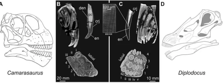
CT scans reveal that each premaxillary tooth family of
Camarasaurus(Fig. 2a, Movies S1 and S2) includes one functional and up to three replacement teeth, whereas each premaxillary tooth family ofDiplodocus(Fig. 2d, Movie S3) includes up to one functional and five replacement teeth. Incremental lines of von Ebner visible in thin section (Figs. 1–2; Table 1) indicate that each premaxillary tooth ofCamarasaurustook over ten months to form (ca. 1 tooth/315 days), whereas eachDiplodocustooth took only six months to form (ca. 1 tooth/185 days). Average tooth replacement rate inCamarasauruswas one tooth per two months (ca. 1 tooth/62 days), whereas it was about one tooth per month inDiplodocus(ca. 1 tooth/35 days).
Functional premaxillary teeth of theCamarasaurusandDiplodocus
individuals are 26.5 cm3 and 1.7 cm3 in volume, respectively. Tooth crowns of theCamarasaurus and Diplodocus individuals are 15.7 cm3and 1.5 cm3in volume, respectively. We estimate total functional (i.e., erupted) tooth volume across the dentition to be 1,272 cm3 in Camarasaurus and 69 cm3 in Diplodocus. When
measuring only tooth crowns, these values are 754 cm3 and 63 cm3, respectively. The volumetric tooth replacement rate was
about 10 times greater in Camarasaurus (1272 cm3/62
days = 20.5 cm3/day) than in Diplodocus (69 cm3/35
days = 2.0 cm3/day). The volumetric crown replacement rate was about seven times greater in Camarasaurus (754 cm3/62
days = 12.2 cm3/day) than in Diplodocus (63 cm3/35
days = 1.8 cm3/day).
Premaxillary tooth crowns ofCamarasaurusindividuals have ca. 1.0 mm-thick enamel on both the labial and lingual surfaces of the teeth; in contrast, the enamel ofDiplodocusis thinner overall (ca. 0.5 mm) and is slightly asymmetrical, with the enamel on the labial face of the tooth about 125–150% the enamel thickness on the lingual face (Figs. 1–2, Table 3).
Our non-invasive approach to estimating tooth replacement rate allowed us to evaluate a broader spectrum of sauropodo-morphs. Tooth length and age are strongly related in both
CamarasaurusandDiplodocus(R2.0.95), but the equations describ-ing these relationships differed between the taxa (see Raw Data S1). We evaluated the performance of our estimation method by estimating tooth replacement rate inCamarasaurusandDiplodocus, taxa for which replacement rate is known. For a given tooth and its successor, our method of estimating both formation time and replacement rate was generally accurate to within one week. When successive replacement estimates are averaged for several teeth in Figure 1. Dental histology of the sauropod dinosaursCamarasaurusandDiplodocus.Thin sections ofCamarasaurus(A,C) andDiplodocus (B,D) premaxillary teeth showing incremental lines of von Ebner (white arrowheads) in dentin. Teeth are oriented with their long axis horizontal and the occlusal surface directed to the right.Ashows the tip of tooth 3iii ofCamarasaurus, andBshows the tip of tooth 4iv ofDiplodocus.CandDare enlarged images of one ‘limb’ of tooth 3ii and 4iii, respectively. Abbreviations:edj, enamel-dentin junction;en, enamel;pc, pulp cavity. [planned for page width].
a single jaw element, the estimates are off by one day at most. Because we were only able to measure the length of one tooth and its successor for each of the non-histologically sampled taxa (aside from the case of Nigersaurus), we expect that our estimates are accurate for those taxa to within one week.
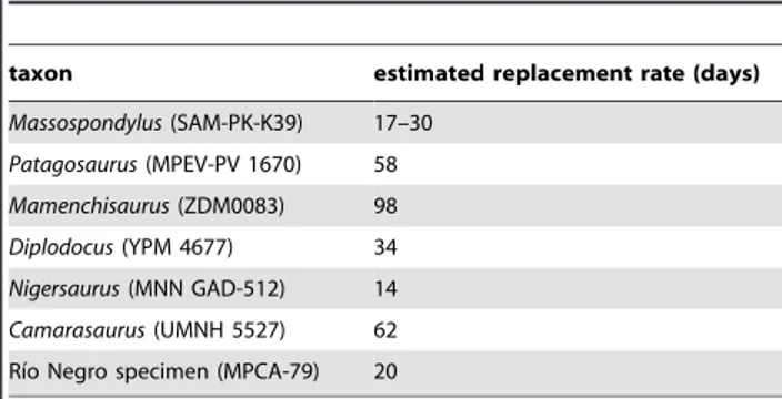
The initial increase in tooth size and crown breadth that occurred near the base of Sauropoda was accompanied by a reduction in tooth replacement rate (as estimated by replacement tooth length). The much smaller teeth of basal sauropodomorphs like Massospondylus and Patagosaurus formed and replaced faster than did the larger teeth of basal sauropods likeMamenchisaurus
(Fig. 3; Table 2). Derived broad-crowned taxa (e.g.,Camarasaurus) exhibited a higher replacement rate than non-neosauropods like
Mamenchisaurusand matched the rate in the much smaller-toothed
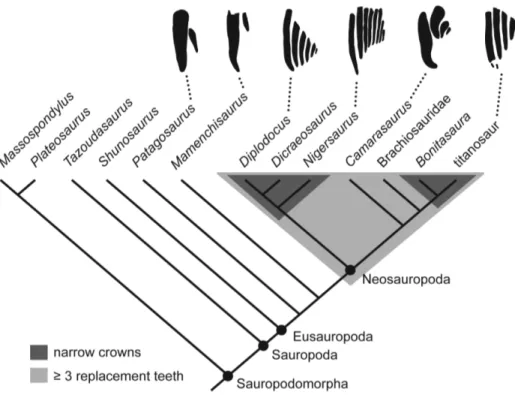
Patagosaurus,but did not achieve rates as high as those observed in the smaller-toothed Massospondylus. Non-neosauropods exhibit a maximum of two replacement teeth per alveolus, whereas neosauropods exhibit three to nine ([6,30], Fig. 3). Within
Neosauropoda, diplodocoids and titanosaurs independently
achieved higher tooth replacement rates than basal neosauropods (Fig. 3). The highly specialized diplodocoidNigersaurusis estimated to have replaced each tooth as often as once every 14 days, twice as fast as previous estimates [30 days, [6]] and by far the highest rate for any dinosaur. The discrepancy between our estimate and a previous one for Nigersaurusis potentially explainable by the fact that the histology-based replacement rate previously reported for
Nigersaurus [6] was based on transverse thin sections of teeth. Transverse sections likely yield inaccurate replacement rates due to the limited number of incremental lines of von Ebner exposed in any given transverse plane.
Discussion
In both Camarasaurus and Diplodocus, a volume equivalent to approximately one tooth is replaced across the dentition every 1–2
days (20.5 cm3/day and 2.0 cm3/day, respectively). These taxa are characterized by different styles of forming and replacing dentition:
Camarasaurus has larger teeth that are replaced less frequently, whereas Diplodocus has smaller teeth that are replaced more frequently. Even withCamarasaurus’ lower tooth replacement rates, both sauropods exhibit tooth replacement rates on par with or higher than those of non-sauropod dinosaurian herbivores (i.e., hadrosaur-oid and ceratopsian ornithischians at 50–83 days; Table 4).
The enamel of Camarasaurus is roughly symmetrical labiolin-gually, in contrast to the slightly asymmetrical enamel ofDiplodocus. The enamel of the diplodocoidNigersaurusis highly asymmetrical, with enamel on the labial side up to ten times thicker than on the lingual side [5,6,31]. Labiolingually asymmetrical enamel appears to characterize several diplodocoids, and extremely asymmetric enamel characterizesNigersaurusor a slightly more inclusive clade [6]. Labiolingually asymmetrical enamel, reduced crown volume, increased replacement rate, and the development of tooth batteries evolved independently in two other dinosaur clades: iguanodon-tian ornithopods [32] and ceratopsian marginocephalians [33]. The repeated evolution of these features together may represent an adaptation to herbivory at large body size and within the context of polyphyodonty, though several important differences in the evolution of these features exist as well [5].
Sauropods were obligate herbivores, but their antecedents were omnivorous [34–37]. The origin and early evolution of sauropods involved increases in tooth volume [10] and body size [38]. Although herbivory and extremely large body size persisted among the vast majority of sauropods, multiple lineages drastically reduced the volume of functional crowns [10]. The repeated independent evolution of narrow crowns suggests that they conferred an adaptive advantage over broad crowns during the second half of sauropod evolution. By the Late Cretaceous, only narrow-crowned sauropod taxa remained [10,39]. Additionally, following the disappearance of diplodocoids from the fossil record in the early Late Cretaceous, tooth crowns in titanosauriforms decreased in volume and breadth until they were similar in size and shape to those of diplodocoids [10]. Sauropods with broad-crowned teeth (e.g.,Camarasaurus) evolved tooth replacement rates on par with those of ornithischian herbivores that persisted into the latest Cretaceous, and each Camarasaurus tooth was more Table 1.Tooth formation time (days) and replacement rate (1
tooth/X days) inDiplodocus(YPM 4677) andCamarasaurus (UMNH 5527).
Diplodocus tooth family
1 2 3 4
tooth position i – 187 – 183
ii 176 – 178 145
iii 141 – 144 113
iv – – 110 –
v – – 67 –
average replacement rate 35 – 37 35
Camarasaurus tooth family
1 2 3 4
tooth position i – – – 315
ii – – 208 253
iii – – – 190
iv – – – 130
average replacement rate – – – 62
Abbreviations: UMNH, Utah Museum of Natural History, Salt Lake City, USA; YPM, Yale Peabody Museum, New Haven, USA.
doi:10.1371/journal.pone.0069235.t001
Table 2.Estimated tooth formation times (ages) and replacement rates in several sauropodomorphs.
taxon estimated replacement rate (days)
Massospondylus(SAM-PK-K39) 17–30
Patagosaurus(MPEV-PV 1670) 58
Mamenchisaurus(ZDM0083) 98
Diplodocus(YPM 4677) 34
Nigersaurus(MNN GAD-512) 14
Camarasaurus(UMNH 5527) 62
Rı´o Negro specimen (MPCA-79) 20
There is a range forMassospondylustooth replacement rate because estimates were made both using the narrow-crowned and broad-crowned equations. Note the similarity of estimated replacement rates forCamarasaurusand Diplodocuswith histologically obtained rates of 62 and 35 days for these taxa, respectively.
Abbreviations: MNN, Muse´e National du Niger, Niamey, Niger; MPCA, Museo Provincial Carlos Ameghino, Cipolletti, Argentina; SAM, South African Museum, Ikizo Museums, Cape Town, South Africa; UMNH, Utah Museum of Natural History, Salt Lake City, USA; YPM, Yale Peabody Museum, New Haven, USA; ZDM, Zigong Dinosaur Museum, Zigong, China.
doi:10.1371/journal.pone.0069235.t002
resistant to wear than smaller teeth by virtue of their larger volume and thicker enamel. Why then did several neosauropod lineages develop narrow-crowned teeth?
One explanation is that fresh teeth are more effective than worn teeth. Replacing a tooth every month reduces the number of excessively worn crowns in the functional dentition, which prolongs contact with opposing teeth and with food. Additionally, although individual teeth were being replaced more frequently, the smaller crown volume results in a lower rate of mineralized tissue production and loss – narrow-crowned taxa had to recoup around 10% of the crown volume of dental tissue that was required in their larger-crowned relatives per tooth replaced. Furthermore, the narrow crowns ofDiplodocusare made up of a larger proportion of enamel to dentin than the broad crowns ofCamarasaurus. The advent of narrow-crowned dentition therefore enabled the animal to have many more fresh teeth at any given time, while losing far less mineralized tissue. Furthermore, smaller, more slender teeth would have allowed for smaller tooth roots (Fig. 2) and smaller and
lighter cranial bones, resulting in a lighter skull overall. A small head-to-body volume ratio sets sauropods apart from other dinosaurian herbivores [10].
In a finite element analysis of the skull ofDiplodocus, Young et al. [40] identified high stresses at the bases of teeth that would have been incurred during branch stripping or other feeding strategies. In the context of those results, they interpreted high tooth replacement rates inDiplodocusas an adaptation that would have accommodated increased levels of tooth breakage. Aside from concerns that tooth breakage is maladaptive, producing ineffective and infection-prone teeth, we briefly discuss one testable consequence of the Young et al. hypothesis. If Diplodocus and other narrow-crowned sauropods experienced tooth breakage as a result of branch stripping or static biting, then the fossil record should bear evidence of such failure. Although the record of cranial remains is sparse for sauropods, it is relatively good for
Diplodocusand other narrow-crowned sauropods, and we know of no evidence of jaws preserving teeth broken in life.
Rather than being related to high levels of tooth breakage, we propose that increased replacement rates are related to increased wear rates that may have been a consequence of a shift in diet [10,20]. Some narrow-crowned taxa (e.g.,Diplodocus) were likely low-browsers [6,18,20,41,42], a behavior that leads to increased ingestion of abrasive exogenous grit [43]. The sauropod most highly specialized for low browsing,Nigersaurus, also has the highest known replacement rate of any dinosaur. In contrast, sauropods with broader tooth crowns and slower replacement rates, such as
MamenchisaurusandCamarasaurus, are thought to have been mid- to upper-canopy browsers [18,20,39,41,42,44,45], where exogenous grit levels are expected to be lowest.
Conclusions
Tooth replacement rate, size, and shape data indicate that despite their somewhat stereotyped body plan and large body size, sauropod dinosaurs exhibited varied approaches to feeding. The coexisting but morphologically disparate and distantly related Late Figure 2. Tooth replacement in the sauropod dinosaursCamarasaurusandDiplodocus.Reconstructed skulls (A,D) and premaxillary teeth (B,C) ofCamarasaurus(A,B) andDiplodocus(C,D).BandCinclude CT-generated sagittal and transverse sections of premaxillary alveoli and photographs of thin sections ofCamarasaurus(UMNH 5527) andDiplodocus(YMP 4677). Premaxillae show replacement teeth in each of the four alveoli adjacent to the symphysis labelled by their position along the tooth row (1–4) and their position in the replacement sequence at each tooth position (i–v). Sagittal sections inBand Cwere taken at premaxillary tooth position 4 inCamarasaurus and premaxillary tooth position 1 in Diplodocus. The symphysis faces the bottom of the page in transverse sections. Photographs of thin sections ofDiplodocusandCamarasaurusteeth show enamel (en), the pulp cavity (pc), daily-deposited incremental lines of von Ebner (arrowheads mark every other line) in the dentin (den), and the crown-root junction (crj). The 20 mm scale bar is for the premaxilla and tooth images in (B); 10 mm scale bar is for premaxilla and tooth images in (C). Skull reconstructions are from [19,46][planned for page width].
doi:10.1371/journal.pone.0069235.g002
Table 3.Summary of enamel thickness (mm) inDiplodocus (YPM 4677) andCamarasaurus(UMNH 5527) at different tooth developmental stages (i–v).
Diplodocus i ii iii iv v
average thickness 0.45 0.45 0.31 0.23 0.08
labial/lingual 1.25 1.41 1.53 1.19 1.12
Camarasaurus i ii iii iv
average thickness 0.97 0.62 0.38 0.12
labial/lingual 0.99 0.93 0.86 1.23
Each value shown is an average of teeth from more than one alveolus at similar stages of development (e.g., ‘ii’ is an average of values for tooth position 2ii, 3ii, 4ii, etc.). See Raw Data S1.
Jurassic sauropodsCamarasaurusandDiplodocusdiffered greatly in their anatomy related to food acquisition:Camarasaurushad a large volume of broad-crowned teeth that were replaced relatively slowly, whereasDiplodocushad a small volume of narrow-crowned teeth that were replaced very quickly. This variety represents a potential factor that allowed multiple gigantic species such as
Camarasaurusand Diplodocusto partition the same ecosystem. The repeated evolution of narrow-crowned teeth in sauropods appears to have been accompanied by an increase in tooth replacement rate, which would have equipped these forms with less worn teeth over their lifetimes and allowed their skulls to be lighter.
Supporting Information
Raw Data S1 Microsoft Excel spreadsheet containing tooth volumes, crown volumes, enamel thicknesses, tooth lengths, and estimation method for tooth replacement rate for various sauropodomorphs investigated in this study.
(XLSX)
Movie S1 CT-generated movie of the premaxilla ofCamarasaurus
(UMNH 5527) in mesiodistal view (see separate.mov file) (MOV)
Movie S2 CT-generated movie of the premaxilla ofCamarasaurus
(UMNH 5527) in apicobasal view (see separate.mov file). (MOV)
Movie S3 CT-generated movie of the premaxilla ofDiplodocus
(YPM 4677), with bone rendered transparent and teeth opaque (see separate.avi file).
(WMV)
Figure 3. Cladogram of sauropodomorphs showing the optimization of key features related to elevated tooth replacement rates.
The light gray field indicates taxa that have at least three replacement teeth at each tooth position; dark gray field encapsulates taxa that have narrow tooth crowns. Silhouettes along the top of the cladogram show the number and size of replacement teeth in one tooth position. These include (from left to right):Patagosaurus(MPEF-PV 1670),Mamenchisaurus[47],Diplodocus(this study),Nigersaurus[Sereno, Wilson, Witmer, Whitlock, Maga, Ide and Rowe, unpublished data],Camarasaurus(this study), and the Rı´o Negro titanosaur (MPCA-79) [48]. Number of replacement teeth is unknown in Brachiosauridae, but the taxon is optimized to have had at least three. Cladogram based on [30] with the addition ofTazoudasaurus[49] andBonitasaura[50]. [planned for column width].
doi:10.1371/journal.pone.0069235.g003
Table 4.Tooth replacement rates (days) for archosaurs.
taxon
tooth replacement rate (days)
Archosauria
crocodiliform 105
Dinosauria
Ornithischia
Triceratops 83
Hadrosauridae
Maiasaura 58
Edmontosaurus 50
Prosaurolophus 81
Saurischia
Sauropoda
Camarasaurus 62
Diplodocus 35
Nigersaurus 14–30
Theropoda
Tyrannosaurus 777
‘albertosaur’ 454
Deinonychus 290
Data for sauropods are from this study; other data are from [6,12]. doi:10.1371/journal.pone.0069235.t004
Acknowledgments
We thank J. Gauthier, W. Joyce, D. Brinkman (YPM), M. Getty (UMNH), P. M. Sander (U. Bonn), R. Schoch (Staatliches Museum fu¨r Naturkunde) and R. Smith (Iziko South African Museum) for loan assistance and permission to sample specimens. Thanks to W. Sanders (University of Michigan Museum of Paleontology) for preparation, Dr. C. Blane, K. Schwarz, and K. Barber (Department of Radiology, University of Michigan Medical Center) for access and assistance with CT scanning. We thank Paul Upchurch (University College London) and Paul Tafforeau
(European Synchrotron Radiation Facility) for critical comments that improved the manuscript.
Author Contributions
Conceived and designed the experiments: MDD J. A. Whitlock J. A. Wilson. Performed the experiments: MDD J. A. Whitlock KMS. Analyzed the data: MDD J. A. Whitlock KMS DCF J. A. Wilson. Contributed reagents/materials/analysis tools: MDD KMS DCF J. A. Wilson. Wrote the paper: MDD J. A. Whitlock KMS DCF J. A. Wilson.
References
1. Owen-Smith RN (1992) Megaherbivores: the influence of very large body size on ecology. Cambridge: Cambridge University Press. 388 p.
2. Sues H-D, Reisz RR (1998) Origins and early evolution of herbivory in tetrapods. Trends in Ecology and Evolution 13: 141–145.
3. Erickson GM, Krick BA, Hamilton M, Bourne GR, Norell MA, et al. (2012) Complex dental structure and wear biomechanics in hadrosaurid dinosaurs. Science 338: 98–101.
4. Teaford FT, Smith MM, Ferguson MWJ (2000) Development, Function and Evolution of Teeth. Cambridge, UK: Cambridge University Press. 314 p. 5. Sereno PC, Wilson JA (2005) Structure and evolution of a sauropod tooth
battery. In: Curry Rogers KA, Wilson JA, editors. The sauropods: evolution and paleobiology. Berkeley: University of California Press. 157–177.
6. Sereno PC, Wilson JA, Witmer LM, Whitlock JA, Maga A, et al. (2007) Structural extremes in a Cretaceous dinosaur. PLoS ONE 2: e1230. 7. Richman JM, Whitlock JA, Abramyan J (2013) Regeneration of reptile teeth. In:
Huang GT-J, Thesleff I, editors. Stem cells in craniofacial development, regeneration and repair. Hoboken: Wiley-Blackwell. 135–151.
8. White TE (1959) The endocrine glands and evolution, no. 3: os cementum, hypsodonty, and diet. Contributions from the Museum of Paleontology, University of Michigan 13: 211–265.
9. Damuth J, Janis CM (2011) On the relationship between hypsodonty and feeding ecology in ungulate mammals, and its utility in palaeoecology. Biological Reviews 86: 733–758.
10. Chure DJ, Britt BB, Whitlock JA, Wilson JA (2010) First complete sauropod dinosaur skull from the Cretaceous of the Americas and the evolution of sauropod dentition. Naturwissenschaften 97: 379–391.
11. Stro¨mberg CAE (2006) Evolution of hypsodonty in equids: testing a hypothesis of adaptation. Paleobiology 32: 236–258.
12. Erickson GM (1996) Incremental lines of von Ebner in dinosaurs and the assessment of tooth replacement rates using growth line counts. Proceedings of the National Academy of Sciences 93: 14623–14627.
13. Erickson GM (1996) Daily deposition of dentine in juvenile Alligator and assessment of tooth replacement rates using incremental line counts. Journal of Morphology 228: 189–194.
14. Ganse B, Stahn A, Stoinski S, Suthau T, Gunga HC (2011) Body mass estimation, thermoregulation and cardiovascualr physiology of large sauropods. In: Klein N, Remes K, Sander PM, editors. Biology of the sauropod dinosaurs: understanding the life of giants. Bloomington: Indiana University Press. 105–115.
15. Paladino FV, Spotila JR, Dodson P (1997) A blueprint for giants: modeling the physology of large dinosaurs. In: Farlow JO, Brett-Surman MK, editors. The Complete Dinosaur. Bloomington: Indiana University Press. 491–504. 16. Spotila JR, Lommen PW, Bakken GS, Gates DM (1973) A mathematical model
for body temperatures of large reptiles: implications for dinosaur biology. The American Naturalist 107: 391–404.
17. Upchurch P, Barrett PM, Dodson P (2004) Sauropoda. In: Weishampel DB, Dodson P, Osmo´lska H, editors. The Dinosauria, Second Edition. Berkeley: University of California Press. 259–324.
18. Upchurch P, Barrett PM (2000) The evolution of sauropod feeding mechanisms. In: Sues HD, editor. Evolution of Herbivory in Terrestrial Vertebrates – Perspectives from the Fossil Record. Cambridge: Cambridge University Press. 79–122.
19. Wilson JA, Sereno PC (1998) Early evolution and higher-level phylogeny of sauropod dinosaurs. Journal of Vertebrate Paleontology Society of Vertebrate Paleontology Memoir 1–68.
20. Whitlock JA (2011) Inferences of diplodocoid (Sauropoda: Dinosauria) feeding behavior from snout shape and microwear analyses. PLoS ONE 6(4): e18304. 21. Rasband WS (1997–2009) ImageJ. Bethesda: US National Institutes of Health. 22. Abramoff M, Magelhaes P, Ram S (2004) Image processing with ImageJ.
Biophotonics International 11: 36–42.
23. Rountrey A (2009) Life histories of juvenile wooly mammoths from Siberia: stable isotope and elemental analyses of tooth dentin. Ann Arbor: University of Michigan.
24. Carlson SJ (1990) Vertebrate dental structures. In: Carter J, editor. Skeletal Biomineralization: Patterns, Processes and Evolutionary Trends. New York: Van Nostrand Reinhold. 531–536.
25. Smith T, Reid DJ, Sirianni J (2006) The accuracy of histological assessments of dental development and age at death. Journal of Anatomy 208: 125–138.
26. Fitzgerald C (1998) Do enamel microstructures have regular time dependencies? Conclusions from the literature and a large scale study. Journal of Human Evolution 35: 371–386.
27. Scheyer TM, Moser M (2011) Survival of the thinnest: rediscovery of Bauer’s (1898) ichthyosaur tooth sections from Upper Jurassic lithographic limestone quarries, south Germany. Swiss Journal of Geosciences 104 (Suppl 1): S147–S157. 28. Hughes SW (2006) Archimedes revisited: a faster, better, cheaper method of accurately measuring the volume of small objects. Physics Teacher 40: 468–474. 29. Holland WJ (1924) The skull ofDiplodocus. Memoirs of the Carnegie Museum 9:
379–403.
30. Wilson JA (2002) Sauropod dinosaur phylogeny: critique and cladistic analysis. Zoological Journal of the Linnean Society 136: 217–276.
31. Sereno PC, Beck AL, Dutheil DB, Larsson HCE, Lyon GH, et al. (1999) Cretaceous sauropods from the Sahara and the uneven rate of skeletal evolution among dinosaurs. Science 286: 1342–1347.
32. Norman DB (2004) Basal Iguanodontia. In: Weishampel DB, Dodson P, Osmo´lska H, editors. The Dinosauria, Second Edition. Berkeley: University of California Press. 413–437.
33. Dodson P, Currie PJ (1990) Neoceratopsia. In: Weishampel DB, Dodson P, Osmo´lska H, editors. The Dinosauria, First Edition. Berkeley: University of California Press. 593–618.
34. Martı´nez RN (2009) Adeopapposaurus mognai, gen. et sp. nov (Dinosauria: Sauropodomorpha), with comments on adaptations of basal sauropodomorphs. Journal of Vertebrate Paleontology 29: 142–164.
35. Martı´nez RN, Alcober OA (2009) A basal sauropodomorph (Dinosauria: Saurischia) from the Ischigualasto Formation (Triassic, Carnian) and the early evolution of Sauropodomorpha. PLoS ONE 4: e4397.
36. Barrett PM (2000) Prosauropod dinosaurs and iguanas: speculations on the diets of extinct reptiles. In: Sues HD, editor. Evolution of herbivory in terrestrial vertebrates. Cambridge: Cambridge University Press. 42–78.
37. Barrett PM, Upchurch P (2007) The evolution of feeding mechanisms in early sauropodomorph dinosaurs. Special Papers in Palaeontology 77: 91–112. 38. Yates AM (2004)Anchisaurus polyzelus(Hitchcock): the smallest known sauropod
dinosaur and the evolution of gigantism among sauropodomorph dinosaurs. Postilla 230: 1–58.
39. Barrett PM, Upchurch P (2005) Sauropodomorph diversity through time: paleoecological and macroevolutionary implications. In: Wilson JA, Curry Rogers KA, editors. The sauropods: evolution and paleobiology. Berkeley: University of California Press. 125–152.
40. Young MT, Rayfield E, J., Holliday CM, Witmer LM, Button DJ, et al. (2012) Cranial biomechanics ofDiplodocus(Dinosauria, Sauropoda): testing hypotheses of feeding behaviour in an extinct megaherbivore. Naturwissenschaften 99: 637–647. 41. Stevens KA, Parrish JM (1999) Neck posture and feeding habits of two Jurassic
sauropod dinosaurs. Science 284: 798–800.
42. Stevens KA, Parrish JM (2005) Neck posture, dentition, and feeding strategies in Jurassic sauropod dinosaurs. In: Tidwell V, Carpenter K, editors. Thunder– lizards: the sauropodomorph dinosaurs. Bloomington: Indiana University Press. 212–232.
43. Ungar PS (1996) Dental microwear of European Miocene catarrhines: evidence for diets and tooth use. Journal of Human Evolution 31: 335–366.
44. Fiorillo AR (1991) Dental microwear on the teeth ofCamarasaurusandDiplodocus: implications for sauropod paleoecology. In: Kielan–Jaworowska Z, Heintz N, Nakrem H, editors. Fifth Symposium on Mesozoic Terrestrial Ecosystems and Biota. Oslo: Paleontological Museum, University of Oslo. 23–24.
45. Fiorillo AR (1998) Dental microwear patterns of the sauropod dinosaurs
Camarasaurus and Diplodocus: evidence for resource partitioning in the Late Jurassic of North America. Historical Biology 13: 1–16.
46. Whitlock JA (2011) A phylogenetic analysis of the Diplodocoidea (Saurischia: Sauropoda). Zoological Journal of the Linnean Society 161: 872–915. 47. Ouyang H, Ye Y (2002) The first mamenchisaurian skeleton with complete skull:
Mamenchisaurus youngi. Chengdu: Sichuan Science and Technology Press. 111 pp. p. 48. Coria RA, Chiappe LM (2001) Tooth replacement in a sauropod premaxilla from the Upper Cretaceous of Patagonia, Argentina. Ameghiniana 38: 463–466. 49. Allain R, Aquesbi N (2008) Anatomy and phylogenetic relationships of
Tazoudasaurus naimi (Dinosauria, Sauropoda) from the late Early Jurassic of Morocco. Geodiversitas 30: 345–424.


