Fernanda Veronese OLIVEIRA(a)
Thiago José DIONÍSIO(b)
Lucimara Teixeira NEVES(b)
Maria Aparecida Andrade Moreira MACHADO(a)
Carlos Ferreira SANTOS(b)
Thais Marchini OLIVEIRA(a)
(a)Department of Pediatric Dentistry,
Orthodontics and Community Health, Bauru School of Dentistry, University of São Paulo, Bauru, SP, Brazil.
(b)Department of Biological Sciences, Bauru School of Dentistry, University of São Paulo, Bauru, SP, Brazil.
Amelogenin gene influence on enamel
defects of cleft lip and palate patients
Abstract: The aim of this study was to investigate the occurrence of mutations in the amelogenin gene (AMELX) in patients with cleft lip and palate (CLP) and enamel defects (ED). A total of 165 patients were divided into four groups: with CLP and ED (n=46), with CLP and with-out ED (n = 34), withwith-out CLP and with ED (n = 34), and withwith-out CLP or ED (n = 51). Genomic DNA was extracted from saliva followed by conducting a Polymerase Chain Reaction and direct DNA sequencing of exons 2 through 7 of AMELX. Mutations were found in 30% (n = 14), 35% (n = 12), 11% (n = 4) and 13% (n = 7) of the subjects from groups 1, 2, 3 and 4, respectively. Thirty seven mutations were detected and distrib-uted throughout exons 2 (1 mutation – 2.7%), 6 (30 mutations – 81.08%) and 7 (6 mutations – 16.22%) of AMELX. No mutations were found in exons 3, 4 or 5. Of the 30 mutations found in exon 6, 43.34% (n = 13), 23.33% (n = 7), 13.33% (n = 4) and 20% (n = 6) were found in groups 1, 2, 3 and 4, respectively. c.261 C > T (rs2106416), a silent mutation, was
detected in 26 subjects, and found more signiicantly (p = 0.003) in pa-tients with CLP (groups 1 and 2 – 23.75%), compared with those with-out CLP (groups 3 and 4 – 8.23%). In the groups withwith-out ED, this silent
mutation was also found more signiicantly (p = 0.032) among subjects with CLP (17.65% in group 2), compared with those without CLP (7.8% in group 4). In conclusion, this study suggested that AMELX may be a candidate gene for cleft lip and palate.
Keywords: Amelogenin; Dental Enamel; Cleft Lip; Cleft Palate.
Introduction
Enamel defects (ED) arise from disturbances during tooth formation,
and cause an altered development or calciication of the organic matrix.1,2
These defects in enamel may be located in a single tooth and may affect several teeth or the entire dentition. Depending on the intensity of the causative agent, their severity may range from a moderate defect to a complete failure in enamel formation.
According to the literature, there are some genes involved in the for-mation of dental enamel, i.e.: amelogenin (AMELX), enamelin (ENAM), kallikrein-4 (KLK-4), matrix metalloprotease-20 (MMP-20), ameloblastin3
and more recently DLX3,4 FAM83H,5,6,7 WDR725 and SLC4A4.8 AMELX
encodes a member of the amelogenin family of extracellular matrix pro-tein and has an important role in biomineralization during tooth enamel development.9 The X-linked amelogenin gene is located on the
Xp22.31-Declaration of Interests: The authors certify that they have no commercial or associative interest that represents a conflict of interest in connection with the manuscript.
Corresponding Author: Carlos Ferreira Santos E-mail: [email protected]
DOI: 10.1590/1807-3107BOR-2014.vol28.0035 Epub XXX XX, 2014
Submitted: Jan 16, 2014
Accepted for publication: May 06, 2014 Last revision: Jul 23, 2014
p22.1 chromosome, and is also known as AMG, AI1E, AIH1, ALGN, AMGL and AMGX.9
Although the defects in enamel formation are not a public health problem, they may cause severe esthetic alterations and compromise tooth enamel structure. Severe forms may lead to early enamel loss, consequently resulting in tooth wear and impaired functioning. Additionally, the relation-ship between ED and tooth caries is well estab-lished. Less mineralized enamel or enamel with an irregular surface may become more susceptible to tooth caries development.10,11,12,13,14,15
Some studies on cleft lip and palate (CLP) subjects report a high prevalence of tooth anomalies when com-pared with the general population.16,17 These enamel
alterations have been frequently and mainly found in the maxillary permanent central incisors adjacent to the clefts. Although these defects are present in the primary dentition, they are more prevalent in the permanent dentition. These anomalies seem to be determined embryologically, and occur at differ-ent stages of tooth developmdiffer-ent. Reports in literature show that CLP subjects have defects in tooth enamel formation, and that the intensity seems to depend on the cleft severity.16,17 Therefore, the aim of this study
was to investigate the occurrence of mutations in the AMELX gene in patients with CLP and ED.
Methodology
The Institutional Review Board of our institution approved the protocol of this study (process #57/2010) regarding ethical issues. The parents or guardians of the children received detailed information dur-ing the pretreatment screendur-ing period, concerndur-ing the procedures involved in the study, and signed informed consent forms.
Study population
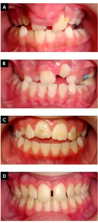
The study population was composed of 165 nonsyndromic subjects with no interfamilial rela-tionship of gender, between the ages of 6 and 15 years, and both with and without ED, ranging from hypomineralization to hypoplasia in permanent maxillary central incisors. They were divided into four groups: Group 1 - with CLP and ED (n = 46; Figure 1A); Group 2 - with CLP and without ED (n
= 34; Figure 1B); Group 3 - without CLP and with ED (n=34; Figure 1C) and Group 4 - without CLP or ED (n=51; Figure 1D).
D C B A
D C B A
Genomic DNA extraction, PCR and direct DNA sequencing
Saliva samples were collected from all subjects, and the genomic DNA was extracted from these samples with the InstaGeneTM Matrix Kit (732-6030,
Bio-Rad Laboratories, Hercules, USA), according to the manufacturer’s standards and based on a pre-vious study.18 A Polymerase Chain Reaction (PCR)
was then conducted in a thermocycler (Veriti 9902, Applied Biosystems, Carlsbad, USA), followed by direct DNA sequencing (3130xl Genetic Analyzer, 4352715, Applied Biosystems, Carlsbad, USA) of the
codiier areas (exons 2, 3, 4, 5, 6 and 7 AMELX). The
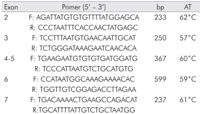
forward and reverse primers, as well as the PCR con-ditions, are listed in Table 1.
Analysis of the sequences obtained
The sequences obtained were analyzed by SeqS-cape Software® 2.6 (Applied Biosystems, Carlsbad,
USA). Mutations found in sequences using the
for-ward primer were conirmed by the sequencing using
the reverse primer.
Each variation of the nucleotide sequence
identi-ied in the sequencing was described using the den
Dunnen and Antonarakis19 nomenclature system.
In order to ind the variations, the bases were num
-bered as of the irst methionine (ATG) of the protein
resulting from this gene.
A search was performed at Blast20 and dbSNP21
databases to determine whether the alterations found
represented polymorphisms. Speciic programs were
used, such as Ensembl,22 to check if mutations found
in the present study had been previously cataloged. The power test was used to determine the number of patients, and a minimum of 30 patients per group was established. A statistical power of 80% and 95%
of conidence were used. Data were submitted to sta -tistical analysis using the Fisher’s exact test.
Statis-tical signiicance was established at 5%. StatisStatis-tical
analysis was performed with STATISTICA (version 11.0, StatSoft Inc., Tulsa, USA).
Results
In relation to the different groups, mutations were found in 30% (n = 14), 35% (n = 12), 11% (n = 4) and 13% (n = 7) of the subjects from groups 1, 2, 3 and 4, respectively. Thirty seven mutations were detected and dis-tributed throughout exons 2 (1 mutation – 2.7%), 6 (30 mutations – 81.08%) and 7 (6 mutations – 16.22%) of AMELX. No mutations were found in exons 3, 4 and 5.
O f t h e 3 0 m u t a t i o n s f o u n d i n e x o n 6, 43.34% (n = 13), 23.33% (n = 7), 13.33% (n = 4) and 20% (n = 6) were found in groups 1, 2, 3 and 4, respectively. c.261C>T (rs2106416), which is a silent mutation, was detected in 26 subjects, and signif-icantly more were found (p = 0.003) in patients with CLP (groups 1 and 2 – 23.75%), compared with those without CLP (groups 3 and 4 – 8.23%). In the groups without ED, this silent mutation was also found more significantly (p = 0.032) among subjects with CLP (17.65% in group 2), compared with those without CLP (7.8% in group 4).
Aside from this single nucleotide polymorphism
(SNP), ive other mutations that lead to an amino
acid substitution (Table 2) were found, one for exon 2 (c.34G>R) and four for exon 6 (c.245T>W, c.362A>G, c.420C>M and c.482C>R).
Table 2 illustrates the mutations found in the present study.
Discussion
Enamel development involves the expression of multiple genes needed to control the complex process of mineralization. Mutations in enamel proteins and protease genes have been associ-ated with ED.3,23,24,25,26 The cause of ED could be a
Table 1. Sequence of forward and reverse primers designed for exons 2, 3, 4,5, 6 and 7 of AMELX and PCR conditions
Exon Primer (5’ – 3’) bp AT
2 F: AGATTATGTGTGTTTTATGGAGCA 233 62˚C R: CCCTAATTTCACCAACTATGAGC
3 F: TCCTTTAATGTGAACAATTGCAT 250 57˚C
R: TCTGGGATAAAGAATCAACACA
4-5 F: TGAAGAATGTGTGTGATGGATG 367 60˚C R: TCCCATTAATGTCTGCATGTG
6 F: CCATAATGGCAAAGAAAACAC 599 59˚C
R: TGGTTGTCGGAGACCTTAGAA
7 F: TGACAAAACTGAAGCCAGACAT 237 61˚C
R:TGCATTTTATTGTCTGCTAATGG
Table 2. Distribution of the mutations found in the four groups studied
Group N Exon Mutation Electropherogram Amino acid
1 1 2 c.34G>R (A/G) (hetero) p.12G>R (Gly>Arg)*
7 6 c.261C>T (homo) p.87H>H (His>His)
6 6 c.261C>Y (C/T) (hetero) p.87H>H (His>His)
(14)
2 2 6 c.261C>T (homo) p.87H>H (His>His)
4 6 c.261C>Y (C/T) (hetero) p.87H>H (His>His)
1 6 c.420C>M (A/C) (hetero) p.140P>P (Pro>Pro)
5 7 off transcript
---(12)
3 1 6 c.362A>R (A/G) (hetero) p.121H>R (His>Arg)*
1 6 c.261C>T (homo) p.87H>H (His>His)
2 6 c.261C>Y (C/T) (hetero) p.87H>H (His>His)
(4)
4 4 6 c.261C>Y (C/T) (hetero) p.87H>H (His>His)
1 6 c.482 A>R (A/G) (hetero) p.161H>R (His>Arg)*
1 6 c.245T>W (A/T) (hetero) p.82V>E (Val>Glu)*
1 7 off transcript
---(7)
Total 37
genetic disorder linked to specific and nonspecific diseases during odontogenesis with the mineral metabolism, especially calcium phosphate. Some researchers suggest that CLP could be related to these enamel alterations, in which case a direct relationship would exist between defect appear-ance and cleft presence.16,17 Considering that ED
could be related to different mutations located in several genes involved in tooth enamel formation, and that the amelogenin gene forms the skeleton for the mineralization and the formation of the enamel crystals,3 the investigation of genes that
transcribe the main proteins and proteases of enamel is essential to gaining a better understand-ing of these alterations. AMELX is composed of seven exons and six introns, and alternative splic-ing results in three different transcripts, accord-ing to Ensembl.22 In the present study, the exons
participating in the transcription of AMELX were sequenced (exons 2-7).
Previous studies have revealed that AMELX is associated with amelogenesis imperfecta (AI),27,28
caused by a mutation in the genes critical to normal enamel formation.27 A mutated AMELX produces
an altered amelogenin polypeptide resulting in AI.29 A study3 evaluated AMELX in mouse models
to gain an understanding of the pathogenesis of AI. The authors asserted that ED is poorly under-stood and that many studies regarding genes in
human tooth development are signiicantly lim -ited.3 Thus, since AMELX encodes a protein that
plays a key role in the organization and structure of the enamel, it is the key player for organizing and structuring this highly mineralized tissue.30
Another study reported two SNPs in the AMELX coding sequences in humans. rs2106416 is a C>T silent substitution in amino acid 87 (His>His), It is located in exon 6, and was also found in the present study (Table 2). The other SNP, rs6639060, is also a C>T silent substitution in amino acid 152 (Leu>Leu),30 and was not detected in the present
study. Nonetheless, it is known that the central region of AMELX(exon 6), although highly vari-able in mammalian evolution, is highly preserved in humans.30
Association and comparison among groups are pertinent in the present study. The mutations found were distributed in all groups studied. Our results suggest that the presence of CLP signifi-cantly increases the frequency of mutations in AMELX, as compared with the presence of ED, since 70.2% of the mutations were found in CLP groups (groups 1 and 2), whereas 29.7% were found in ED groups (groups 3 and 4). CLP may be involved in a broader dysmorphic spectrum of anomalies17 − 81.08% of all mutations were found
in exon 6 of AMELX. A single mutation common to all groups was c.261C>T mutation, SNP rs2106416, located in region NM_006125.2 of chromosome X, representing 70.2% of all mutations detected. This SNP changes the C ancestral allele into the T mutant allele. In 1,000 genomes allele frequen-cies, the T mutant allele is present in 17% of all populations, whereas in the American population it is present in 12%.26 In our study, this mutant
allele was found in 15.7% of the sample (26 SNPs in 165 individuals; Table 2).
This is the first report demonstrating that AMELX may be a candidate gene for CLP. Further studies are necessary to investigate mutations and polymorphisms, including SNPs, in the codifying and splicing of candidate gene areas for defects in enamel formation. This will contribute to elu-cidating the consequences of these mutations in the proteins, as regards tooth enamel develop-ment, and determining why CLP is more related to mutations than ED is.
Conclusion
In conclusion, the present study suggested that AMELX may be a candidate gene for cleft lip and palate.
Acknowledgments
The authors would like to acknowledge the assistance of all the volunteers and the financial support of The São Paulo Research Foundation
(Fundação de Amparo à Pesquisa do Estado de São
Paulo - FAPESP; scholarship to FVO process #
1. Robinson C, Brookes SJ, Shore RC, Kirkham J. The develop-ing enamel matrix: nature and function. Eur J Oral Sci. 1998 Jan;106(Suppl 1):282-91.
2. Smith CE. Cellular and chemical events during enamel matu-ration. Crit Rev Oral Biol Med. 1998 Apr;9(2):128-61. 3. Wright JT, Hart TC, Hart PS, Simmons D, Suggs C, Daley B,
et al. Human and mouse enamel phenotypes resulting from mutation or altered expression of AMEL, ENAM, MMP20 and KLK4. Cells Tissues Organs. 2009 Dec;189(1-4):224-9. 4. Stephanopoulos G, Garefalaki ME, Lyroudia K. Genes and
related proteins involved in amelogenesis imperfecta. J Dent Res. 2005 Dec;84(12):1117-26.
5. Wright JT, Torain M, Long K, Seow K, Crawford P, Aldred MJ, et al. Amelogenesis imperfecta: genotype-phenotype studies in 71 families. Cells Tissues Organs. 2011 Aug;194(2-4):279-83. 6. Haubek D, Gjørup H, Jensen LG, Juncker I, Nyegaard M,
Børglum AD, et al. Limited phenotypic variation of hypo-calcified amelogenesis imperfect in a Danish five-generation family with a novel FAM83H nonsense mutation. Int J Pae-diatr Dent. 2011 Nov;21(6):407-12.
7. Lee SK, Lee KE, Jeong TS, Hwang YH, Kim S, Hu JC, et al. FAM83H mutations cause ADHCAI and alter intracellular protein localization. J Dent Res. 2011 Mar;90(3):377-81. 8. Urzúa B, Ortega-Pinto A, Morales-Bozo I, Rojas-Alcayaga G,
Cifuentes V. Defining a new candidate gene for amelogen-esis imperfecta: from molecular genetics to biochemistry. Biochem Genet. 2011 Feb;49(1-2):104-21.
9. ncbi.nlm.nih.gov [homepage on the Internet]. Maryland, USA: Online Mendelian Inheritance in Man; Johns Hopkins Uni-versity School of Medicine Available; 2012. [cited 2013 Nov 4]. Available from: http://www.nlm.nih.gov/omim/300391/. 10. Gasse B, Grabar S, Lafont AG, Quinquis L, Opsahl
Vi-tal S, Davit-Béal T, et al. Common SNPs of AmelogeninX (AMELX) and dental caries susceptibility. J Dent Res. 2013 May;92(5):418-24.
11. Olszowski T, Adler G, Janiszewska-Olszowska J, Safranow K, Kaczmarczyk M. MBL2, MASP2, AMELX, and ENAM gene polymorphisms and dental caries in Polish children. Oral Dis. 2012 May;18(4):389-95.
12. Bailleul-Forestier I, Molla M, Verloes A, Berdal A. The genetic basis of inherited anomalies of the teeth. Part 1: clinical and molecular aspects of non-syndromic dental disorders. Eur J Med Genet. 2008 Jul-Aug;51(4):273-91.
13. Parapanisiou V, Gizani S, Makou M, Papagiannoulis L. Oral health status and behaviour of Greek patients with cleft lip and palate. Eur Arch Paediatr Dent. 2009 Jun;10(2):85-9. 14. Hong L, Levy SM, Warren JJ, Broffitt B. Association between
enamel hypoplasia and dental caries in primary second mo-lars: a cohort study. Caries Res. 2009 Oct;43(5):345-53. 15. Kang SW, Yoon I, Lee HW, Cho J. Association between
AMELX polymorphisms and dental caries in Koreans. Oral Dis. 2011 May;17(4):399-406.
16. Maciel SP, Costa B, Gomide MR. Difference in the prevalence of enamel alterations affecting central incisors of children with complete unilateral cleft lip and palate. Cleft Palate Craniofac J. 2005 Jul;42(4):392-5.
17. Freitas JA, Neves LT, Almeida AL, Garib DG, Trindade-Suedam IK, Yaedú RY, et al. Rehabilitative treatment of cleft lip and palate: experience of the Hospital for Rehabilitation of Craniofacial Anomalies/USP (HRAC/USP) - Part 1: overall aspects. J Appl Oral Sci. 2012 Feb;20(1):9-15.
18. Sakai VT, Campos MR, Machado MAAM, Lauris JR, Greene AS, Santos CF. Prevalence of four putative periodontopathic bacteria in saliva of a group of Brazilian children with mixed dentition: 1-year longitudinal study. Int J Paediatr Dent. 2007 May;17(3):192-9.
19. den Dunnen JT, Antonarakis SE. Mutation nomenclature extensions and suggestions to describe complex mutations: a discussion. Hum Mutat. 2000 Jan; 15(1):7-12.
20. ncbi.nlm.nih.gov [homepage on the Internet]. Maryland, USA: BLAST Assembled RefSeq Genomes. 2012 [cited 2012 Jul 4]. Available from: http://www.ncbi.nlm.nih.gov/blast/. 21. ncbi.nlm.nih.gov [homepage on the Internet]. Maryland,
USA: dbSNP Short Genetic Variations. 2012 [cited 2012 Jul 4]. Available from: http://www.ncbi.nlm.nih.gov/snp/. 22. ensembl.org [homepage on the Internet]. Cambridgeshire,
UK : Ensembl Project Human (Homo sapiens). Download Human genome sequence; 2011 [cited 2012 Jul 4]. Available from: http://www.ensembl.org/Homo_sapiens/Info/Index/. 23. Hu JC, Yamakoshi Y, Yamakoshi F, Krebsbach PH, Simmer
JP. Proteomics and genetics of dental enamel. Cells Tissues Organs. 2005 Apr;181(3-4):219-31.
24. Hu JC, Simmer JP. Developmental biology and genet-ics of dental malformations. Orthod Craniofac Res. 2007 May;10(2):45-52.
25. Kapadia H, Mues G, D’Souza R. Genes affecting tooth mor-phogenesis. Orthod Craniofac Res. 2007 Nov;10(4):237-44. 26. Sawada T, Sawada T, Sekiguchi H, Yamashita H, Shintani
S, Yanagisawa T. Histological and immunohistochemical analyses of molar tooth germ in enamelin-deficient mouse. Acta Histochem. 2011 Sep;113(5):542-6.
27. Wright JT. The molecular etiologies and associated pheno-types of amelogenesis imperfecta. Am J Med Genet A. 2006 Dec 1;140(23):2547-55.
28. Hu JC, Chan HC, Simmer SG, Seymen F, Richardson AS, Hu Y, et al. Amelogenesis imperfecta in two families with defined AMELX deletions in ARHGAP6. PLoS One. 2012 Dec;7(12):e52052.
29. Lau EC, Slavkin HC, Snead ML. Analysis of human enamel genes: insights into genetic disorders of enamel. Cleft Palate J. 1990 Apr;27(2):121-30.
30. Richard B, Delgado S, Gorry P, Sire JY. A study of poly-morphism in human AMELX. Arch Oral Biol. 2007 Nov;52(11):1026-31.