Vol.48, n. 4 : pp. 599-610, July 2005
ISSN 1516-8913 Printed in Brazil
BRAZILIAN ARCHIVES OF
BIOLOGY AND TECHNOLOGY
A N I N T E R N A T I O N A L J O U R N A L
Anatomy of the Pericarp and Seed-coat of Lithraea
molleoides (Vell.) Engl. (Anacardiaceae) with Taxonomic
Notes
Sandra Maria Carmello-Guerreiro
1*and Adelita Aparecida Sartori Paoli
2 1Departamento de Botânica; Instituto de Biologia; Universidade Estadual de Campinas - UNICAMP; C. P. 6109; 13083-970; Campinas - SP - Brasil. 2Departamento de Botânica; Instituto de Biociências; Universidade Estadual Paulista - UNESP; C. P. 199; 13506-900; Rio Claro - SP - Brasil
ABSTRACT
The aim of the present work was to record anatomical data for the fruit and seed of Lithraea molleoides (Vell.) Engl, and compare the results with those for L. brasiliensis and the genera Schinus and Rhus. The L. molleoides fruit was a drupe with a friable and lignified exocarp. The mesocarp was parenchymatous with large secretory canals associated with vascular bundles. The endocarp consisted of four layers: an outer layer of polyhedral cells with prismatic crystals of calcium oxalate, and three inner layers of sclereids in a palisade arrangement. The ovule was anatropous, unitegmic, and crassinucelate. In the chalazal region, a cup-like zone of tanniniferous parenchymal cells formed the hypostase. The developing seed had a circinotropous–like shape, that originated through curvature of the long, coarse funicle that surrounded the tegument and embryo sac. The ripe seed was endotestal with bar-like thickenings or pittings in the cell walls.
Key words: Anacardiaceae, anatomy, pericarp, seed-coat
*
Author for correspondence
INTRODUCTION
The Anacardiaceae was currently divided into five tribes (Anacardieae, Spondiadeae, Rhoeae, Dobineeae and Semecarpeae), but this supra-generic classification was still controversial. Morphological, anatomical and rbcL sequence analyses by Terrazas and Chase (1996) suggested that two large clades should be recognized. One of these was monophyletic and contained members of the Spondiadeae group, while the second contained Anacardieae and Rhoeae. Although floral features had been used extensively to separate the taxa within the Anacardiaceae, Terrazas and Chase (1996)
suggested that anatomical features of the endocarp and wood characters were more useful.
The tribe Rhoeae contained the genera Rhus,
Schinus, Pistacia, Astronium, Schinopsis, Lithraea, Myracrodruon and others. Based on an analysis of
the structure of the pericarp in 29 genera, Wannan and Quinn (1990) identified two basic types of endocarp- Spondias-type and Anacardium-type - in the Anacardiaceae. The tribe Rhoeae could be divided into three groups A, B and C based on endocarp structure. Groups A and B were classified as Anacardium–type and group C as Spondias-type. The genera Rhus, Schinus, Schinopsis, Lithraea and
Myracrodruon were classified in group A, in which
and seeds of Lithraea brasiliensis, Pienaar and Von Teichman (1998) concluded that this species ought to be included in the genus Rhus under the name Rhus brasiliensis, a combination that, until publication of the foregoing article, was not valid.
In this report, we describe new anatomical data for the fruit and seed of a Brazilian species of
Lithraea, L. molleoides, and compare the results
with those for L. brasiliensis and the genera
Schinus and Rhus. Currently, only four species
of Lithraea were known worldwide, with L.
molleoides and L. brasiliensis being native to
Brazil (Engler, 1876; Barkley, 1962).
MATERIALS AND METHODS
Fruits of Lithraea molleoides (Vell.) Engl. at different stages of development were collected from specimens grown in the Botanical Garden of the Instituto de Biociências, UNESP, Botucatu, SP, Brazil. The morphological and anatomical studies were done using fresh material and material fixed in FAA (Johansen, 1940). The morphological characteristics of the fruits were described and illustrated using fruits sampled from at least five trees. The nomenclature used here was based on Radford et al. (1974) for the formed of the fruits, Spjut (1994) for the type of the fruit, and Roth (1977) for the pericarp layers.
Permanent slides were prepared from fixed samples embedded in plastic resin (Historesin, Leica) using the technique described by Gerrits (1991). The embedded material was glued to blocks of wood with epoxy-glue. Sections were cut on a rotary microtome with type C steel knives. The sections were stained with 0.05% toluidine blue in acetate buffer with pH 4.7 (O’Brien et al., 1964) and mounted in synthetic resin.
For histochemical tests, the sections of fresh material were treated with a) an aqueous solution of FeCl3 to detect phenolic compounds, b) phloroglucinol-HCL for lignified walls (Sass,
Morphology of the fruit and seed
The fruit was a white-greyish, smooth globose drupe 4-8mm in diameter, with a friable exocarp when ripe. The secretory mesocarp was dark and joined to the stone endocarp. The seed, one per fruit, was laterally compressed (Fig. 7) and derived from an anatropous ovule (Fig. 1) inserted on the basal-lateral side of the ovary. The seed-coat was membranaceous, smooth, and light yellow with a dark brown patch. As the seed grew, the long, curved, coarse funicle stick to the tegument of the seed (Figs. 2-7). The chalazal region and the funicle were seen externally as a dark brown patch in the ripe seed.
Ovary
The ovary was uniloculate with only one ovule (Fig. 1). The outer epidermis, which was uniseriate was covered with a cuticle and has stomata. The epidermal cells were radially elongated, with evident nuclei (Figs. 8 and 9). The ovarian mesophyll was formed by parenchyma cells and secretory canals associated with vascular bundles. The parenchyma cells close to the canals contained phenolic compounds (Figs. 8 and 9)
The inner epidermis of the ovary was multilayered with periclinal divisions that formed a pluristratified endocarp (sensu stricto) (Figs. 10-12). The layer of cells covering the locule had a cuticle and was the first to elongate radially (Fig. 12).
The structure of the pericarp
Longitudinal and transversal sections of young fruits showed the following anatomical features:
a) An outer epidermis with radially-elongated cells (Fig. 13);
b) A parenchymatous central zone characterized by secretory canals associated with vascular bundles arranged compactly, next to the other, in one layer, and a large number of parenchyma cells with phenolic compounds surrounding the secretory canals (Fig. 13); and
The outer epidermis form the exocarp (sensu
stricto) which, in the ripe fruit, lignified and
became friable (Figs. 13 and 14 ).
During the development of the fruit, the mesocarp consisted of a parenchymatous zone, secretory canals and vascular bundles and underwent very few changes, most of which involved an increase in the number of cells and in the size of the secretory canals (Figs. 15-17). The presence of parenchyma cells with phenolic compounds (Figs. 16 and 17) and canals with an amber colored secretion (Fig. 17) was characteristic in this species. In the ripe fruit, the mesocarp was secretory, became black and detaches from the exocarp. The separation of the exocarp and mesocarp occurred between the epidermis and the first layer of mesocarp cells (Fig. 14).
The endocarp consisted of four layers of cells, that were fully derived from the inner epidermis of the ovary wall and formed the endocarp sensu
stricto (Figs. 10-12). In ripe fruit, the endocarp
consisted of an outer layer of small polyhedric cells with prismatic crystals of calcium oxalate followed by three layers of sclereids in palisade in which the second layer had smaller cells than the first and third layers (Figs. 18-20). In ripe fruit, the four layers of cells that formed the endocarp became thicker and the walls became lignified (Figs. 19 and 20).
The seed
The ovule was anatropous, unitegmic and crassinucelate, with an evident dorsal raphe and a coarse funicle inserted in a basal-lateral position in the ovary wall (Figs.1 and 8). The developing seed had a circinotropous–like shape originates through the curvature of the long, coarse funicle that surrounded the tegument and embryo sac (Figs. 2-7, 21 and 26)
In the micropyle region, the tegument was formed by 5-12 layers of cells (Figs. 22 and 23). Tanniniferous deposits were present in the outer epidermis (Fig. 23). In the inner epidermis the cells were small, with a dense cytoplasm and were arranged compactly (Fig. 23). In the anti-raphe region, the tegument was formed by 4-5 layers of cells (Figs. 24 and 25). The outer epidermis cells were radially elongated and those of the inner epidermis were small and cubic, with a dense cytoplasm, and were arranged compactly. In the chalazal region, a very characteristic cup-like zone of tanniniferous parenchyma cells partially surrounded the nucellus and the embryo sac to formed the hypostase (sensu
lato) (Figs. 21, 24, 26 and 27). The funicle was
coarse, with a vascular bundle that was surrounded by a sheath of phenolic compound cells (Fig. 27) and grew towards the micropyle to formed a funicular obturator (Fig. 21 and 26).
In a further stage of development, the single tegument or testa in the micropyle and anti-raphe region had 4-12 layers of cells. The outer epidermis of the testa was formed by large polyhedral radially elongated cells containg phenolic compounds (Fig. 28). The inner epidermis of the testa has small, cubic cells with evident nuclei and a dense cytoplasm (Fig. 28). The mesophyll had a loose fitting and were compressed in some places. Immediately below the tegument, a few cell layers of the nucellus were compressed. The endosperm had several layers of cells with an evident nucleus and dense cytoplasm in the peripheral embryo sac (Figs. 25, 27and 28) and was quite vacuolated in the center. The ripe seed was exalbuminous, but showed the remainder of the nucellus and endosperm.
In the raphe-chalazal region, the hypostase contained cells with phenolic compounds and druses of calcium oxalate (Fig. 30).
Figures 1-7 - 1. Longitudinal section of a flower with an anatropous ovule inserted on the basal-lateral side
of the ovary. 2. Longitudinal section of a very young seed. 3-7. Stages of seed development. (CA=chalaza; EP=epidermis; FN=funicle; HP=hypostase; RA=raphe; TE=testa; SP=sepal; PE=petal; OV=ovule; OB=obturator; VB=vascular bundle; RC=receptacle)
PE
VB
OB
RC
VB
FN
RA
CA
EP
HP
1
2
3
4
5
6
7
OV
CA
RA
Figures 8-14 - 8. Longitudinal section of an ovary. (OM=ovarian mesophyll) (scale bar=250µm). 9-13. Transversal
secction of a young fruit. 9.Uniseriate exocarp (EX) and mesocarp (ME) (scale bar= 50µm). 10. Mesocarp (ME) with secretory canals (SC) and the multilayered endocarp (EN) (scale bar=50µm). 11. Endocarp in division (arrows) (scale bar=50µm). 12. Multilayered young endocarp (EN) (scale bar=50µm). 13. Pericarp. (SC=secretory canal; EN=endocarp; EX=exocarp; short arrow=stoma; white arrows=phenolic compounds) (scale bar=50µm). 14. Detail of the mature exocarp. White arrow showed the region of separation between the exocarp and mesocarp (scale bar= 125µm).
OM
8
9
EX
ME
ME
SC
10
EN
12
EX
11
SC
EN
1314
Figures 15-20 - 15. Transversal section of a ripe fruit (scale bar=1000µm). 16. Pericarp in longitudinal section
(scale bar=150µm). 17. Mesocarp with phenolic compounds and secretory canals with dense secretion and multilayered endocarp (scale bar=500µm). 18. Four layers of endocarp without lignification (scale bar=125µm). 19. Three outer layers of ripe endocarp. The arrow showed the crystalliferous layer and two palisade lignified layers (★★★★ and ✱✱✱✱) (scale bar=50µm). 20. Lignified
inner layer of ripe endocarp (scale bar=50µm).EN=endocarp; SC=secretory canal.
15
18
16
17
19
20
SC
SC
EN
Figures 21-26 - 21. Longitudinal section of the circinotropous-like shape of a young seed. (VB=vascular bundle)
(scale bar=500µm). 22. Detail of the micropyle region of a young seed. (TE=testa; ES=Embryo sac) (scale bar=125µm). 23. Testa in the micropyle region. The black arrow showed the outer epidermis of the testa with phenolic compounds and the white arrow showed the inner epidermis of the testa (scale bar=80µm). 24. The anti-raphe (AR) region indicated with arrow; hypostase (HP) and embryo sac (ES)(scale bar=125µm). 25. Detail of the layers of the anti-raphe region. (TE=testa; NU=nucellus; ED=endosperm) (scale bar=50µm). 26. Longitudinal section of a young seed showing a well-developed hypostase (arrow); endosperm (ED) and heart-shaped embryo (scale bar=100µm).
21 24
22
25
23
26
VB
AR
ES
HP
TE
ES
TE
NU
Figures 27-32 - 27. Chalazal region showing the endosperm (ED), nucellus (NU), hypostase (HP), vascular bundle
(VB) (scale bar=250µm). 28. Longitudinal section of the anti-raphe region (TE=testa, NU=nucellus; ED=endosperm) (scale bar=40µm). 29. Longitudinal section of the ripe seed. The arrow indicates the vestigial funicular aril (scale bar=100µm). 30. Raphe-chalazal region of the ripe seed. Arrows show druses (EM=embryo) (scale bar=50µm). 31. Anti-raphe region. The arrow indicates secondarily thickened, lignified and abundantly pitted walls. (FA=vestigial funicular aril; EM=embryo) (scale bar=250µm). 32. Remains of the testa (black arrow) and pitted walls (white arrow) of the inner epidermis of the testa (scale bar=50µm).
VB
HP
ED
TE
NU
ED
EM
FA
27
28
29
30
31
The seed-coat was formed by waste from the testa, funicle, raphe-chalazal region and the hypostase (Fig. 30), with the testa cells appearing as flattered, non specialized cells (Figs. 30 and 32). The cells of the inner epidermis of the testa were small, secondarily thickened, lignified and had extensively pitted walls (Figs. 31 and 32), indicating an endostestal status. In some parts of the anti-raphe, only the endotesta covered the embryo (Fig.32).
In the ripe seeds, the only multiplicative region was the raphe-chalazal, with the micropyle and the anti-raphe regions showing little stratification (Figs. 29-32).
DISCUSSION
In the Anacardiaceae, the structure of the pericarp was an important diagnostic character, especially at the generic level. According to Wannan and Quinn (1990), the endocarp of the Anacardiaceae could be divided into two basic types: the Spondias-type, in which the sclereids were irregularly guided, and the
Anacardium-type, in which they were regularly guided. Lithraea, Rhus and Schinus all had an Anacardium-type endocarp.
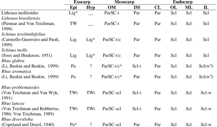
The structures of the exo-, meso- and endocarp of Rhus, Schinus and Lithraea showed many resemblances and were summarized in Table 1. The exocarp in L. molleoides and in L.
brasiliensis (Pienaar and Von Teichman, 1998)
were formed only by the outer epidermis. In ripe fruit, the cells had a thick non-lignified but very strongly cutinized wall that was separated at the mesocarp, which made it friable. In some species of Schinus, such as S. terebinthifolius (Carmello-Guerreiro and Paoli, 2002) and S.
molle (Soos and Hausknost, 1951), the exocarp
was formed by the epidermis plus the hypoderm, and became lignified, friable and separeted from the mesocarp. In Rhus lancea (Von Teichman and Robbertse, 1986; Von Teichman,1989) and in R. problematodes (Von Teichman and Van Wyk, 1991), the exocarp consisted of the outer epidermis, the hypoderm and a viariable number of mesocarp layers. In R. lancea, the hypoderm showed cells with thick, lignified walls, while in
Rhus problematodes they were not lignified. In Rhus (Toxicodendron) diversiloba (Copeland
and Doyel, 1940), the exocarp was papery, but its was unclear how many layers constitute the
exocarp. The exocarp of Rhus aromatica and R.
glabra was formed by the outer epidermis (Li et al.,
1999).
Schinus and Lithraea had a friable exocarp that
separeted from the mesocarp. In Rhus glabra and R.
aromatica, the single-layerd exocarp and the outer
mesocarp (with parenchyma cells) were considered as “papery fruit peel” that separeted from the rest of the fruit (vascular bundles and inner mesocarp) (Li et
al., 1999). It was unclear whether other Rhus species
had a the presence of friable exocarp and if this separeted from the mesocarp.
In all of the species studied in these three genera, the mesocarp was formed largely by parenchyma cells, tannic idioblasts and secretory canals associated with the vascular bundle. However, in Rhus lancea (Von Teichman and Robbertse, 1986; Von Teichman, 1989), R. problematodes (Von Teichman and Van Wyk, 1991), R. glabra and R. verniciflua (Brizicky, 1963; Li et al., 1999), theinner part of the mesocarp was sclerified and could be a part of the endocarp. In
Lithraea molleoides, L. brasiliensis (Pienaar and
Von Teichman, 1998), Schinus terebinthifolius (Carmello-Guerreiro and Paoli, 2002) and S. molle (Soos and Hausknost, 1951), no sclerification of the inner part of the mesocarp was observed..
The endocarp could be formed by the inner epidermis of the ovary and its immediate derivates, in which case it was referred to as sensu stricto; if the endocarp also include layers of the mesocarp then it was referred to as sensu lato (Roth, 1977). In
Lithraea molleoides, L. brasiliensis (Pienaar and
Von Teichman, 1998) and Schinus terebinthifolius (Carmello-Guerreiro and Paoli, 2002) the endocarp was of the sensu stricto type since was formed by four layers fully derived from the inner epidermis and consisted of an outermost crystalliferous layer and three inner layers formed by sclereids in palisad; the latter layers covered the locule.
In Rhus lancea (Von Teichman and Robbertse, 1986; Von Teichman, 1989), R. problematodes (Von Teichman and Van Wyk, 1991), R. glabra and
R. verniciflua (Brizicky, 1963; Li et al., 1999), the
endocarp was sensu lato since the sclerified portion of the mesocarp was part of the endocarp. Only in
R. aromatica was the inner mesocarp
parenchymatous and the endocarp was defined as
sensu stricto (Li et al., 1999).
The inner part of the endocarp, or endocarp sensu
stricto, developed from the inner epidermis of the
macrosclereids in contact with the locule had wavy radial walls, except in R. glabra and R.
aromatica (Li et al., 1999). This feature was not
observed in any species of Lithraea or Schinus studied so far.
The seeds of Rhus, Schinus and Lithraea were considered to be partially pachychalazal (Von Teichman, 1991; Carmello-Guerreiro and Paoli, 1999) since the chalazal part of the seed-coat was smaller than the tegument part, and was characterized by an endotegmen with a thickened, lignified cell wall and bar-like or pitted thickenings that prevent the collapse of the cells (Piennar and von Teichman 1998). The partial pachychalaza observed in these genera was easily distinguished externally by that dark-brown color seen in the seed-coat. A tanniferous hypostase
The ovules of L. molleoides and R. problematodes were basal but in Schinus they were apical (Carmello-Guerreiro and Paoli, 1999). Rhus and Schinus had an anatropous ovule (McNair, 1921; Kelkar, 1958; Von Teichman, 1991; Von Teichman and Van Wyk, 1991). L. molleoides also has an anatropous ovule, but after fertilization the funicle grew, curves and surrounded the tegument and embryo sac to give the young seed a circinotropous-like shape. In addition, the ovule of L. molleoides was unitegmic whereas in the species of Rhus and Schinus mentioned above the ovule was bitegmic.Pienaar and Von Teichman (1998) described the ovule of L. brasiliensis as bitegmic.
Table 1 - Comparison of the anatomical characters of the exo-, meso- and endocarp in Lithraea, Schinus and Rhus
species.
Exocarp Mesocarp Endocarp
Epi Hyp OM IM CL OL ML IL
Lithraea molleoides Lig* __ ParSC-t Par Par Scl Scl Scl
Lithraea brasiliensis
(Piennar and Von Teichman, 1998)
TW __ ParSC-t Par Par Scl Scl Scl
Schinus terebinthifolius
(Carmello-Guerreiro and Paoli, 1999)
Lig Lig* ParSC-t/c Par Par Scl Scl Scl
Schinus molle
(Soos and Houknost, 1951) Lig Lig* ParSC-t/c Par Par Scl Scl Scl
Rhus glabra
(Li, Baskin and Baskin, 1999) Pa ? ParSC-t/c* Scl-t Par Scl Scl Scl(w?)
Rhus aromatica
(Li, Baskin and Baskin, 1999) Pa ? ParSC-t/c* Par Par Scl Scl Scl(w?)
Rhus problematodes
(Von Teichman and Van Wyk, 1991)
TWt TWt ParSC-scl Scl-t Par Scl Scl Scl-w
Rhus lancea
(Von Teichman and Robbertse, 1986; Von Teichman, 1989)
TWt TWt ParSC-scl Scl-t Par Scl Scl Scl-w
Rhus diversiloba
(Copeland and Doyel, 1940) Pa* ? ParSC-scl Par Par Scl Scl Scl-w
As shown for several species of Rhus (McNair, 1921; Kelkar, 1958; Von Teichman, 1991; Von Teichman and Van Wyk, 1991) and for S. molle (Soos and Hausknost, 1951; Copeland, 1959) and S.
terebinthifolius (Carmello-Guerreiro and Paoli,
1999), the ovules of Rhus and Schinus showed greater similarity among themselves than do those of Rhus compared to Lithraea. In Schinus, as in
Rhus, the ovule was bitegmic, with the outer
tegument being much longer than the inner one. In L. molleoides, the bar-like thickenings or pittings on the cell walls of the inner epidermis of the testa characterized it as an endotestal seed, as indicated by Corner (1976). This type of thickening has been a very common feature of different species of Anacardiaceae, in the endotesta and endotegmen.
CONCLUSION
Lithraea and Schinus differed Rhus in the structure
of their endocarp. In the first two genera, the endocarp was of the sensu stricto type and consists of four layers of sclereids derived from the inner epidermis of the ovary. In Rhus, the endocarp was sensu lato type since it had layers of the mesocarp as well as the four layers from the inner epidermis. In addition to these differences, the sclereids in contact with the locule had wavy walls in Rhus and straight walls in Schinus and
Lithraea.
Further anatomical and taxonomic studies were needed to solve persisting problems regarding the generic limits in this family. In particular the types of the ovules and the number of teguments need to studied in detail in the three genera. Based on the findings of this study, it was concluded that L.
brasiliensis should not be included in the genus Rhus, contrary to the suggestion of Piennar and
Von Teichman (1998).
ACKNOWLEDGEMENTS
This work was supported by of CAPES and FAPESP.
RESUMO
O fruto de Lithraea molleoides (Vell.) Engl. é uma drupa globosa, branca-acinzentada, lisa, com exocarpo friável e lignificado quando maduro. O mesocarpo é parenquimático com grandes canais secretores associados aos feixes vasculares. O endocarpo é composto de quatro camadas: na camada mais externa as células são poliédricas, com cristais prismáticos de oxalato de cálcio, e nas três camadas internas as células são esclereides em paliçada. O envoltório da semente é membranáceo, liso, amarelo-pálido com uma mancha marrom escura. O óvulo é anátropo, unitegumentado, crassinucelado, inserido em posição basal-lateral. O funículo é crasso e cresce em direção à micrópila formando o obturador funicular. Na região calazal uma zona com células parenquimáticas de conteúdo tanífero formam a hipóstase. A semente em desenvolvimento apresenta uma forma circinótropa, originada a partir da curvatura do funículo crasso e longo que circunda o tegumento e o saco embrionário. A semente madura é endotestal com as paredes das células da endotesta espessadas e lignificadas em forma de barras ou pontuações.
REFERENCES
Barkley, F. A. (1962), Anacardiaceae: Rhoideae:
Lithraea. Phytologia, 8, 329-365.
Brizicky, G. K. (1963), The genera of Sapindales in the southeastern United States. J. Arnold Arbor., 44, 462-501.
Carmello-Guerreiro, S. M. and Paoli, A. A. S. (1999), Morfologia e anatomia da semente de Schinus
terebinthifolius Raddi, (Anacardiaceae), em
desenvolvimento. Revta.brasil. Bot., 22, 91-98. Carmello-Guerreiro, S. M. and Paoli, A. A. S. (2002),
Ontogeny and structure of the pericarp of Schinus
terebinthifolius Raddi (Anacardiaceae). Braz. Arch. Biol. Tech., 45, 73-79.
Copeland, H. F. (1959), The reproductive structures of
Schinus molle (Anacardiaceae). Madrõno, 15, 14-24.
Copeland, H. F. and Doyel, B. E. (1940), Some features of the structure of Toxicodendron diversiloba. Am. J.
Bot., 27, 932-939.
Corner, E. J. H. (1976), The Seeds of Dicotyledons. Cambridge : Cambridge University Press. 2 v. Engler, A. (1876), Anacardiaceae. In: Martius, C. P. F.
York : McGraw-Hill.
Kelkar, S. S. (1958), Embryology of Rhus mysurensis Heyne. J. Indian Bot. Soc., 37, 114-122.
Li, X.; Baskin, J. M. and Baskin, C. C. (1999), Pericarp ontogeny and anatomy in Rhus aromatica Ait. and
Rhus glabra L. (Anacardiaceae). J. Torrey Botan. Soc., 126, 279-288.
MacNair, J. B. (1921), The morphology and anatomy of
Rhus diversiloba. Am. J. Bot. 8, 179-191.
O'Brien, T. P.; Feder, N. and McCully, M. E. (1964), Polychromatic staining of plant cell walls by toluidine blue O. Protoplasma, 59, 368-373.
Pienaar, M. E. and Von Teichman, I. (1998). The generic position of Lithraea brasiliensis Marchand (Anacardiaceae): evidence from fruit and seed structure. Bot. J. Linn. Soc., 126, 327-337.
Radford, A. E.; Dickinson, W. C.; Massey, J. R. and Bell, C. R. (1974), Vascular Plant Systematics. New York : Harper and Row.
Roth, I. (1977), Fruits of Angiosperms. Berlin : Gebrüder Borntraeger.
Sass, J. E. (1951), Botanical Microtechnique. 3rd ed. Ames : State Press.
Soos, E. and Hausknost, M. (1951), Die Früchte von
Schinus molle L., dem Pfeffestrauch (Anacardiaceen). Scientia Pharmaceutica, 19, 213-219.
Spjut, R. W. (1994), A systematic treatment of fruit types. Mem. N. Y. Bot. Gard., 70, 1-82.
Bot., 55, 383-384.
Von Teichman, I. (1991), Ontogeny of the seed-coat of
Rhus lancea L. fil., and pachychalazy in the
Anacardiaceae. Bot. J. Linn. Soc., 107, 35-47.
Von Teichman, I. and Robbertse, P. J. (1986), Development and structure of the pericarp and seed of Rhus lancea L. fil. (Anacardiaceae), with taxonomic notes. Bot. J. Linn. Soc., 93, 291-306. Von Teichman, I. and Van Wyk, A. E. (1991),
Taxonomic position of Rhus problematodes (Anacardiaceae): evidence from fruit and seed structure. S. Afr. J. Bot., 57, 29-33.
Von Teichman, I. and Van Wyk, A. E. (1994), Structural aspects and trends in the evolution of recalcitrant seeds in dicotyledons. Seed Sci. Res. 4, 225-239.
Wannan, B. S. and Quinn, C. J. (1990), Pericarp structure and generic affinities in the Anacardiaceae.
Bot. J. Linn. Soc., 103, 225-252.