Experimental Evidence of Biological
Interactions among Different Isolates of
Trypanosoma cruzi
from the Chaco Region
Paula G. Ragone1,2*, Cecilia Pérez Brandán1,2, Mercedes Monje Rumi1,2, Nicolás Tomasini1,2, Juan J. Lauthier1,2, Rubén O. Cimino3, Alejandro Uncos2,
Federico Ramos2, Anahí M. Alberti D´Amato1,2, Miguel A. Basombrío2, Patricio Diosque1,2
1Unidad de Epidemiología Molecular, Instituto de Patología Experimental, CONICET (Consejo Nacional de Investigaciones Científicas y Técnicas), Universidad Nacional de Salta, Salta-Capital, Argentina,2Instituto de Patología Experimental, CONICET (Consejo Nacional de Investigaciones Científicas y Técnicas), Universidad Nacional de Salta, Salta-Capital, Argentina,3Cátedra de Química Biológica, Facultad de Ciencias Naturales, Universidad Nacional de Salta, Salta-Capital, Argentina
Abstract
Many infectious diseases arise from co-infections or re-infections with more than one geno-type of the same pathogen. These mixed infections could alter host fitness, the severity of symptoms, success in pathogen transmission and the epidemiology of the disease. Trypa-nosoma cruzi, the etiological agent of Chagas disease, exhibits a high biological variability often correlated with its genetic diversity. Here, we developed an experimental approach in order to evaluate biological interaction between threeT.cruziisolates belonging to different Discrete Typing Units (DTUs TcIII, TcV and TcVI). These isolates were obtained from a re-stricted geographical area in the Chaco Region. Different mixed infections involving combi-nations of two isolates (TcIII + TcV, TcIII + TcVI and TcV + TcVI) were studied in a mouse model. The parameters evaluated were number of parasites circulating in peripheral blood, histopathology and genetic characterization of each DTU in different tissues by DNA hybrid-ization probes. We found a predominance of TcVI isolate in blood and tissues respect to TcIII and TcV; and a decrease of the inflammatory response in heart when the damage of mice infected with TcVI and TcIII + TcVI mixture were compared. In addition, simultaneous presence of two isolates in the same tissue was not detected. Our results show that biologi-cal interactions between isolates with different biologibiologi-cal behaviors lead to changes in their biological properties. The occurrence of interactions among different genotypes ofT.cruzi observed in our mouse model suggests that these phenomena could also occur in natural cycles in the Chaco Region.
a11111
OPEN ACCESS
Citation:Ragone PG, Pérez Brandán C, Monje Rumi M, Tomasini N, Lauthier JJ, Cimino RO, et al. (2015) Experimental Evidence of Biological Interactions among Different Isolates ofTrypanosoma cruzifrom the Chaco Region. PLoS ONE 10(3): e0119866. doi:10.1371/journal.pone.0119866
Academic Editor:Herbert B. Tanowitz, Albert Einstein College of Medicine, UNITED STATES
Received:September 1, 2014
Accepted:January 16, 2015
Published:March 19, 2015
Copyright:© 2015 Ragone et al. This is an open access article distributed under the terms of the
Creative Commons Attribution License, which permits unrestricted use, distribution, and reproduction in any medium, provided the original author and source are credited.
Data Availability Statement:All relevant data are within the paper and its Supporting Information files.
Funding:This manuscript was supported by funding agency Argentina: Agencia Nacional de Promoción Científica y Tecnológica (http://www.agencia.mincyt. gob.ar), through a grant given to Dr. Patricio Diosque (PICT-2012-2174). The funders had no role in study design, data collection and analysis, decision to publish, or preparation of the manuscript.
Background
Advances in molecular typing techniques show that many infectious diseases may arise from co-infections or re-infections with more than one genotype of the same pathogen. In these mixed in-fections the co-infecting parasites may be interacting among each other within the same host deter-mining host fitness, severity of disease symptoms, parasite transmission successful rate and
epidemiology of the disease [1]. Various mechanisms can cause interactions between parasite species or among different genotypes of the same species within an individual host. For example, parasites can infect the same target site within a host and directly interact among each other by interference competition, or indirectly by resources competition or via the host immune system [2].
In general, biological interactions between protozoan parasites have been divided into two main groups: those who involved parasites belonging to the same species and the ones that occur between closely to distantly related different species [3]. In this sense, several research studies have reported mixed infections inLeishmania spp[4,5,6],Plasmodium spp[7,8,9], Try-panosoma bruceiandTrypanosoma congolense[10,11,12].
Trypanosoma cruziis the etiologic agent of Chagas disease, an illness that affects several million people in Latin America and still remains an important public health problem in certain endemic areas of Argentina. This parasite shows a high genetic variability which has been the basis to clas-sify it into six Discrete Typing Units (DTUs), TcI to TcVI [13]. In addition to this genetic diversi-ty,in vitroandin vivo T.cruziinfection models showed a high biological variability among different genotypes ofT.cruzi[14,15,16,17,18,19]. Although it is supposed that genetic and bio-logical diversities of the parasite are essential to determine the clinical course of Chagas disease, specific associations between particular clinical manifestations and a determined lineage have not been clearly demonstrated [20]. Furthermore, the host genetics and its ability to establish an im-mune response to control the infection are very important in the outcome of the disease [21].
The consequences of mixed infections by differentT.cruziDTUs have been studied in animal models using different laboratory strains. It has been demonstratedin vivothat the tissue tropism of oneT.cruzigenotype could change in the presence of another genotype of a different DTU [22,23]. Even more, the histopathological damage and the intensity of the inflammatory process resulting of these co-infections also present remarkable variations [24,25]. Other studies involvingT.cruzi
mixed infections showed that the parasite load in peripheral blood could be altered either increasing or decreasing according to the co-infecting strains [26,27,28]. Even the outcome of specific chemo-therapy has been proven to be altered by these events of concomitant infection byT.cruzi[26,29].
In several geographical areas of the Southern Cone of America, the occurrence of natural mixed infections by different genotypes ofT.cruzihave been widely reported in humans [20,30,31,32,33], in wild and domestic animals [34,35] and in the vectorTriatoma infestans[34,36].
In a previous work we described the different biological properties displayed by three select-ed isolates obtainselect-ed from the Chaco region of Argentina and belonging to DTUs TcIII, TcV and TcVI [17]. These isolates have the particularity of circulating sympatrically in a restricted geographical area; therefore, here we describe the biological outcome resulting ofin vivo exper-imental dual-mixed infections with theseT.cruzistrains. Our working hypothesis is the exis-tence of biological interactions among differentT.cruziisolates in the vertebrate host.
Methods
Ethics statement
Trypanosoma cruzi
isolates
DifferentTrypanosoma cruziisolates were examined in the present work. These isolates were obtained from Las Leonas settlement (W 61° 39’8.7”, S 27° 01’49”), located in the South-west of Chaco Province, Argentina. The protocol of obtaining samples was approved by the Bioeth-ics Committee of the Faculty of Health Sciences of the National University of Salta, Argentina (Resolution N°052–10). The inhabitants signed an informed consent form before sampling at each house. This study did not involve endangered or protected species.
Parasites were recovered from the feces of either, naturally infectedTriatoma infestansor insects used for xenodiagnosis of mammalian hosts. The isolates were identified as DTUs TcIII (LL051-P24), TcV (LL014–1) and TcVI (LL040–1) by Multilocus Sequence Typing (MLST) technique, using a typing scheme proposed by Lauthier and cols, [38]. These parasites were maintained in a vector transmission model developed by Ragone and cols, (2012). Hereafter, each isolates will be named according to the corresponding DTU.
Experimental infection in mice
Six groups of 4 male C57BL/6J mice (one month old) were inoculated by intraperitoneal (i.p.) route with parasites recovered from the feces of infected insects and each infected group was followed during 30 days after infection. Prior infection, the feces were visualized microscopical-ly, in order to distinguish epimastigotes, metacyclic and intermediate forms (parasites whose morphology is intermediate between epimastigotes and tripomastigotes); according to Kollien and cols. [39]. The final inoculation dose was adjusted according to the amount of metacyclic and intermediate forms. For single infections with TcIII, TcV or TcVI, 104parasites were inoc-ulated per mouse. Instead, 5x10³parasites from each isolate were inoculated for dual mixed in-fections (TcIII + TcV, TcIII + TcVI and TcV + TcVI). We decided to maintain constant the final dose used in both, single and dual-mixed infections, in order to avoid possible differences due to the final number of inoculated parasites. For this reason, we examined in previous assays if the two doses, 104and 5x10³parasite/mouse of each isolate show differences in the biological parameters to be studied. No statistical differences were observed between these doses (data not shown). Control groups inoculated with Phosphate Buffer Saline (PBS) were included in the experiment. Animal care guidelines adopted by the Health Sciences Faculty, National Uni-versity of Salta, Argentina, were strictly followed.
Biological parameters evaluated
Parasitemia. Fresh blood from inoculated animals was collected in heparinized glass
capil-lary pipettes by sectioning the tail tip under slight anesthesia. Ten microliters (μl) of blood
were placed between slide and cover slip and the number of parasites per 100 fields was re-corded microscopically (400X) at different time points.
Histopathology. Animals were sacrificed at 30 days post infection (dpi) by exposure to
halothane, and cardiac and skeletal muscle samples were collected. Tissue samples were divided into two parts; one was stored at -80°C for DNA extractions and the other part was fixed in 10% formalin and processed using routine histological techniques. Serial histological sections (3 to 5μm thick), were stained with hematoxylin-eosin and observed under microscope (50,
Detection of
T
.
cruzi
DNA in blood and tissue samples
Peripheral blood was obtained at 30 dpi from each animal (350μl) and mixed with 700μl of
guanidine buffer. DNA extractions were performed from 100μl of the mixture
blood-guani-dine buffer by using the phenol-chloroform method. DNA extractions from a skeletal and car-diac muscle sample (obtained at 30 dpi) were performed using a kit (PureLink Genomic DNA Kit, Invitrogen). Subsequently, PCR amplification of 330 bp corresponding to minicircle hy-pervariable regions (mHVR) was performed using primers 121 (5’-AATAATGTACGGG(T/ G)GAGATGCATGA-3’) and 122 (5’-GGTTCGATTGGGGTTGGTGTAATATA-3’) [40]. Amplification was performed in a MJR PTC-100 thermocycler (MJ Research, Watertown, MA, USA). The reaction products were visualized in a 2% agarose gel stained with ethidium bromide.
Identification of
T
.
cruzi
Discrete Typing Units in biological samples
Detection of each isolate was carried out by hybridization with specific mHVR-kDNA non-ra-dioactive probes in blood and tissues samples taken at 30 dpi. The probes were constructed using DNA from isolates LL051-P24 (TcIII), LL055R3cl2 (TcV) and CL-Brener (TcVI). The primers for probe construction were CV1 (5’-GATTGGGGTTGGAGTACTAT-3’) and CV2 (5’-TTGAACGGCCCTCCGAAAAC-3’) which produced a 290-bp fragment. Restriction sites for Sau96I and ScaI which allow elimination of the minicircle constant region of these PCR fragments were included in the sequence of these primers [41]. PCR fragments were further digested with the restriction endonucleases obtaining a 250-bp final product. The specificity of each generated DNA probe was evaluated in our laboratory by Southern blot analysis against different 330-bp mHVR PCR products of severalT.cruziisolates belonging to the dif-ferent DTUs [42]. Briefly, Southern blot analysis was performed using 10μl of each 121–122
PCR product. Samples were subjected to electrophoresis, transferred to Hybond N + nylon membranes (Roche Diagnostics) and cross-linked with UV light to fix the DNA. The mem-branes were pre-hybridized for at least 30 minute at 42°C and individually hybridized with the generated probes. Labeling of the probe and DNA hybridization were performed according to the protocol supplied with the PCR-DIG DNA-labeling and detection kit (Roche Applied Sci-ence, USA).
For assessing the limit of detection of the technique,T.cruziDNA of the different isolates involved in the study was analyzed individually and combined. The limit of the technique in detecting single infections was 0.1 fg/μl for TcIII and TcV DTUs, and 0.5 fg/μl for TcVI DTU.
Whereas that in the mixed infections we evaluated different proportions of one isolate com-bined with other isolate, from 0.1 fg/μl to 0.9 fg/μl. As a result of this experiment, the detection
limit for TcV was 0.1 fg/μl in the TcIII + TcV and TcV + TcVI mixtures; while the detection
limit for TcIII was 0.5 fg/μl in TcIII + TcV and TcIII+TcVI. Finally the detection limit for
TcVI in the mixtures TcV+TcVI and TcIII+TcVI was 0.5 fg/μl.
Statistical analysis
Results
Predominance of the more virulent isolate in peripheral blood and DTU
detection by mHVR-kDNA hybridization
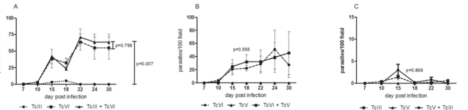
As in a previous study Ragone and cols. [17] in this work a marked difference was well estab-lished respecting the parasitemia between the three different isolates. The TcV isolate presented non-detectable parasitemia in fresh blood mounts. However, PCR assays corroborated infec-tion by this isolate. On the other hand, circulating parasites in peripheral blood were detected in mice inoculated with TcIII and TcVI, being the parasitemia of TcVI significantly higher than the one obtained by TcIII (p = 0,013). When co-infection models (TcIII + TcVI, TcIII + TcV and TcV + TcVI) were considered, the pattern of parasitemia was the one corresponding to the more virulent DTU in all cases (Figs. 1A-C). In the co-infection involving TcIII + TcVI and TcV + TcVI, the parasitemia was the one described for TcVI alone. In addition, in the mix-ture TcIII + TcV, a behavior equal to TcIII was observed. As expected, there were
non-significant differences between TcVI vs. TcIII + TcVI and TcVI vs. TcV + TcVI; neither between TcIII vs. TcIII + TcV. No statistical comparison between TcV vs. TcIII + TcV or TcV + TcVI was carried out since no circulating parasites were detected for TcV isolate.
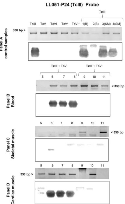
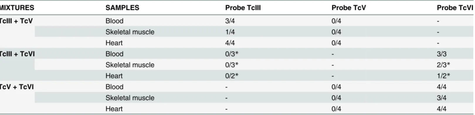
Finally, in blood samples collected at 30 days post-infection we applied specific mHVR-kDNA hybridizations to determine the circulating DTU in single and dual infections. TcVI was identified in all the infection formulas involved (TcIII + TcVI and TcV + TcVI mixtures) (Fig. 2B), while TcIII was only detected in the mixture TcIII + TcV (Fig. 3B). Even though no visible parasites were detected for TcV, positive PCR could be obtained (Fig. 4); however, no hybridization signal for TcV was detected in the mixture TcV + TcVI. In addition, for the blend TcV + TcIII, only TcIII could be detected (Fig. 3B).Table 1shows the number of mice with positive signal for each specific probe.
Histopathological damage and its association with the different infecting
DTUs
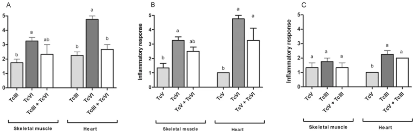
When single infections were considered, different patterns of tissue damage were observed. TcVI isolate induced significantly more histological lesions in skeletal muscle (p = 0.0026) and heart (p<0.0001) than TcIII and TcV. In addition, TcIII produced significantly more damage in heart than TcV (p<0.0001). Briefly, TcVI induced severe damage in heart and moderate damage in skeletal muscle; additionally amastigote nests and fiber homogenizations were found in both tissues in animals infected with this isolate. On the other hand, TcIII induced mild-moderate lesions in heart and skeletal muscle and no amastigote nests or cellular alter-ation were found; while TcV induced only mild lesions in the analyzed tissues.
However, in our co-infecting models a moderate damage in heart and skeletal muscle was found in the mixtures TcIII + TcVI and TcV + TcVI, while in mice infected with TcIII + TcV, mild histological lesions were observed in both tissues. In addition, amastigote nests were found in heart and skeletal muscle of animals infected with TcVI + (TcIII or TcV).
statistical differences between single and mixed infections were detected; however, the lesions induce by the mixture were significantly different than the induce by TcV alone (p = 0,001) (Fig. 5C).
In skeletal muscle, the intensity of the lesions found in mice infected with TcIII + TcVI and TcV + TcVI mixtures was intermediate respect to each single infection; however, no statistical differences were detected (Fig. 5A and 5B). In TcIII + TcV co-infection the damage in skeletal muscle was equal to the one detected in single infections with TcIII or TcV (Fig. 5C).
Heart and muscle tissues were analyzed by PCR and posterior DTU specific hybridization assays.T.cruziDNA was detected in both, skeletal and cardiac muscle, in all experimental groups. In addition, we found the presence of the isolates TcVI or TcIII in the mixed infections (Fig. 2C and DandFig. 3C and D). Again, although TcV probe worked correctly (Fig. 4), no hybridization against positive PCR samples of animals infected with TcV + TcIII or TcV + TcVI mixtures was observed. DTU detection by hybridization tests is summarized inTable 1.
Discussion
Here, we developed an experimental approach in order to evaluate if mixed infections involv-ingT.cruziisolates, belonging to different DTUs and collected from a restricted area in the Chaco Region of Argentina, display evidence of biological interaction in a mouse model. In this work we analyzed the parasitemia and histological damage in heart and skeletal muscle of C57BL/6J mice infected with a combination of the different isolates analyzed. The intrinsic properties of the isolates under study were as follow: TcVI isolate induces high parasitemia, se-vere histological damage in heart and moderate lesions in skeletal muscle. On the other hand, the TcIII isolate induces less parasitemia than TcVI, and mild-moderate histological damage in heart and skeletal muscle. Finally TcV isolate induces sub-patent parasitemia and mild lesions in the analyzed tissues. When TcVI was combined, either with TcIII or TcV, only the parasite-mia pattern of TcVI was observed, suggesting that TcVI isolate predominates over TcIII and TcV isolates. This result was supported by the fact that only DNA of TcVI was detected in the co-infection models involving this isolate and TcIII or TcV. Similar results were obtained for the mixture TcIII + TcV, where the observed parasitemia pattern corresponded to the TcIII pattern and only DNA of TcIII was detected by the probes. This result could be due to several factors: the survival of one isolate in peripheral blood could be related to different mechanisms associated with its ability to escape from the host immune system [28], or due to a selective process within the host cells in favor of a given DTU [43]. On the other hand, in a previous work we reported that the isolates TcIII and TcVI induce a higher serological response than TcV [17]; in consequence we believe that in a mixed infection event between TcV + (TcIII or
Fig 1. Parasitemia in peripheral blood of singles and mixed infections.This variable was measured by microscopic observation, of animals inoculated with single and mixed isolates. (A) Shows difference between TcIII vs TcVI vs TcIII + TcVI, (B) TcVI vs TcV + TcVI and (C) TcIII vs TcIII + TcV. The parasitemia of TcV isolate was sub-patent.
TcVI), the isolate TcV may be susceptible to the immunological response induced by TcIII or TcVI. Therefore, TcV availability in the host would be reduced, or even eliminated from the host, and not detected at least by the technique herein applied.
When cardiac muscle samples of mice infected with the mixtures TcIII + TcVI and TcV + TcVI were analyzed; only presence of TcVI isolate was confirmed by hybridizations assays.
Fig 2. Southern blot analyses using the TcVI (CL-Brener) probe.Each panel shows the electrophoretic pattern of minicircle regions and kDNA transferred to a nylon membrane. Panel A, lanes TcIII, TcV, TcVI, TcV*and TcVI*correspond to DNA of parasite culture from LL051-P24 (DTU TcIII), LL055R3cl2 (DTU TcV), CL-Brener (DTU TcVI), LL014–1 (DTU TcV*) and LL040–1 (DTU TcVI*) respectively; lane 1–4: blood (B),
skeletal muscle (SM) and heart (H) samples of mouse infected with TcVI isolate. The asterisk as superscript of the DTU indicates DNA sample from culture of the same inoculated isolate. Panel B, C and D: blood, skeletal muscle and cardiac muscle, respectively, of animals infected with TcV + TcVI (Lane 5–8) and TcIII +
TcVI (Lane 9–11).
However, cardiac lesions induced by TcIII + TcVI mixture were the same that the induced by TcIII isolate alone, suggesting that the presence of TcIII, perhaps in an early infection, modifies the alterations that TcVI alone could cause in the co-infected mice, in spite of the TcIII isolate is not detected in this mixture. On the other hand, skeletal muscle samples of animals
co-Fig 3. Southern blot analyses using the TcIII (LL051-P24) probe.Each panel shows the electrophoretic pattern of minicircle regions and kDNA transferred to a nylon membrane. Panel A: lane TcIII, TcV, TcVI, TcV* and TcVI*correspond to DNA of parasite culture from LL051-P24 (DTU TcIII), LL055R3cl2 (DTU TcV), CL-Brener (DTU TcVI), LL014–1 (DTU TcV*) and LL040–1 (DTU TcVI*) respectively; and lane 1–4: blood (B)
and skeletal muscle (SM) samples of mouse infected with TcIII isolate. The asterisk as superscript of the DTU indicates DNA sample from culture of the same inoculated isolate. Panel B, C and D: blood, skeletal muscle and cardiac muscle, respectively, of animals infected with TcIII + TcV (Lane 5–8) and TcIII + TcVI (Lane
9–11).
infected with TcVI + (TcV or TcIII) showed intermediate values of damage compared to single infections, indicating that the combination of two isolates could modify the expected lesions in this tissue. These results could be due to the ability of each isolate to infect cells since it is known thatT.cruziis capable of infecting a wide variety of host cells, but the persistence of this parasite in cardiac and skeletal muscles depends on its ability to enter in the host cell and in the interaction with this [44]. On the other hand, it has been reported that the co-infections be-tween different strains ofT.cruzitrigger both protective inflammatory immunity and regulato-ry immune mechanisms that attenuate the damage in heart in a mouse model [25].
Based on these observations we evidence biological interaction in our murine co-infection model. However, it is important to note that the prevalence of either one isolate or the another could vary according to the infection period in which blood samples are taken [25,28,45] as well as the analyzed tissue [22].
Fig 4. Southern blot analyses using the TcV (LL055R3cl2) probe.Electrophoretic pattern of minicircle regions and kDNA transferred to a nylon membrane. Lane TcIII, TcV, TcVI, TcV*and TcVI*correspond to DNA of parasite culture from LL051-P24 (DTU TcIII), LL055R3cl2 (DTU TcV), CL-Brener (DTU TcVI), LL014–
1 (DTU TcV) and LL040–1 (DTU TcVI), respectively; and lane: 1–6 blood (B) skeletal muscle (SM) and
cardiac muscle (H) of mouse infected with TcV isolate.
doi:10.1371/journal.pone.0119866.g004
Table 1. mHVR-kDNA hybridization results for the detection of individual DTUs in mixed infections.
MIXTURES SAMPLES Probe TcIII Probe TcV Probe TcVI
TcIII + TcV Blood 3/4 0/4
-Skeletal muscle 1/4 0/4
-Heart 4/4 0/4
-TcIII + TcVI Blood 0/3* - 3/3
Skeletal muscle 0/3* - 2/3*
Heart 0/2* - 1/2*
TcV + TcVI Blood - 0/4 4/4
Skeletal muscle - 0/4 3/4
Heart - 0/4 4/4
The values correspond to mice with positive hybridization for a specific probe in relation to the total of mice inoculated with the mixture. *indicate one or two sample lost.
In spite of several studies demonstrating the presence of biological interactions within mam-malian hosts [23,25,27,28,46], these works analyzed strains ofT.cruzifrom very different geo-graphical regions. In the present work, the threeT.cruziisolates studied were obtained in the same temporal period, in the same geographical area and simultaneously introduced into the animal model.
Is important to note that the DTUs studied in this work have epidemiological relevance, since the TcV and TcVI are more prevalent in domestic cycles [34,42,47,48], whereas the TcIII DTU was found in domestic animals [34]. It is clear that the extrapolation of co-infection results obtained from animal models to the natural occurrence of the phenomena in other hosts should be carefully considered. We emphasize the predominance of TcVI in the mixture TcV + TcVI since this mixture was found in human blood samples in endemics areas of Argentina [42,49]. Many studies have shown that the biological differences between closely related DTUs are smaller than the biological differences among genetically divergent DTUs [14,15,19,27]. How-ever, an interesting question that emerges from this work is why two genetically close strains of
T.cruzi, as TcV and TcVI, exhibit opposite biological properties. Even the biological interac-tion between them could lead to the predominance of one isolate (TcVI) over the other (TcV), at least during the acute phase and in our specific experimental model. In this sense we believe that the interaction with the host immune response, as well as the mechanisms related to the regulation of the acute inflammatory response and the proteomic expression of different DTUs could also be contributing to the pathology of each isolate and its interactions.
Conclusions
The results presented in this work show that biological interactions among different genotypes ofT.cruzifrom the Chaco Region do occur in our experimental model. Consequently, our re-sults demonstrate that the biological interaction betweenT.cruzistrains in a mammal host is phenomenon that could be occurring in natural cycles. However, to examine this hypothesis field studies involving natural hosts should be performed.
Acknowledgments
We are grateful to Marcela Portelli for technical help in the histological preparations, María Celia Mora for assistance in animal care and Dr. Julio Nasser for contributions to this study.
Fig 5. Histological damage in mice infected with single and mixed infections.Inflammatory response observed in skeletal muscle and heart samples of experimental groups inoculated with: (A) TcIII, TcVI or TcIII + TcVI, (B) TcV, TcVI or TcV + TcVI and (C) TcIII, TcV or TcIII + TcV. Different letters above error bars indicate statistical difference (p<0.05).
Author Contributions
Conceived and designed the experiments: PGR CPB MAB PD. Performed the experiments: PGR CPB MMR NT JJL RC AU FR AMAD. Analyzed the data: PGR. Contributed reagents/ materials/analysis tools: PD. Wrote the paper: PGR CPB PD.
References
1. Pedersen AB, Fenton A (2007) Emphasizing the ecology in parasite community ecology. Trends Ecol Evol 22: 133–139. PMID:17137676
2. Telfer S, Birtles R, Bennett M, Lambin X, Paterson S, et al. (2008) Parasite interactions in natural popu-lations: insights from longitudinal data. Parasitology 135: 767–781. doi:10.1017/S0031182008000395
PMID:18474121
3. Cox FE (2001) Concomitant infections, parasites and immune responses. Parasitology 122 Suppl: S23–38. PMID:11442193
4. Bastrenta B, Mita N, Buitrago R, Vargas F, Flores M, et al. (2003) Human mixed infections of Leishman-iaspp. andLeishmania-Trypanosoma cruziin a sub Andean Bolivian area: identification by polymerase chain reaction/hybridization and isoenzyme. Mem Inst Oswaldo Cruz 98: 255–264. PMID:12764443 5. Mendes DG, Lauria-Pires L, Nitz N, Lozzi SP, Nascimento RJ, et al. (2007) Exposure to mixed
asymp-tomatic infections withTrypanosoma cruzi,Leishmania braziliensisandLeishmania chagasiin the human population of the greater Amazon. Trop Med Int Health 12: 629–636. PMID:17445130 6. Veland N, Valencia BM, Alba M, Adaui V, Llanos-Cuentas A, et al. (2013) Simultaneous infection with
Leishmania (Viannia) braziliensisandL.(V.) lainsoniin a Peruvian patient with cutaneous leishmaniasis. Am J Trop Med Hyg 88: 774–777. doi:10.4269/ajtmh.12-0594PMID:23382155
7. Bell AS, de Roode JC, Sim D, Read AF (2006) Withhost competition in genetically diverse malaria in-fections: parasite virulence and competitive success. Evolution 60: 1358–1371. PMID:16929653 8. Dinko B, Oguike MC, Larbi JA, Bousema T, Sutherland CJ (2013) Persistent detection ofPlasmodium
falciparum,P.malariae,P.ovale curtisiandP.ovalewallikeri after ACT treatment of asymptomatic Gha-naian school-children. Int J Parasitol Drugs Drug Resist 3: 45–50. doi:10.1016/j.ijpddr.2013.01.001
PMID:24533292
9. Flores-Mendoza C, Fernandez R, Escobedo-Vargas KS, Vela-Perez Q, Schoeler GB (2004) Natural Plasmodiuminfections in Anopheles darlingi andAnopheles benarrochi(Diptera: Culicidae) from east-ern Peru. J Med Entomol 41: 489–494. PMID:15185955
10. Balmer O, Tostado C (2006) New fluorescence markers to distinguish co-infectingTrypanosoma brucei strains in experimental multiple infections. Acta Trop 97: 94–101. PMID:16212925
11. Ezeokonkwo RC, Ezeh IO, Onunkwo JI, Obi PO, Onyenwe IW, et al. (2010) Comparative haematologi-cal study of single and mixed infections of mongrel dogs withTrypanosoma congolenseand Trypano-soma brucei brucei. Vet Parasitol 173: 48–54. doi:10.1016/j.vetpar.2010.06.020PMID:20638796 12. Omeje JN, Anene BM (2012) Comparative serum biochemical changes induced by experimental
infec-tion ofT.bruceiandT.congolensein pigs. Vet Parasitol 190: 368–374. doi:10.1016/j.vetpar.2012.07.
008PMID:22858639
13. Zingales B, Miles MA, Campbell DA, Tibayrenc M, Macedo AM, et al. (2012) The revisedTrypanosoma cruzisubspecific nomenclature: rationale, epidemiological relevance and research applications. Infect Genet Evol 12: 240–253. doi:10.1016/j.meegid.2011.12.009PMID:22226704
14. Dos Reis D, Monteiro WM, Bossolani GD, Teston AP, Gomes ML, et al. (2012) Biological behaviour in mice ofTrypanosoma cruziisolates from Amazonas and Parana, Brazil. Exp Parasitol 130: 321–329.
doi:10.1016/j.exppara.2012.02.016PMID:22406038
15. dos Santos DM, Talvani A, Guedes PM, Machado-Coelho GL, de Lana M, et al. (2009)Trypanosoma cruzi: Genetic diversity influences the profile of immunoglobulins during experimental infection. Exp Parasitol 121: 8–14. doi:10.1016/j.exppara.2008.09.012PMID:18848935
16. Lisboa CV, Pinho AP, Monteiro RV, Jansen AM (2007)Trypanosoma cruzi(kinetoplastida Trypanoso-matidae): biological heterogeneity in the isolates derived from wild hosts. Exp Parasitol 116: 150–155.
PMID:17274984
17. Ragone PG, Perez Brandan C, Padilla AM, Monje Rumi M, Lauthier JJ, et al. (2012) Biological behavior of differentTrypanosoma cruziisolates circulating in an endemic area for Chagas disease in the Gran Chaco region of Argentina. Acta Trop 123: 196–201. doi:10.1016/j.actatropica.2012.05.003PMID:
18. Revollo S, Oury B, Laurent JP, Barnabe C, Quesney V, et al. (1998)Trypanosoma cruzi: impact of clon-al evolution of the parasite on its biologicclon-al and medicclon-al properties. Exp Parasitol 89: 30–39. PMID:
9603486
19. Toledo MJ, de Lana M, Carneiro CM, Bahia MT, Machado-Coelho GL, et al. (2002) Impact of Trypano-soma cruziclonal evolution on its biological properties in mice. Exp Parasitol 100: 161–172. PMID:
12173401
20. del Puerto R, Nishizawa JE, Kikuchi M, Iihoshi N, Roca Y, et al. (2010) Lineage analysis of circulating Trypanosoma cruziparasites and their association with clinical forms of Chagas disease in Bolivia. PLoS Negl Trop Dis 4: e687. doi:10.1371/journal.pntd.0000687PMID:20502516
21. Macedo AM, Machado CR, Oliveira RP, Pena SD (2004)Trypanosoma cruzi: genetic structure of popu-lations and relevance of genetic variability to the pathogenesis of chagas disease. Mem Inst Oswaldo Cruz 99: 1–12. PMID:15486627
22. Andrade LO, Machado CR, Chiari E, Pena SD, Macedo AM (1999) Differential tissue distribution of di-verse clones ofTrypanosoma cruziin infected mice. Mol Biochem Parasitol 100: 163–172. PMID:
10391378
23. Franco DJ, Vago AR, Chiari E, Meira FC, Galvao LM, et al. (2003)Trypanosoma cruzi: mixture of two populations can modify virulence and tissue tropism in rat. Exp Parasitol 104: 54–61. PMID:12932760 24. Andrade SG, Campos RF, Sobral KS, Magalhaes JB, Guedes RS, et al. (2006) Reinfections with
strains ofTrypanosoma cruzi, of different biodemes as a factor of aggravation of myocarditis and myosi-tis in mice. Rev Soc Bras Med Trop 39: 1–8. PMID:16501758
25. Rodrigues CM, Valadares HM, Francisco AF, Arantes JM, Campos CF, et al. (2010) Coinfection with differentTrypanosoma cruzistrains interferes with the host immune response to infection. PLoS Negl Trop Dis 4: e846. doi:10.1371/journal.pntd.0000846PMID:20967289
26. Martins HR, Silva RM, Valadares HM, Toledo MJ, Veloso VM, et al. (2007) Impact of dual infections on chemotherapeutic efficacy in BALB/c mice infected with major genotypes ofTrypanosoma cruzi. Antimi-crob Agents Chemother 51: 3282–3289. PMID:17638698
27. Martins HR, Toledo MJ, Veloso VM, Carneiro CM, Machado-Coelho GL, et al. (2006)Trypanosoma cruzi: Impact of dual-clone infections on parasite biological properties in BALB/c mice. Exp Parasitol 112: 237–246. PMID:16406355
28. Sales-Campos H, Kappel HB, Andrade CP, Lima TP, Mattos ME Jr., et al. (2014) A DTU-dependent blood parasitism and a DTU-independent tissue parasitism during mixed infection ofTrypanosoma cruziin immunosuppressed mice. Parasitol Res 113: 375–385. doi:10.1007/s00436-013-3665-z
PMID:24178748
29. Toledo MJ, Bahia MT, Carneiro CM, Martins-Filho OA, Tibayrenc M, et al. (2003) Chemotherapy with benznidazole and itraconazole for mice infected with differentTrypanosoma cruziclonal genotypes. Antimicrob Agents Chemother 47: 223–230. PMID:12499195
30. Burgos JM, Diez M, Vigliano C, Bisio M, Risso M, et al. (2010) Molecular identification ofTrypanosoma cruzidiscrete typing units in end-stage chronic Chagas heart disease and reactivation after heart trans-plantation. Clin Infect Dis 51: 485–495. doi:10.1086/655680PMID:20645859
31. Coronado X, Zulantay I, Albrecht H, Rozas M, Apt W, et al. (2006) Variation inTrypanosoma cruziclonal composition detected in blood patients and xenodiagnosis triatomines: implications in the molecular epidemiology of Chile. Am J Trop Med Hyg 74: 1008–1012. PMID:16760511
32. Diez C, Lorenz V, Ortiz S, Gonzalez V, Racca A, et al. (2010) Genotyping ofTrypanosoma cruzi subli-neage in human samples from a North-East Argentina area by hybridization with DNA probes and spe-cific polymerase chain reaction (PCR). Am J Trop Med Hyg 82: 67–73. doi:
10.4269/ajtmh.2010.09-0391PMID:20064998
33. Garcia A, Ortiz S, Iribarren C, Bahamonde MI, Solari A (2014) Congenital co-infection with different Try-panosoma cruzilineages. Parasitol Int 63: 138–139. PMID:24422218
34. Cardinal MV, Lauricella MA, Ceballos LA, Lanati L, Marcet PL, et al. (2008) Molecular epidemiology of domestic and sylvaticTrypanosoma cruziinfection in rural northwestern Argentina. Int J Parasitol 38: 1533–1543. doi:10.1016/j.ijpara.2008.04.010PMID:18585717
35. Rozas M, Botto-Mahan C, Coronado X, Ortiz S, Cattan PE, et al. (2007) Coexistence ofTrypanosoma cruzigenotypes in wild and periodomestic mammals in Chile. Am J Trop Med Hyg 77: 647–653. PMID:
17978065
36. Bosseno MF, Yacsik N, Vargas F, Breniere SF (2000) Selection ofTrypanosoma cruziclonal genotypes (clonet 20 and 39) isolated from Bolivian triatomines following subculture in liquid medium. Mem Inst Oswaldo Cruz 95: 601–607. PMID:10998206
38. Lauthier JJ, Tomasini N, Barnabe C, Rumi MM, D'Amato AM, et al. (2012) Candidate targets for Multilo-cus Sequence Typing ofTrypanosoma cruzi: validation using parasite stocks from the Chaco Region and a set of reference strains. Infect Genet Evol 12: 350–358. doi:10.1016/j.meegid.2011.12.008
PMID:22210092
39. Kollien AH, Schaub GA (2000) The development ofTrypanosoma cruziin triatominae. Parasitol Today 16: 381–387. PMID:10951597
40. Gomes ML, Macedo AM, Vago AR, Pena SD, Galvao LM, et al. (1998)Trypanosoma cruzi: optimization of polymerase chain reaction for detection in human blood. Exp Parasitol 88: 28–33. PMID:9571142 41. Veas F, Breniere SF, Cuny G, Brengues C, Solari A, et al. (1991) General procedure to construct highly
specific kDNA probes for clones ofTrypanosoma cruzifor sensitive detection by polymerase chain re-action. Cell Mol Biol 37: 73–84. PMID:2059987
42. Rumi MM, Perez Brandan C, Gil JF, D'Amato AM, Ragone PG, et al. (2013) Benznidazole treatment in chronic children infected withTrypanosoma cruzi: serological and molecular follow-up of patients and identification of Discrete Typing Units. Acta Trop 128: 130–136. doi:10.1016/j.actatropica.2013.07.
003PMID:23880286
43. Pena DA, Eger I, Nogueira L, Heck N, Menin A, et al. (2011) Selection of TcIITrypanosoma cruzi popu-lation following macrophage infection. J Infect Dis 204: 478–486. doi:10.1093/infdis/jir292PMID:
21742848
44. Andrade LO, Andrews NW (2005) TheTrypanosoma cruzi-host-cell interplay: location, invasion, reten-tion. Nat Rev Microbiol 3: 819–823. PMID:16175174
45. D'Avila DA, Macedo AM, Valadares HM, Gontijo ED, de Castro AM, et al. (2009) Probing population dy-namics ofTrypanosoma cruziduring progression of the chronic phase in chagasic patients. J Clin Microbiol 47: 1718–1725. doi:10.1128/JCM.01658-08PMID:19357212
46. Lo Presti MS, Esteves BH, Moya D, Bazan PC, Strauss M, et al. (2014) CirculatingTrypanosoma cruzi populations differ from those found in the tissues of the same host during acute experimental infection. Acta Trop 133: 98–109. doi:10.1016/j.actatropica.2014.02.010PMID:24560963
47. Corrales RM, Mora MC, Negrette OS, Diosque P, Lacunza D, et al. (2009) Congenital Chagas disease involvesTrypanosoma cruzisub-lineage IId in the northwestern province of Salta, Argentina. Infect Genet Evol 9: 278–282. doi:10.1016/j.meegid.2008.12.008PMID:19162237
48. Diosque P, Barnabe C, Padilla AM, Marco JD, Cardozo RM, et al. (2003) Multilocus enzyme electro-phoresis analysis ofTrypanosoma cruziisolates from a geographically restricted endemic area for Cha-gas' disease in Argentina. Int J Parasitol 33: 997–1003. PMID:13129520