58
Pakistan Veterinary Journal
ISSN: 0253-8318 (PRINT), 2074-7764 (ONLINE)Accessible at: www.pvj.com.pk
Molecular Characterization of
Staphylococcus aureus
Isolated from Meat and Their Antibiotic
Resistance Profiles
Ziad W Jaradat1, Yaser H. Tarazi2* and Qotaibah O. Ababneh3 1
Department of Biotechnology and Genetic Engineering, Faculty of Science and Arts, 2Department of Basic Veterinary Medical Sciences, Faculty of Veterinary Medicine, Jordan University of Science and Technology, P. O. Box 3030 Irbid, 22110, Jordan; 3Department of Biophysics and Biochemistry, Texas A&M University, College Station, TX, USA *Corresponding author: [email protected]
A R T I C L E H I S T O R Y A B S T R A C T
Received: Revised: Accepted:
October 18, 2012 May 01, 2013 June 16, 2013
The aims of this study were to characterize S. aureus isolates from different meat sources in Jordan and study their genetic relationship using PCR-RFLP in addition to their antibiotic resistance profiles. Thirty S. aureus isolates were identified and confirmed by PCR techniques. The isolates from goat and camel meats were sensitive to the majority of the tested antibiotics. Plasmid profiling revealed that 26 isolates contained at least one plasmid with no correlation between the number of plasmids and the resistance profiles. PCR-RFLP of the coagulase gene (coa) classified the isolates to several clusters upon digestion with Alu I or Cfo I restriction enzymes. This study concluded that the 30 S. aureus isolates were genetically diverse and comprised heterogeneous population with 7 genotypes at both 33.1 and 51.2 similarity levels.
©2013 PVJ. All rights reserved
Key words:
Antibiotics Coagulase gene Meat
RFLP S. aureus
To Cite This Article: Jaradat ZW, YH Tarazi and QO Ababneh, 2014. Molecular characterization of Staphylococcus aureus isolated from meat and their antibiotic resistance profiles. Pak Vet J, 34(1): 58-62.
INTRODUCTION
Staphylococcus aureus has been implicated in a variety of infections in human and animals (Bartlett and Hulten, 2010; Gu et al., 2013; Khan et al., 2013). The treatments of such infections become difficult due the emergence of multidrug resistant strains such as methicillin resistant S. aureus (MRSA) MRSA and S. aureus is also considered as a major food borne pathogen (Hennekinne et al., 2010; Kenar et al., 2012). In Jordan, Disease Control Directorate/Ministry of Health reported that staphylococcal food poisoning during seven years (1999-2005), was implicated in 176, 83, 172, 133, 103, 278, and 90 cases, respectively. Relatively high numbers of staphylococcal food poisoning in Jordan stressed the need to study and characterize S. aureus isolated from locally produced meat. Therefore, the detection and typing of S. aureus isolates and understanding their antibiotic susceptibility are essential to maintain rigor in quality control within the food processing environment. Traditional methods of S. aureus typing have been replaced by molecular typing (Sabat et al.,2006). In this study, RFLP of the coagulase gene was performed, as it is cheap and easy to proceed, and has high level of reproducibility (Hookey et al., 1998; Morandi et al., 2010). Typing the S. aureus isolates by RFLP enables our
understanding of their genetic structure and improve our capability of tracing the origin of S. aureus outbreaks. Therefore, the aim of this study was to characterize S. aureus isolates from meat of domestic animals used in Jordan; camels, beef, sheep and goats by PCR-RFLP of the coagulase gene, the isolates were tested for the presence of methicillin resistance genes and the antimicrobial susceptibility patterns.
MATERIALS AND METHODS
Sampling, processing and isolation of S. aureus: Two hundred raw meat samples were collected from camels, beef, sheep and goats during 2009 from abattoirs of four governorates in Jordan. Samples were processed as previously reported (Al-Tarazi et al., 2009). All samples were cultured on Baird-Parker agar base (BPA) supplemented with egg yolk-tellurite emulsion (Oxoid UK). Presumptive S. aureus isolates were further identified using Microbact Staph 12S system (Oxoid, UK).
Antibiotic test: Agar disk diffusion test of Penicillin (P; 10U), Oxacillin (OX; 1µg), Cefoxitin (FOX; 10µg), Gentamicin (GN; 10µg), Tobramycin (TOB; 10µg), Amikacin (AK; 30µg), Teicoplanin (TEC; 30 µg), Ciprofloxacin (CIP; 5µg), Chloramphenicol (C; 30µg),
Erythromycin (E; 15µg), Tetracycline (TE; 15µg), Trimethoprim/Sulphamethoxazole (SXT; 1.25/23.75µg), Amoxicillin-clavulanic acid (AMC.20/10µg), Azithromycin
(AZD; 15µg), Moxifloxacin (MXF; 5µg), Clindamycin (DA; 2µg), Rifampicin (RA; 5µg), Quinupristin/ Dalfopristin (QA; 15µg), Linezolid (LZD; 30µg) and Vancomycin (VA; 30µg) was carried out using the Clinical Laboratory Standard Institute guidelines, supplement M2-A9 (CLSI, 2008).
S. aureus identification and confirmation of Mec A and
Coa genes by PCR: DNA from the isolates was extracted using the Wizard Genomic DNA purification kit (Promega, USA) and the purity of the extracted DNA was tested by running samples on agarose gel electrophoresis and pure DNA stored in -80°C. Thermal cycling for PCR amplification was performed as described in Table 2.
DNA restriction endonuclease analysis of the PCR-amplified coagulase gene: Restriction analysis was conducted as described by Hookey et al. (1998) and Plasmid profiling of the isolateswas performed using the Wizard Plus SV minipreps DNA purification system (Promega, USA).
RFLP cluster analysis of the isolates: RFLP banding patterns of the restriction coa PCR-products for the 30 S. aureus isolates were examined by estimating the size of each fragment on the electrophoresis gel and bands were scored, with the data coded as a factor of 1 or 0, representing the presence or absence of a restriction band, respectively. A similarity matrix among S. aureus isolates was produced using the Jaccard coefficient. A dendrogram showing the genetic relatedness among the isolates was constructed from the resulting data using SPSS version 15 (SPSS Inc. 1989-2007). The cutoff for the dendrogram was selected based on the average of mean similarity matrix.
RESULTS AND DISCUSSION
Isolation and enumeration of S. aureus: Thirty of 200 meat samples revealed presence of PCR-confirmed S. aureus (Table 1). Those isolates were then tested for antibiotic sensitivity, plasmid profile and cluster analysis to understand their genetic relatedness using Alu I and Cfo I restriction enzyme analysis. The combination of phenotypic and genotypic characterization is often effective in tracing a particular isolate incriminated in an outbreak. Genomic fingerprinting is necessary for classifying multiple isolates into groups with similar or close similarity indices (Sabour et al., 2004). In this work, phenotypic and genotypic properties and relatedness of the 30 S. aureus isolates were investigated.
Testing of antibiotic resistance by disk diffusion and PCR methods: Thirty S. aureus isolates were tested for sensitivity to commonly used antibiotics. All the tested isolates were completely sensitive to six antibiotics (OX, GN, AK, TOB, AZM and C) (Table 1). Also the isolates showed high susceptibility (>90%) to Qd, RA, MET, LZD and DA antibiotics. This indicates the lower frequency of using these antibiotics for therapy or as animal feed
additive. In contrast, the majority of the isolates (87 and 80%) were resistant to penicillin and amoxicillin-clavulanic acid, respectively, highlighting the frequent use of these antibiotics for animals. This appears to be in agreement with other report (Guven et al., 2010).
Among the 30 isolates, one isolate was sensitive to all the antibiotics tested and five other isolates exhibited resistance to two, while the rest exhibited resistance to more than two antibiotics. These results are similar to those published by Andrew et al. (2011). In this study, when analyzing multidrug resistance patterns of antibiotics to each animal species, it appeared that isolates of beef meats exhibited the highest resistance; one isolates resistance to 11 and another to 8 antibiotics, followed by two isolates from camel and goat's meat which was resistant to 8 and 7 antibiotics, respectively (Table 1).
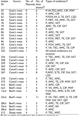
Table 1: Source of the PCR S. aureus confirmed isolates, number of plasmids, number of antibiotics each isolate resistant to and types of these antibiotics.
Isolate ID
Source No. of
Plasmids No. of Anti- biotics@
Types of antibiotics$
55 Goat’s meat 0 6 P,VA,TEC,AMC, CIP, MXF
56 Goat’s meat 0 3 P, AMC, CIP
62 Goat’s meat 2 7 P,FOX,VA, E, TE, SXT, LZD
66 Goat’s meat 2 6 P, MET, VA, AMC, TE, SXT
76 Goat’s meat 2 3 P, AMC, SXT
79 Goat’s meat 0 4 AMC, TE, CIP, SXT
83 Goat’s meat 2 2 P, SXT
84 Goat’s meat 1 4 P, AMC, TE, SXT
92 Goat’s meat 1 3 P, AMC, SXT
94 Goat’s meat 2 7 P, FOX, MET, TE, CIP, SXT
96 Goat’s meat 3 2 P, AMC
99 Goat’s meat 1 5 P, FOX, AMC, TE, SXT
251 Camel’s meat 4 6 P, VA, TEC, AMC, TE, CIP
252 Camel’s meat 1 0 All tested antibiotics are
sensitive
254 Camel’s meat 1 6 P, TEC, AMC, TE, CIP, SXT
258 Camel’s meat 3 2 VA, SXT
259 Camel’s meat 1 5 P, AMC, TE, CIP, DA
263 Camel’s meat 0 2 CIP, SXT
266 Camel’s meat 1 4 P, AMC, CIP, SXT
267 Camel’s meat 1 8 P,AMC, E,TE, CIP, DA, SXT,
LZD
270 Camel’s meat 1 3 P, TEC, AMC,
272 Camel’s meat 1 6 P, AMC, TE, CIP, MXF, SXT
4cd Beef’s meat 1 3 P, AMC, CIP,
6cd Beef’s meat 7 6 P, VA, AMC, E, CIP, MXF
12cd Beef’s meat 1 8 P,VA,TEC, AMC, E,TE, CIP,
MXF
22cd Beef’s meat 1 11 P, VA, TEC, AMC, E, TE, CIP,
MXF, DA, SXT, QD
28cd Beef’s meat 1 5 P, AMC, TE, CIP, SXT
33cd Beef’s meat 2 3 P, AMC, RA
38cd Beef’s meat 0 2 P, AMC
512 Sheep meat 4 3 P, TEC, AMC
@: the number of antibiotics the isolate is resistant to $: the types of the antibiotics the isolates are resistant to
Table 2: Oligonucleotide primer pairs and PCR running conditions used for S. aureus
Gene Primers 5’…….3’ Amplification conditions size (bp) Product Reference
Temp Time No. of Cycles
Thermonuclease (nuc)
Pri-1
5’GCGATTGATGGTGATACGGTT 3’ Pri-2
5’ AGCCAAGCCTTGACGAACTAAAGC 3’
94°C 5 min 1 270 Pinto et al. (2005)
94°C 30 s 35
55°C 45 s
72°C 45 s
72°C 10 min 1
Methicillin resistance (mecA)
mecA F
5-'GTAGAAATGACTGAACGTCCGATGA 3'
mecA R 5'CCAATTCCACATTGTTTCGGTCTAA 3'
94°C 4 min 1 310 Geha et al. (1994)
94°C 45 s 35
50°C 45 s
72°C 60 s
72°C 2 min 1
Coagulase (coa)
Coa F
5'-ATA GAG ATG CTG GTA CAG G-3’ Coa R
5'-GCT TCC GAT TGT TCG ATG C-3’
94°C 45 s 1 Variablea Hookey et al. (1998)
94°C 20 s 35
57°C 15 s
72°C 15 s
72°C 2 min 1
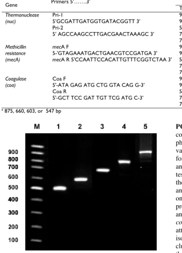
a 875, 660, 603, or 547 bp
Fig. 1: Agarose gel electrophoresis of amplified PCR fragments of coagulase gene for S. aureus sub species aureus isolates. Lane 1: three isolates of 500 bp, lane 2: fifteen isolates of 600 bp, lane 3: nine isolates of 660 bp, lane 4: two isolates of 800 bp, lane 5: one isolate of 850 bp.
this study, there was no relationship between these two facts which appears to be in agreement with results reported by Yamazumi et al. (2001), where the difference in phenotypic and genotypic methicillin resistance might be due to heterogeneous expression of mec A gene by Staphylococcus strains.
Plasmid Profiling: When the 30 S. aureus isolates were tested for the presence of plasmids, 5 isolates did not contain any plasmid although they exhibit resistance to some antibiotics indicating that the resistance genes might be located on the chromosomal genome of these isolates. Also, the majority (14 isolates) contained only 1 plasmid. Moreover, the isolate that contained the highest number of plasmids (7) exhibited resistance to only 6 antibiotics while an isolate showed resistance to 11 different antibiotics contained only 1 plasmid highlighting the lack of correlation between the number of plasmids and the antibiotic resistance profiles (r2 = 0.003) (Table 1). These results are supported by a study of Dharmalingam et al. (2003) who reported no correlation between the presence of the plasmids in Helicobacter pylori isolates and their antibiotic resistance. These results indicated the inadequacy of plasmid analysis to obtain any results on the antibiotic resistance profiles of S. aureus isolates.
PCR-RFLP analysis of coagulase gene: Production of coagulase by S. aureus isolates is considered an important phenotypic feature and is used to type this pathogen. The variability of the 3’ end region of the coa gene is the basis for typing which is used to trace pathogens among animals or humans (da Silva and da Silva, 2005). Upon testing the isolates for the presence of coa gene by PCR, the size of the amplified genes were 500, 600, 660, 800 and 850 bp in 3, 15, 9, 2 and 1 isolate, respectively, with one amplicon for each isolate (Fig. 1). This indicates the presence of the same allelic forms of coa gene (da Silva and da Silva, 2005). In this study, PCR products of the coa gene were digested with CfoI and Alu I enzymes in an attempt to confirm the resulting RFLP patterns for these isolates. Both enzymes generated the same number of clusters but differ in the distribution of the isolates among the clusters in that, clusters appeared to be identical after digestion with one enzyme are different when digested with the other enzyme (Fig. 2 and 3).
Among the clusters, cluster A for both enzymes shared 6 isolates and cluster B for Alu I enzyme shared 12 isolates with cluster C of the Cfo I enzymes. These results differ from a study conducted by Hookey et al., (1998) who reported very similar clusters obtained for the S. aureus isolates digested with Alu I and Cfo I enzymes.
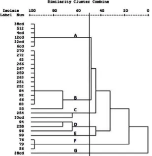
Dendrogram was constructed on the basis of similarity index among S. aureus isolates using Alu -generated RFLP (Fig. 2). A 33.1% cutoff value gave 3 major clusters with 26 isolates while 4 isolates formed single clusters. Cluster C contains 15 isolates (9 from goats and 6 from camel meat) with similarity range of 0.36-1.0. The genotype C cluster was predominant with three sub clusters, sub cluster I for isolates from camel’s meat, sub-clusters II and III form goat meat collected from two different locations suggesting the existence of the same clones in different geographical regions.
Cluster B contained only 3 isolates (similarity level 1.0) from camel meat suggesting that these isolates are indistinguishable or have been derived from similar clones or represent the same strain. Whereas, all meat samples 7 beef's and 3 sheep’s formed one cluster (A) with a similarity ranged from 0.5-1.0. Genotypes D, E, F and G contain one isolate in each with minimal similarity.
contaminating strains are predominant in slaughterhouses in Jordan and animals are moved easily before being slaughtered. In addition, there were some isolates belonging to the same cluster although isolated from animals in different places. This substantiates the existence of certain strains of S. aureus that might have spread among the animals in these places (Aires-de-Sousa et al., 2007).
The dendrogram was constructed on the basis of the similarity index among S. aureus isolates using Cfo I
Fig. 2: Dendrogram derived from Jaccard Coefficient Cluster analysis based on combined similarity matrix obtained from Coagulase RFLP-
using Alu I restriction enzyme showing genetic relatedness among S.
aureus isolates from meat in Jordan. The scale at the top shows the
similarity index. The cutoff was set at 33.1% similarity. Clusters are numbered by capital letter A-G. Numbers at the left side of the dendrogram denote the isolate identification numbers.
Fig. 3: Dendrogram derived from Jaccard Coefficient Cluster analysis based on combined similarity matrix obtained from Coagulase RFLP-
using Cfo I restriction enzyme showing genetic relatedness among S.
aureus isolates from meat in Jordan. The scale at the top shows the
similarity index. The cutoff was set at 51.2% similarity. Clusters are numbered by capital letter A-G. Numbers at the left side of the dendrogram denote the isolate identification numbers.
RFLP Fig. 3. A 51.2% cutoff value gave 7 distinct clusters. Cluster A contained 6 indistinguishable isolates (similarity of 1.0), 5 of them from beef meat, 1 isolate from sheep meat. The high rate of similarity within a particular group (same abattoir) indicates cross contamination among the isolates at some point during processing (van Loo et al.,2007). Cluster B contained 14 isolates (similarity 0.5-1.0) of which 6 isolates from goat meat that was obtained from 3 different abattoirs while the other 8 isolates from camels meat that was obtained from the same abattoir. It is noteworthy that all the isolates but one (isolate no. 55) were indistinguishable with 100% similarity when digested with Cfo I while they were not identical when digested with Alu I enzyme. The similarity in the genotypes of some of the isolates necessitates the use of two enzymes to resolve differences among isolates. Genotypes C and D contained mixtures of the isolates from both camels and goats meat with cluster D contains 3 isolates (similarity 0.7-0.8) one from camels and two from goats meat while cluster C containing two isolates sharing 0.7% similarity. Cluster F contained 3 isolates (similarity 1.0) all from goat’s meat. Clusters E and G contained one isolate each (no. 99 and no. 28 cd, respectively). Only isolate (no. 28 cd) was not digested by the Cfo I enzyme. Typing these isolates by RFLP using two restriction enzymes revealed that the isolates were genetically diverse and comprise a heterogeneous population with 7 genotypes at 33.1% and 51.2% similarity levels after restriction with Alu I and Cfo I enzymes, respectively.
Conclusion: The presence of multidrug resistant S. aureus isolated mainly from beef’s meat indicating the misuse of the antibiotics, and that aminoglycopeptides are the antibiotics of choice for treatment of a multi-resistant S. aureus infection in animals. RFLP of the coa gene offers a good discriminatory power in typing S. aureus isolates collected from different geographical regions in Jordan. The data presented would help food and animal health workers to enhance safety precautions before slaughtering and while handling meat originated from different animals.
Acknowledgment: This research was supported by
Deanship of Research at the Jordan University of Science and Technology (Grant no. 215/2007).
REFERENCES
Aires-De-Sousa M, CER Parente, OV Motta, ICF Bonna, DA Silva and H
De Lencastre, 2007. Characterization of Staphylococcus aureus
isolates from buffalo, bovine, ovine and caprine milk samples collected in Rio de Janeiro State, Brazil. Appl Environ Microbiol, 73: 3845-3849.
Anand KB, P Agrawal, S Kumar and KKapila,2009. Comparison of
cefoxitin disc diffusion test, oxacillin screen agar, and PCR for mec A gene for detection of MRSA. Indian J Med Microbiol, 27: 27-29. Andrew EW, T Contente-Cuomo, J Buchhagen, CM Liu, L Watson, K
Pearce, JT Foster, J Bowers, EM Driebe, DM Engelthaler, PS Keim and LB Price1, 2011. Multidrug-resistant Staphylococcus aureus in US meat and poultry. Clin Infect Dis, 52: 1227-1230.
Bartlett AH and KG Hulten, 2010. Staphylococcus aureus pathogenesis; secretion systems, adhesins, and invasins. Pediatr Infect Dis J, 29: 860-861.
Da Silva ER and N Da Silva, 2005.Coagulase gene typing of S. aureus
isolated from cows with mastitis in southeastern Brazil. Can J Vet Res, 69: 260-264.
Dharmalingam S, UA Rao, G Jayaraman and SP Thyagarajan, 2003. Relationship of plasmid profile with the antibiotic sensitivity pattern of helicobacter pylori isolates from peptic ulcer disease patients in Chennai. Indian J Med Microbiol, 21: 257-261. Geha DJ, JR Uhl, CA Gustaferro and DH Persing, 1994. Multiplex PCR
for identification of methicillin-resistant staphylococci in the clinical laboratory. J Clin Microbiol 32: 1768-1772.
Gu CQ, XY Hu, CQ Xie, WP Zhang, DH Wang, Q Zhou and GF Cheng, 2013. Observation on arthritis in broiler breeder chickens experimentally infected with Staphylococcus aureus. Pak Vet J, 33: 195-199.
Guven K, MB Mutlu, A Gulbandilar and P Cakir, 2010. Occurrence and
characterization of Staphylococcus aureus isolated from meat and
dairy product consumed in Turkey. J Food Safety, 30: 196-212. Hennekinne JA, A Ostyn, F Guillier, S Herbin, AL Prufer, S Dragacci,
2010. How should staphylococcal food poisoning outbreaks be characterized?. Toxins (Basel), 2: 2106-16.
Hookey JV, JF Richardson and BD Cookson, 1998. Molecular typing of
Staphylococcus aureus based on PCR restriction fragment length
polymorphism and DNA sequence analysis of coagulase gene. J Clin Microbiol 36: 1083-1089.
Kenar B, Y Kuyucuoğlu and E Șeker, 2012. Antibiotic susceptibility of coagulase-negative staphylococci isolated from bovine subclinical mastitis in Turkey. Pak Vet J, 32: 390-393.
Khan A, R Hussain, MT Javed and F Mahmood, 2013. Molecular analysis of virulent genes (coa and spa) of Staphylococcus aureus involved in natural cases of bovine mastitis. Pak J Agric Sci, 50: 739-743. Morandi S, M Brasca, R Lodi, L Brusetti, C Andrighetto, and A
Lombardi, 2010. Biochemical profiles, restriction fragment length polymorphism (RFLP), random amplified polymorphic DNA (RAPD) and multilocus variable number tandem repeat analysis (MLVA) for typing Staphylococcus aureus isolated from dairy products. Res Vet Sci, 88: 427-435.
Pinto B, E Chenoll and R Aznar, 2005. Identification and typing of
food-borne Staphylococcus aureus by PCR based techniques. Syst Appl
Microbiol, 28: 340-352.
Sabat A, N Malachowa, J Miedzobrodzki and W Hryniewicz, 2006. Comparison of PCR-based methods for typing Staphylococcus aureus isolates. J Clin Microbiol, 44: 3804-3807.
Sabour PM, JJ Gill, D Lepp, JC Pacan, R Ahmed, RI Dingwel, and K Leslie, 2004. Molecular typing and distribution of Staphylococcus
aureus isolates in Eastern Canadian dairy herds. J Clin Microbiol,
42: 3449-3455.
Van Loo IHM, BMW Diederen, PHM Savelkoul, JHC Woudenberg, R Roosendaal, A Van Belkum, NL Toom, C Verhulst, PHJ Van Keulen, and JAJW Kluytmanis, 2007. Methicillin-resistant
Staphylococcus aureus in meat products, the Netherlands. Emerg
Infect Dis, 13: 1753-1755.