A Newly Emergent Turkey Arthritis Reovirus
Shows Dominant Enteric Tropism and
Induces Significantly Elevated Innate
Antiviral and T Helper-1 Cytokine Responses
Tamer A. Sharafeldin1,3*, Sunil K. Mor1, Nader M. Sobhy1, Zheng Xing2,4, Kent M. Reed2, Sagar M. Goyal1, Robert E. Porter1
1Department of Veterinary Population Medicine and Minnesota Veterinary Diagnostic Laboratory University of Minnesota, St. Paul, MN, 55108, United States of America,2Department of Veterinary and Biomedical Sciences, University of Minnesota, St. Paul, MN, 55108, United States of America,3Department of Pathology, Faculty of Veterinary Medicine, Zagazig University, Zagazig, 44519, Egypt,4School of Medicine, Nanjing University, Nanjing, 210093, China
*shara022@umn.edu
Abstract
Newly emergent turkey arthritis reoviruses (TARV) were isolated from tendons of lame 15-week-old tom turkeys that occasionally had ruptured leg tendons. Experimentally, these TARVs induced remarkable tenosynovitis in gastrocnemius tendons of turkey poults. The current study aimed to characterize the location and the extent of virus replication as well as the cytokine response induced by TARV during the first two weeks of infection. One-week-old male turkeys were inoculated orally with TARV (O’Neil strain). Copy numbers of viral genes were estimated in intestines, internal organs and tendons at½, 1, 2, 3, 4, 7, 14 days Post inoculation (dpi). Cytokine profile was measured in intestines, spleen and leg tendons at 0, 4, 7 and 14 dpi. Viral copy number peaked in jejunum, cecum and bursa of Fabricius at 4 dpi. Copy numbers increased dramatically in leg tendons at 7 and 14 dpi while minimal copies were detected in internal organs and blood during the same period. Virus was detected in cloacal swabs at 1–2 dpi, and peaked at 14 dpi indicating enterotropism of the virus and its early shedding in feces. Elevation of IFN-αand IFN-βwas observed in intes-tines at 7 dpi as well as a prominent T helper-1 response (IFN-γ) at 7 and 14 dpi. IFN-γand IL-6 were elevated in gastrocnemius tendons at 14 dpi. Elevation of antiviral cytokines in intestines occurred at 7dpi when a significant decline of viral replication in intestines was observed. T helper-1 response in intestines and leg tendons was the dominant T-helper response. These results suggest the possible correlation between viral replication and cyto-kine response in early infection of TARV in turkeys. Our findings provide novel insights which help elucidate viral pathogenesis in turkey tendons infected with TARV.
OPEN ACCESS
Citation:Sharafeldin TA, Mor SK, Sobhy NM, Xing Z, Reed KM, Goyal SM, et al. (2015) A Newly Emergent Turkey Arthritis Reovirus Shows Dominant Enteric Tropism and Induces Significantly Elevated Innate Antiviral and T Helper-1 Cytokine Responses. PLoS ONE 10(12): e0144085. doi:10.1371/journal. pone.0144085
Editor:Pierre Roques, CEA, FRANCE
Received:August 10, 2015
Accepted:November 12, 2015
Published:December 11, 2015
Copyright:© 2015 Sharafeldin et al. This is an open access article distributed under the terms of the Creative Commons Attribution License, which permits unrestricted use, distribution, and reproduction in any medium, provided the original author and source are credited.
Data Availability Statement:All relevant data are within the paper and its Supporting Information files.
Funding:This work was supported by Rapid Agriculture Response Fund, University Of Minnesota (RARF), https://www.maes.umn.edu/research/rapid-agto SG.
Introduction
Turkey reoviruses are associated with a variety of turkey enteric diseases [1,2,3,4,5,6,7,8] and experimentally were found to replicate in intestines and bursa of Fabricius by 2–5 days post inoculation (dpi) [9]. These viruses induced atrophy of the bursa of Fabricius and lympho-cytic depletion in both the spleen and bursa of Fabricius of experimentally infected 3-day-old specific pathogen free (SPF) and commercial turkey poults [10,11].
Reoviruses were first isolated from ruptured tendons of turkeys with tenosynovitis/arthritis in 1980s [12,13]. However, Koch’s postulates were not fulfilled because reovirus strains isolated from turkeys with tenosynovitis/arthritis did not induce tenosynovitis/arthritis when inoculated into footpads of 1-day-old poults [14]. Recently, newly emergent turkey reoviruses were isolated from gastrocnemius and digital flexor tendons of 15 to 18-week-old lame turkeys in the Mid-western USA and these viruses were shown to be genetically distinct from chicken reoviruses (CARV [15]). These newly emergent viruses, tentatively called turkey arthritis reoviruses (TARV), showed a unique ability, unlike turkey enteric reoviruses (TERV) and CARV, to induce histologic tenosynovitis by 4 weeks post challenge in 1-week-old turkey poults inoculated via the oral, intratracheal and footpad routes [16]. Specifically, TARV-O’Neil induced the high-est histopathologic inflammation score in tendon sheath compared with other TARV strains and avian reoviruses as early as 1–2 weeks PI [16]. Surprisingly, in this work, no reovirus related lameness was observed in the infected turkeys up to 4 weeks PI. In another work [17], the previ-ous results were confirmed when TARV-O’Neil via oral route induced histologic inflammation at 4 weeks of age (3 weeks PI) and lameness was first displayed at 8 weeks of age (7 weeks PI). From these two studies, the established experimental model considered histologic tenosynovitis as an acceptable early endpoint instead of late endpoint (Lameness) for the future experiments.
According to these results, the aim of the present study was to characterize the early patho-genesis and the resulting cytokine profile of TARV infection in turkey poults to understand reovirus- host interaction and the role of immune response in viral pathogenesis.
Materials and Methods
Poults
One-day-old male turkey poults (n = 80) were purchased from a commercial turkey hatchery. Birds were divided into two groups of 40 each and placed in air filtered isolators supplied with food and waterad-libitum. Five birds were bled upon arrival and serum was tested for reovirus
antibodies using an avian reovirus ELISA kit (IDEXX, Westbrook, ME). In addition, fecal sam-ples were collected from ten poults and tested for presence of reovirus by real-time reverse transcription-polymerase chain reaction (rRT-PCR) [18].
Virus
The TARV-O’Neil strain isolated in 2011 from gastrocnemius/digital flexor tendons of lame turkeys in Minnesota, was obtained from Dr. Jack Rosenberger, AviServe, Newark, DE. The virus was propagated and titrated on QT-35 cells. Briefly, 300μL of virus stock was inoculated
on complete monolayer of 175cm2flask. After adsorption for 1 hr at 37°C, MEM with 4% DHS (donor horse serum) and antibiotics was added and incubated at 37°C. Flask was observed daily to check for CPE (cytopathic effect). Flask was frozen after observing 80% CPE. After three freeze-thaw cycles, the infected cell culture suspension was centrifuged and supernatant was collected. Virus was then titrated on QT-35 cells to a titer of 105.5TCID50/ml. TARV-O’
Experimental design
One group of poults (n = 40) was inoculated orally at 7 days of age with 0.2 ml of 105.5TCID50/
ml of TARV-O’Neil and the second group (n = 40) was inoculated with 0.2 ml of virus-free MEM. The two groups of male poults were kept in two separate air filtered isolators and were supplied food ad libitum. Five birds from each group were euthanized by pentobarbital injec-tion at 0, 1/2, 1, 2, 3, 4, 7 and 14 days post inoculainjec-tion (dpi) and samples were collected as described below. The study was designed to be terminated at 2 weeks PI based on the finding that TARV-O’Neil induced significantly higher tenosynovitis histologic scores (Early end point) compared with other TARV strains and avian reoviruses as early as 1–2 weeks PI [16]. Procedures for housing, inoculation and euthanasia of birds were approved by the Institutional Animal Care and Use Committee (IACUC), University of Minnesota (Protocol No.
1205A14203)
Histopathology
Samples from intestines (duodenum, jejunum and cecum), bursa of Fabricius, heart, liver, spleen, kidney, and intertarsal joint with gastrocnemius tendon were collected and fixed in 10% neutral-buffered formalin. Tissues were trimmed, processed, embedded in paraffin, sectioned at 5 microns, and stained with hematoxylin and eosin (H&E) for histologic examination.
Viral gene copy numbers
Duodenum, jejunum, cecum, bursa of Fabricius, cloacal swab, heart, liver, spleen, kidney and gastrocnemius, and digital flexor tendons were collected and 100 mg of each tissue sample was homogenized in Hanks’balanced salt solution (HBSS) containing 2% donor horse serum. The homogenate was then centrifuged at 1500 g for 20 min and the supernatant was subjected to RNA extraction using QIAamp Viral RNA mini kit (Qiagen, Valencia, CA). Swabs were treated as tissue samples. RNA was extracted using TRIZOL RNA extraction protocol (Life Technolo-gies, Carlsbad, CA) from 200μl sample of whole blood (with anticoagulant). Copy numbers of
the S4 gene were then determined by a previously developed quantitative RT-PCR method spe-cific for turkey reovirus S4 gene [18]. It has been reported that one TCID50of TARV-MN4 was
equivalent to11.6± 0.2RNA copies of the S4 gene [18].
Cytokine profile
Samples of intestines (duodenum, jejunum and cecum), spleen, and tendons at 0, 4, 7 and 14 dpi were tested for the presence of mRNA of eleven cytokines including proinflammatory cyto-kines [Interleukin 6 (IL-6), lipopolysaccharide-induced tumor necrosis-alpha factor (LITAF)]; antiviral cytokines [Interferon-α(IFN-α), IFN-β]; IL-2; T helper1 (Th1) (IFN-γ, IL-12); T helper 2 (Th2) (IL-4, IL-5); and T helper 17 (Th17) IL-17. The housekeeping gene [Glyceralde-hyde 3-phosphate dehydrogenase (GAPDH)] was used to calibrate the reactions. GAPDH was reported to be a stable housekeeping gene in intestines of turkeys [19]. Segments of duodenum, jejunum, cecum, spleen, and leg tendons (gastrocnemius and digital flexor) were collected, immersed in RNA Later (Life Technologies) and kept frozen at -20 C until use. For RNA extraction, 100mg of tissue was homogenized with RLT lysis buffer in tubes with ceramic beads and allowed to settle for 10–15 min. The supernatant was subjected to total RNA extraction using RNeasy mini kit (Qiagen, Valencia, CA). RNA was subjected to reverse transcription using Primscript RT Master Mix (TAKARA BIO, Otsu, Shiga, Japan). Resulting DNA product was analyzed by PCR using SYBR1
sequences (Table 1). The PCR reactions included the following stages; holding stage (50°C for 60 sec and 95°C for 30 sec), PCR cycling stage (95°C for 5 sec and 60°C for 30 sec) up to 40 cycles and melting curve stage (95°C for 15 sec, 60°C for 60 sec and 95°C for 15 sec). Relative expression levels were calculated using the 2–ΔΔCTmethod whereΔΔCT= (ΔCTtarget cytokine
gene—ΔCTCalibrator (GAPDH))Timex—(ΔCTtarget cytokine gene—ΔCTCalibrator
(GAPDH))Time 0[20]. Analyses were performed in duplicate.
Statistical analysis
Average Ct values were compared between infected and non-infected groups using parametric student’st-test and statistical significant difference was considered at P<0.05. The
non-parametric Mann Whitney U test was used to test for significant differences in the median virus gene copy number between time points in different tissues.
Results
Histopathology
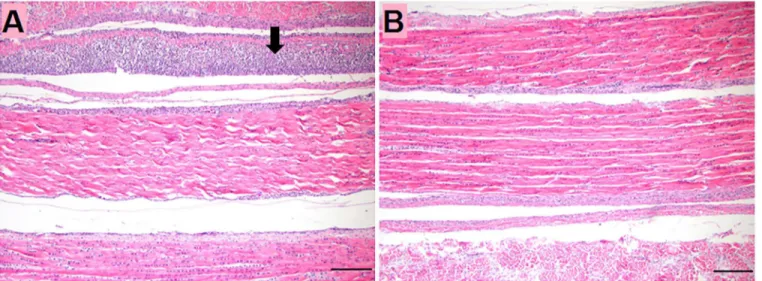
No significant lesions were observed in sections of internal organs, intestines or intertarsal joint and tendons until 14dpi. However, gastrocnemius tendons showed tenosynovitis charac-terized by mild to moderate, diffuse subsynovial infiltration of lymphocytes (Fig 1).
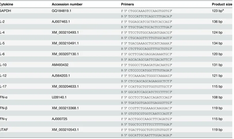
Table 1. List of cytokine genes and the primers used for RT-PCR amplification.
Cytokine Accession number Primers Product size
GAPDH GQ184819.1 F 5’CTGGCAAAGTCCAAGTGGTG3’ 123 bpa
R 5’TCCCATTCTCAGCCTTGACA3’
IL-2 AJ007463.1 F 5’TGGAGCATCGCTATCACCAG3’ 136 bp
R 5’TTGCTGACTGCACTCCTTGA3’
IL-4 XM_003210493.1 F 5’TTCCTGTGGCAAGATGAACG3’ 124 bp
R 5’CTGCAGGTTCTTGTGGCAGT3’
IL-5 XM_003210491.1 F 5’TGACGAAAGCTGCATCAAAA3’ 134 bp
R 5’CTCTTGCCAGGTTTGCTGTG3’
IL-6 XM_003207130.1 F 5’GCTTCGACGAGGAGAAATGC3’ 120 bp
R 5’AGCACAGCGATTCGACATTC3’
IL-10 AM493432 F 5’TGGGCCTGAAGATGACAATG3’ 131 bp
R 5’CTCCCCCATGGCTTTGTAGA3’
IL-12 AJ564203.1 F 5’TCCAAAGACTGGGCCAAAAG3’ 121 bp
R 5’CTCCAGCAGCAGAAGGCTCT3’
IL-17 XM_003204633.1 F 5’CCATTGCTGTTGGTGTTGCT3’ 115 bp
R 5’GGCATCCAGCATCTCCTTTC3’
IFN-α U28140.1 F 5’GCCTCCTCAACCAGATCCAG3’ 108 bp
R 5’TGATGGTGAGGTGAGGGTTG3’
IFN-β XM_003213368.1 F 5’CCGTTCTGGAAAGCAAGGAC3’ 119 bp
R 5’GTGTGCGTGGTCAATCCAGT3’
IFN-γ AJ000725 F 5’ACCTGGCCAAGCTTCAGATG3’ 115 bp
R 5’TGGCTCCTTTTCCTTTTGGA3’
LITAF XM_003210543.1 F 5’TGACTTGGCTGTCGTGTGGT3’ 119 bp
R 5’GGCATTGCAATTTGGACAGA3’
aBase pairs
Virus gene copy numbers (
Fig 2
)
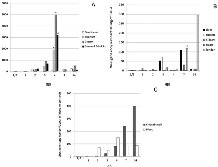
Copies of the S4 gene were detectable at 1–2 dpi in different intestinal segments and bursa of Fabricius. At 2 dpi, median viral gene copy numbers were (240, 140, 235 and 250)/100mg in duodenum, jejunum, cecum and bursa of Fabricius, respectively. Median gene copy numbers peaked at 4 dpi in jejunum, cecum and bursa of Fabricius (1800, 5000 and 3200) copies/100mg, respectively. These values were significantly higher (P<0.05) than values in the same intestinal
segments at other time points before and after 4 dpi. At 7 dpi, gene copy numbers remarkably declined in all intestinal segments followed by slight elevation at 14 dpi, where the median peaked in the duodenum (260 copies/100mg) (Fig 2A).
In liver, kidney, spleen and heart, median gene copy numbers were under 100 copies/100mg at all-time points but were highest at 3 dpi in spleen and liver (53 and 72) copies/100mg and at 7 dpi in liver (110) copies/100mg. Copy number was low in tendon early but showed a dra-matic and significant increase at 7 dpi (118 copies/100mg) (P<0.05). Viral load increased in
tendon and at 14 dpi, reached 295 copies/100mg (Fig 2B).
In blood, median viral gene copy numbers peaked at 4 dpi (150 copies/200μl). Viral load
detected in the cloacal swabs increased at 3 and 4 dpi (25 and 80 copies/100 mg) and reached a peak at 14 dpi (400 copies/100mg) (Fig 2C)
Cytokine profiling
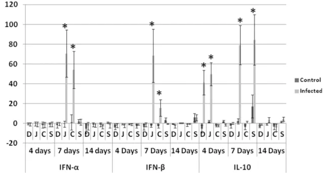
Antiviral and anti-inflammatory cytokines. At 7 dpi, IFN-αand IFN-βwere significantly higher in only the jejunum and cecum of infected groups compared with non-infected control. IL-10 showed significantly higher fold change in infected groups compared with non-infected control in duodenum and jejunum at 4 dpi and in jejunum and spleen at 7 dpi (Fig 3).
T helper 1, 2 and 17. IL-12 showed significantly higher fold change in the infected group compared with non-infected control in jejunum and cecum at 7 dpi while IFN-γincreased sig-nificantly in jejunum at 7 and 14 dpi, spleen at 4, 7 and 14 dpi (Fig 4) and in tendons at 14 dpi (Fig 5). Th2 (IL-4 and IL-5) and Th17 (IL-17) cytokines did not show statistically significant differences between infected and non-infected control groups (Fig 4).
Fig 1. Histologic section in gastrocnemius tendon of turkeys at 14 dpi.(A) TARV-infected turkey at 14 dpi showing subsynovial lymphocytic infiltration (Arrow). (B) Non-infected control at 14 dpi. Bar = 100μm.
Proinflammatory cytokines. Average fold changes of IL-6 were significantly higher in infected group than non-infected control in duodenum and jejunum at 4 and 7 dpi when com-pared with day zero (Fig 6). IL-6 was significantly elevated in tendons of infected birds at 14 dpi. Average fold changes of LITAF were significantly higher in infected groups only in jeju-num and cecum at 7 dpi. IL-2 in duodejeju-num and jejujeju-num of infected birds had a significantly higher fold changes at 4 and 7 dpi.
Discussion
The present work aimed to study tissue distribution and tropism of a newly emergent turkey reovirus associated with tenosynovitis/arthritis (TARV-O’Neil strain). Understanding tropism of this newly emergent virus helps in characterizing pathogenesis in tendons. Characterizing Fig 2. Viral gene copy numbers as determined by RT-PCR.Values represent the medians of five turkeys at each time point; (A) Intestines and bursa of Fabricius. Virus gene copy number significantly (P<0.05) peaked in jejunum, cecum and bursa of Fabricius at 4 dpi and a significantly declined at 7 dpi. (B) Internal organs have minimal gene copy number and a significant elevation (P<0.05) is present in tendons at 7 and 14 dpi. (C) Blood has minimal gene copy number that peaked at 4 dpi. Cloacal swabs, show detectable titer starting at 1–3 dpi and peaked at 14 dpi. Mann Whitney U test.*Significantly (P<0.05) higher than same tissue in other time points (before and after).XSignificantly (P<0.05) higher than same tissue in the proceeding time points (before).
Fig 3. Fold change in antiviral cytokines (IFN-αand IFN-β) and anti-inflammatory IL-10.At 7 dpi IFN-αand IFN-βin jejunum and cecum of infected birds were significantly elevated. IL-10 is significatnly elevated in duodenum and jejunum of infected birds at 4 dpi and in jejunum and spleen of infected birds at 7 dpi.D: duodenum,J: jujenum,C: cecum,Sspleen,Daysrefers to days post inoculation (dpi).*Analyses were performed in duplicate (Five turkeys/group at each time point) and values are mean±3SD. Differences between the infected and non-infected groups within the same organ are significant (P<0.05).
doi:10.1371/journal.pone.0144085.g003
Fig 4. Fold changes in Th1, Th2 and Th17 cytokines observed in turkeys.Significant elevation of Th1 cytokine IFN-γwas observed in the jejunum of infected birds at 7 and 14 dpi and in spleen at 4, 7 and 14 dpi. The other Th1 cytokine, IL-12, shows significant elevation in jejunum and cecum of infected birds at 7 dpi. Th2 (IL-4 and IL-5) and Th17(IL-17) are not significantly different at P<0.05.D: duodenum,J: jujenum,C: cecum,Sspleen,Daysrefers to days post inoculation (dpi). Analyses were performed in duplicate (Five turkeys/group at each time point) and values are mean±3SD.*Differences between infected group and non-infected groups within the same organ are significant (P<0.05).
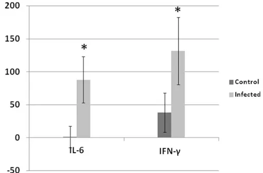
Fig 5. Fold change of IL-6 and IFN-γin tendons at 14 dpi.Both IL-6 and IFN-γwere significantly elevated at 14 dpi in tendons of infected birds compared with non-infected control. Analyses were performed in duplicate (Five turkeys/group at each time point) and values are mean±3SD.*Differences between infected group and non-infected groups within the same organ are significant (P<0.05).
doi:10.1371/journal.pone.0144085.g005
Fig 6. Fold change in proinflammatory cytokines (IL-6 and LITAF) and IL-2.IL-6 and IL-2 are significantly elevated in dudenum and jejunum of infected birds at 4 and 7 dpi. LITAF has significantly higher fold increase in infected birds compared with non-infected control in jejunum and cecum at 7 dpi.D: duodenum,J: jujenum,C: cecum,Sspleen,Daysrefers to number of days post inoculation (dpi). Analyses were performed in duplicate (Five turkeys/group at each time point) and values are mean + 3SD.*Difference between infected group and non-infected groups within the same organ are significant (P<0.05).
the cytokine profile induced by infection enhances our knowledge about the immune response in turkeys against the viral infection.
As demonstrated by rRT-PCR, the intestines (Jejunum and cecum) and bursa of Fabricius are the main sites of viral replication (based on the S4 gene copy number), peaking significantly (P<0.05) at 4 dpi. This technique can detect as few as 10 viral gene copies [18]. These findings
are in agreement with the results of previous studies, which showed that the intestines and bursa of Fabricius were the initial sites of replication (within 2–5 dpi) of several TERVs [9] and chicken reoviruses [21]. Viral gene copy number was low in internal organs and blood com-pared with intestines and bursa of Fabricius while numbers increased significantly (P<0.05) in
gastrocnemius tendon at 7 dpi, peaking at 14 dpi. Although the viral RNA was detected in blood at 2 dpi and peaked at 4 dpi, this low level of viremia was not associated with general sys-temic clinical illness at these time points. Chicken reoviruses have been shown to initially repli-cate in intestines, reach the blood at 2–3 dpi, and subsequently the internal organs within 3–5 dpi [22]. The low viral gene copies in blood in our study explains the absence of early systemic disease induced by TARV-O’Neil and failure of the virus to reach numbers in internal organs as high as in intestines and bursa of Fabricius. Further investigation of virus-host’s cells interac-tion will help understanding the reason of low viral load in blood and internal organs.
Reoviral pathogenesis has not been examined in turkey tendons. Previous studies of chick-ens inoculated with chicken reoviruses, targeted the hock joint, which was reported as an important site for virus replication [23,24,25]. In this report, we showed that the TARV-O’ -Neil strain replicated in tendons and reached peak copy number at 14 dpi. Only at this later time point was lymphocytic tenosynovitis observed in the gastrocnemius tendon sheath of infected birds. There were no lesions in tendons at earlier time points when viral gene copy number was low. These data indicate that inflammation in the tendon sheath was associated with the presence of a detectable virus titer.
Viral gene copy numbers measured in the cloacal swabs were indicative of early viral shed-ding starting at 1–2 dpi and peaking at 14 dpi. This may explain the rapid spread of infection among young birds in the field. In chicken, shedding of reovirus was reported at two weeks PI via oral route [26]. A separate study found shedding to peak at 1–2 weeks PI, and decrease after 3 weeks PI [27,28]. TARV-O’Neil appears to have earlier shedding in turkeys, although this dif-ference may be due to better early detection by the highly sensitive technique (rRT-PCR) [18].
Viral gene copies significantly (P<0.05) peaked at 4 dpi in intestines (jejunum and cecum)
and bursa of Fabricius and then significantly (P<0.05) decreased at 7 dpi. This can be
attrib-uted to the antiviral effect of IFN-αand IFN-β, which were significantly elevated at 7dpi in jeju-num and cecum of infected birds. This suggests that interferons played an important antiviral role in limiting TARV-O’Neil replication in intestines. We analyzed cytokine profile in multi-ple tissues of turkeys infected with the reovirus in order to understand the immunopathogen-esis of the infection. In addition to the previous report of GAPDH stability in intestines of turkeys [19], we analyzed the mRNA expression (CT values) of GAPDH at different time points in each tissue using ANOVA and P values were more than 0.05. Insignificant difference confirmed the stability of GAPDH and its validity to be used as a house keeping gene. The sig-nificant elevation of IL-2 (P<0.05) in intestines of infected groups at 4 and 7 days, suggests
intestinal sections as represented by leukocyte infiltration, dilated blood vessels or exudation, usually associated with elevated proinflammatory cytokines at those time points. Similarly, IL-10 showed statistically significant elevation in intestines and spleen of infected birds at 4 and 7 dpi but apparently was not effective at down regulating Th1 cytokines in infected birds.
Th1 cytokines IFN-γand IL-12 were significantly elevated in intestines and spleen of infected birds while Th-2 (IL-4 and IL-5) and Th-17 (IL-17) did not show significant elevation. This dominant Th1 response in intestines at 7 and 14 dpi excluded the possibility of an immu-noglobulin role during the early course of infection and seemingly limits the possibility of an autoimmune reaction mediated by IL-17 [29]. The elevated IL-10 might be indicative of the activity of regulatory T (T reg) over Th17 which was supported by absence of destructive bone lesions in a long-term pathogenicity trial [17].
Comparing the cytokine response with viral replication and histologic alteration in leg ten-dons helps determine the events during the course of viral infection preceding lesions in leg tendons. Only at 14 dpi, did IL-6 and IFN-γshow significant elevations (P<0.05). This increase
corresponds to subsynovial lymphocytic infiltration in gastrocnemius tendon sheath first observed at 14 dpi. It is likely this lymphocytic infiltration was induced by viral replication reaching a peak at 14 dpi.
In chickens, replication of reoviruses with high multiplication rate was shown to induce sig-nificantly higher production of IL-6, IL-10 and INF-γcompared to those with low multiplica-tion rate [30]. Viral replication was accompanied with inflammation in leg tendons while in intestines, where replication was higher, inflammation was not a factor. Viral replication may be associated with inflammatory cells in GALT and lymphocytes infiltrating the tendon sheath. Little is known about the release of avian reoviruses and its association with cell lysis. Mamma-lian reoviruses may release from infected cells without inducing cell death [31] or may induce apoptosis prior to release [32]. We have not observed any syncytia formation by histologic examination, although avian reovirus is characterized by formation of cell-cell fusion (syncytia formation), mostly mediated by P10 protein [33]. Future studies using electron microscopy, specific immunohistochemistry and transcriptome analysis will be very helpful in determining details of the virus replication cycle including adhesion, assembly and release, as well as the cells where the virus replicates for local and systemic spread.
Conclusions
The newly emergent turkey arthritis reovirus (TARV-O’Neil) is mostly enterotropic with a ten-dency to replicate in tendons later during the course of infection. The enterotropic virus is shed early during the course of infection in feces and evokes a significantly elevated antiviral cyto-kine response in intestines at 7 dpi when the viral replication was significantly decreased. Addi-tionally, viral infection induced a dominant Th1 cytokine response but neither Th2 nor Th17 cytokines was elevated in infected birds during the first 2 weeks of infection. Further research is required to demonstrate viral pathogenesis at a later stage of infection when clinical lameness becomes evident.
Supporting Information
S1 Table. Means (M) and standard deviations (SD) of virus gene copy numbers at different days post inoculation in different organs (Per 100 mg of tissue or 200μl of blood or cloacal swab).
S2 Table. Means and standard deviations of different cytokines fold changes in duodenum (D), jejunum (J), cecum (C), spleen (S) and tendon (T) in infected birds and non infected controls at different time points post inoculation
(DOCX)
Acknowledgments
This study was funded in part by the Rapid Agricultural Response Fund, established by the Minnesota legislature and administered by the University of Minnesota Agricultural Experi-ment Station. We thank Dr. Jack Rosenberger for providing TARV-O’Neil for the study.
Author Contributions
Conceived and designed the experiments: TS RP SG ZX KR. Performed the experiments: TS SM NS RP ZX. Analyzed the data: TS KR ZX. Contributed reagents/materials/analysis tools: TS NS SM ZX RP SG. Wrote the paper: TS SG RP ZX KR.
References
1. Nersessian BN, Goodwin MA, Page RK, Kleven SH. Studies on orthoreoviruses isolated from young turkeys. II. Virus distribution in organs and serological response of poults inoculated orally. Avian Dis. 1985; 29: 963–969. PMID:3833236
2. Heggen-Peay CL, Qureshi MA, Edens FW, Sherry B, Wakenell PS, O’Connell PH, et al. Isolation of areovirus from poult enteritis and mortality syndrome and its pathogenicity in turkey poults. Avian Dis. 2002; 46: 32–47. PMID:11922348
3. Woolcock PR, Shivaprasad HL. Electron microscopic identification of viruses associated with poult enteritis in turkeys grown in California 1993–2003. Avian Dis. 2008; 52: 209–21. PMID:18646448 4. Jindal N, Patnayak DP, Ziegler AF, Lago A, Goyal SM. A retrospective study on poult enteritis
syn-drome in Minnesota. Avian Dis. 2009; 53: 268–275. PMID:19630235
5. Jindal N, Patnayak DP, Chander Y, Ziegler AF, Goyal SM. Detection and molecular characterization of enteric viruses from poult enteritis syndrome in turkeys. Poult. Sci. 2010; 89: 217–226. doi:10.3382/ ps.2009-00424PMID:20075272
6. LojkićI, Biđin M, Biđin Z, Mikec M. Viral agents associated with poult enteritis in Croatian commercial turkey flocks. Acta. Vet. Brno. 2010; 79: 91–98.
7. Calvert AJ. Light Turkey Syndrome: Field Study and Inoculation Trial M.S., University of Minnesota. 2012; 195 pages.
8. Mor SK, Sharafeldin TA, Abin M, Kromm M, Goyal SM, Porter RE et al. The occurrence of enteric viruses in Light Turkey Syndrome. Avian Pathol. 2013; 42: 497–501. doi:10.1080/03079457.2013. 832145PMID:24066896
9. Pantin-Jackwood MJ, Spackman E, Day JM. Pathology and virus tissue distribution of turkey origin reo-viruses in experimentally infected turkey poults. Vet. Pathol. 2007; 44: 185–195. PMID:17317795 10. Spackman E, Pantin-Jackwood MJ, Day JM, Sellers H. The pathogenesis of turkey origin reoviruses in
turkeys and chickens. Avian Pathol. 2005; 34: 291–296. PMID:16147564
11. Day JM, Spackman E, Pantin-Jackwood MJ. Turkey origin reovirus-induced immune dysfunction in specific pathogen free and commercial turkey poults. Avian Dis. 2008; 52: 387–391. PMID:18939624 12. Levisohn S, Gur-Lavie A, Weisman J. Infectious synovitis in turkeys: Isolation of tenosynovitis virus-like
agent. Avian Pathol. 1980; 9: 1–4. PMID:18770233
13. Page RK, Fletcher OJ, Villegas P. Infectious tenosynovitis in young turkeys. Avian Dis. 1982; 26: 924– 927. PMID:7159326
14. Al Afaleq AA, Jones RC. Pathogenicity of three turkey and three chicken reoviruses for poults and chicks with particular reference to arthritis/tenosynovitis. Avian pathol. 1980; 18: 433–440.
15. Mor SK, Sharafeldin TA, Porter RE, Ziegler A, Patnayak DP, Goyal SM. Isolation and characterization of a turkey arthritis reovirus. Avian Dis. 2013; 57: 97–103. PMID:23678736
16. Sharafeldin TA, Mor SK, Bekele AZ, Verma H, Goyal SM, Porter RE. The role of avian reoviruses in tur-key tenosynovitis/arthritis. Avian Pathol. 2014; 43: 371–378. doi:10.1080/03079457.2014.940496
17. Sharafeldin TA, Mor SK, Bekele AZ, Verma H, Noll SL, Goyal SM et al. Experimentally induced lame-ness in turkeys inoculated with a newly emergent turkey reovirus. Vet. Res. 2015; 46: 11. doi:10.1186/ s13567-015-0144-9PMID:25828424
18. Mor SK, Verma H, Bekele AZ, Sharafeldin TA, Porter RE, Goyal SM. One step real-time RT-PCR for the detection of turkey reoviruses. Avian Dis. 2014; 58: 404–407. PMID:25518435
19. Haritova AM. Gastrointestinal tissue expression of villin mRNA in turkeys. BJVM. 2012; 15:13–20.
20. Livak KJ, Schmittgen TD. Analysis of relative gene expression data using real-time quantitative PCR and the 2(-Delta Delta C(T)) Method. Methods 2001; 25: 402–408. PMID:11846609
21. Jones RC, Islam MR, Kelly DF. Early pathogenesis of experimental reovirus infection in chickens. Avian Pathol. 1989; 18: 239–253. PMID:18679857
22. Kibenge FSB, Gwaze GE, Jones RC, Chapman AF, Savage CE. Experimental reovirus infection in chickens: observations on early viraemia and virus distribution in bone marrow, liver and enteric tis-sues. Avian Pathol. 1985; 14: 87–98. PMID:18766901
23. Walker ER, Friedman MH, Olson NO. Electron microscopy study of an avian reovirus that causes arthri-tis. J. Ultrastruct. Molec. Struct. Res. 1972; 41: 67–79.
24. Sahu SP, Olson NO. Comparison of the characteristics of avian reoviruses isolated from the digestive and respiratory tract with viruses isolated from the synoviae. Am. J. Vet. Res. 1975; 36: 847–850. PMID:167621
25. Jones RC, Kibenge FSB. Reovirus-induced tenosynovitis in chickens: the effect of breed. Avian Pathol. 1984; 13: 511–528. PMID:18766865
26. Kibenge FSB, Dhillon AS. A comparison of the pathogenicity of four avian reoviruses in chickens. Avian Dis. 1987; 31: 39–42. PMID:3579791
27. Islam MR, Jones RC. An enzyme-linked immunosorbent assay for measuring antibody titre against avian reovirus using a single dilution of serum. Avian Pathol. 1988; 17: 421–425.
28. Al Afaleq AI, Jones RC. Comparison of single and repeated oral infection of chicks with two avian reovi-ruses. Res. Vet. Sci. 1994; 57: 96–99. PMID:7973100
29. Komatsu N, Takayanagi H. Arthritogenic T cells in autoimmune arthritis. Int. J. Biochem. Cell Biol. 2015; 58: 92–96. doi:10.1016/j.biocel.2014.11.008PMID:25450406
30. Shen P, Yang J, Su B, Lee L. Cytokine mRNA expression in chicken experimentally infected with differ-ent avian reovirus strains. Taiw. Vet. J. 2014; 40: 29–36.
31. Lai CM, Mainou BA, Kim KS, Dermody TS. Directional release of reovirus from the apical surface of polarized endothelial cells. MBio. 2013; 4: e00049–13. doi:10.1128/mBio.00049-13PMID:23572551 32. Forrest JC, Dermody TS. Reovirus receptors and pathogenesis. J. Virol. 2003; 77: 9109–9115. PMID:
12915527