Abstract
Fragment-based approaches are the current standard forde novoprotein structure predic-tion. These approaches rely on accurate and reliable fragment libraries to generate good structural models. In this work, we describe a novel method for structure fragment library generation and its application in fragment-basedde novoprotein structure prediction. The importance of correct testing procedures in assessing the quality of fragment libraries is demonstrated. In particular, the exclusion of homologs to the target from the libraries to cor-rectly simulate ade novoprotein structure prediction scenario, something which surprisingly is not always done. We demonstrate that fragments presenting different predominant pre-dicted secondary structures should be treated differently during the fragment library genera-tion step and that exhaustive and random search strategies should both be used. This information was used to develop a novel method, Flib. On a validation set of 41 structurally diverse proteins, Flib libraries presents both a higher precision and coverage than two of the state-of-the-art methods, NNMake and HHFrag. Flib also achieves better precision and coverage on the set of 275 protein domains used in the two previous experiments of the the Critical Assessment of Structure Prediction (CASP9 and CASP10). We compared Flib li-braries against NNMake lili-braries in a structure prediction context. Of the 13 cases in which a correct answer was generated, Flib models were more accurate than NNMake models for 10.“Flib is available for download at:http://www.stats.ox.ac.uk/research/proteins/
resources”.
Introduction
Fragment-basedde novoprotein structure prediction is the current standard for template-free modelling of proteins. This approach, exemplified by ROSETTA [1,2,3,4], relies on creating a library of fragments extracted from known protein structures. In this context, a structural frag-ment is a continuous subset of the residues of a protein. Such structural fragfrag-ments are usually less than 20 residues long. Each fragment in the library represents a specific position in the se-quence to be modelled (target). The most common technique used to select library fragments is the sequence similarity between the fragment and the region of the target sequence that the OPEN ACCESS
Citation:de Oliveira SHP, Shi J, Deane CM (2015) Building a Better Fragment Library forDe Novo
Protein Structure Prediction. PLoS ONE 10(4): e0123998. doi:10.1371/journal.pone.0123998
Academic Editor:Yang Zhang, University of Michigan, UNITED STATES
Received:November 17, 2014
Accepted:February 25, 2015
Published:April 22, 2015
Copyright:© 2015 de Oliveira et al. This is an open access article distributed under the terms of the
Creative Commons Attribution License, which permits unrestricted use, distribution, and reproduction in any medium, provided the original author and source are credited.
Data Availability Statement:Data have been deposited to Figshare:http://dx.doi.org/10.6084/m9. figshare.1328655.
Funding:SHPO was funded by: Conselho Nacional
fragment represents. Fragments from the library are pieced together in order to generate com-plete models of the target structure. There are many fragments in the library for each position in the target (e.g. ROSETTA's fragment libraries contain 200 fragments per position [1]) and many proteins are hundreds of residues long. In order to explore this large combinatorial space, heuristics are needed [4]. Commonly used heuristics rely on statistical and physical po-tentials to ensure that global structural features of proteins are sustained/respected in the mod-els generated from the combinations of fragments.
A major problem for all fragment-basedde novoapproaches occurs when the fragment li-brary for a given target does not contain good fragments for a particular region. In that case, low accuracy models will be generated regardless of the precision of the potentials being used and regardless of the amount of computation time invested in the modelling routine [5]. For that reason, accurate fragment library generation is crucial to the success of template-free modelling [5].
NNMake [6] is ROSETTA’s method for fragment library generation. NNMake extracts frag-ments from a template database of non-identical high resolution structures (<2.5 Å). It scores every length nine segment of the target sequence exhaustively against all length nine segments within its template database. NNMake's score is based on sequence similarity, predicted sec-ondary structure and predicted torsion angle. A library for the target sequence consists of the 200 top-scoring fragments per target position. The latest version of NNMake was tested on a set of 62 small globular proteins (all shorter than 150 residues in length). In their test proce-dure, the authors removed all sequence homologs. In order to assess the quality of generated fragment libraries, ROSETTA was used to generate decoys for each target. The number of de-coys generated varied according to target length (ranging from 4,065 to 19,183 dede-coys). The av-erage coordinate root-mean square deviation (cRMSD) between the native structure of the target and the top 0.1 percentile models were computed (average of the 62 targets
cRMSD = 3.75 Å). One of the current limitations of NNMake relates to the use of fragment with a fixed length (nine residues long). This may not be ideal as it has been shown that accu-rate structures can be built using fragments as short as four residues [7]. It has also been re-ported that fragments longer than nine residues can generate better results if the modelling routine is adjusted accordingly [8]. Other fragment generation software extracts either longer fragments or fragments with varying lengths [9,10,11,12].
Other fragment library generation software also attempt to increase the amount of local structural information used to generate libraries. For example, FRAGFOLD [13] uses super-secondary structure fragments, which express the relationship between consecutive super-secondary structure elements. FRazor [14] builds on NNMake and incorporates solvent accessibility and contact capacity into its scoring scheme. The authors of FRazor claim that their scoring scheme improves the precision of NNMake libraries by discarding low quality fragments suggesting that a sequence-based score can benefit from additional structural information.
HHFrag [10] selects fragments slightly differently from many other methods: by means of profile hidden markov models (HMM). HHFrag uses the HHpred toolchain [11] to build a profile-HMM of the target sequence and a profile-HMM for each sequence in a pre-defined template database. The template database used by HHFrag is the April, 2010 build of PDBse-lect25 [15], a subset of 4,824 protein chains with less than 25% identity extracted from the PDB [16]. Sequence and predicted secondary structure information are used in the generation of the profile-HMM. The HMM of the target is divided into a set of overlapping HMM fragments of variable length (6−21 residues). Fragment extraction is performed by HMM-HMM alignment
using the HHSearch algorithm [17]. Each of the 6 to 21-long target HMMs is aligned and scored against every HMM profile for the proteins in the template database. All fragments with a probability0.2 are accepted. For positions where a minimum of ten fragments are not
Pharma provided support in the form of salaries for authors [JS], but did not have any additional role in the study design, data collection and analysis, decision to publish, or preparation of the manuscript. The specific roles of these authors are articulated in the‘author contributions’section.
For some target positions, HHFrag does not output any fragments, which can cause difficulties during the modelling step.
A recent fragment library generation programme SAFrag [19] also builds HMM profiles to detect fragment candidates, in an analogous fashion to HHFrag. SAFrag HMM profiles are ex-trapolated from a 27 state structural alphabet. The extracted fragments vary in length from 6 to 27 amino acids. Fragments are scored based on a profile-profile comparison, using the Jensen Shannon divergence. Two different template databases can be used by SAFrag: pdb25 and pdb50. Pdb25 is the same database used by HHFrag, whereas pdb50 imposes a 50% pairwise se-quence identity cutoff. The method was validated on a set of 111 targets [18]. SAFrag reports a higher precision and coverage than HHFrag. However their cutoff for defining a good fragment is less strict (RMSD to native structure<2.0Å). They also allowed homologs and the target structure itself to be included in their template database. Further, the method outputs on aver-age less than two fragments per target position, which suggests that homolog structures are dominating the fragment libraries generated by SAFrag. SAFrag is only available as a web-server.
As described above, different methods diverge in the number of fragments used per posi-tion, in the length of the fragments used, in the selectivity of the template databases from which fragments are extracted, and in the way the extraction is performed. In this work, we in-vestigate these aspects of fragment library generation and how they affect the precision of the library. We also evaluate the impact of including/excluding homologs among the set of known structures that fragments can be extracted to assess its impact on methods such as HHFrag and SAFrag. In a realde novostructure prediction method homologs would not be available, so it is important to exclude those fragments during method training and validation.
Current methods score all types of fragments using the same methodology regardless of the predominant predicted secondary structure of the fragment. Here we analysed the relationship between the predominant predicted secondary structure of fragments and our ability to accu-rately predict fragments for that position. We observed that fragments with a predominant pre-dicted secondary structure (e.g.α- helical fragments) can be predicted more accurately than other types of fragments.
Based on these analyses, we have implemented Flib, a fragment library generation software that exploits the predominant predicted secondary structure of fragments to increase the preci-sion of generated fragment libraries. We have generated fragment libraries for two validation sets: a set of 41 structurally diverse proteins extracted from the PDB (PDB-representative set) and a set of 275 protein domains that were used in CASP9 [20] and CASP10 [21] (CASP set). Fragment libraries generated by Flib were compared with libraries generated by NNMake [6] and HHFrag [10] and found to obtain the best balance between precision and coverage in both test sets.
for 12 of the 41 proteins in our test set. We compared our modelling results against running SAINT2 with NNMake fragment libraries. NNMake libraries generated accurate models for 8 cases. Of the 13 cases for which accurate models were generated using SAINT2, Flib libraries generated more accurate models in 10 cases.
These results indicate that Flib can be used to improve the accuracy ofde novoprotein struc-ture prediction and demonstrate the importance of discriminating between different secondary structure fragments when performing fragment extraction.
Results
Fragment Library Quality Assessment
We assess library quality using two commonly employed metrics: global precision and cover-age. Precision is defined as the number of good fragments divided by the total number of frag-ments in a library (the proportion of good fragfrag-ments in the libraries). Coverage is defined as the number of residues represented by at least one good fragment divided by the number of residues of the target (the proportion of protein residues represented by a good fragment). Dif-ferent methods employ difDif-ferent cutoffs to distinguish between good fragment conformations and bad fragment conformations. Instead of selecting a single cutoff, we have varied the good fragment cutoff between 0.1 to 2.0 Å computing the precision and coverage across the range.
The Rationale Behind Flib
Flib extracts fragments from a database of known structures using a target sequence. A frame-work describing Flib’s pipeline is shown inFig 1.
Template Database Construction
The template database, the initial set of structures from which fragments for libraries will be ex-tracted, can be built in many ways. Regardless of how the template database is built, for testing purposes it is important to remove homologs to the target. We use the Flib template database, in which we impose a 90% sequence identity cutoff and a 5.0Å resolution cutoff, as it proved to lead to the most accurate fragment libraries (seeS1 Fig).
Fragment Extraction
The next step in a fragment library generation pipeline is fragment extraction. Most fragment library generation software methods use an exhaustive search approach [6,7,8,10,19]. We compared the use of an exhaustive approach and a random approach. In our exhaustive ap-proach, every fragment ranging from 6 to 20 residues in the template database is scored against all of the positions of the target. The exhaustive library is composed of the top 1,000 scoring fragments per target position. The score used in Flib is based on a sequence component, a pre-dicted secondary structure component and a prepre-dicted torsion angle component (seeMethods section for more details).
When comparing our three scores (sequence, secondary structure and torsion angle scores) we observed a higher correlation between predicted torsion angle score and fragment RMSD to native structure compared to the other two (S2 Fig). This indicates that the predicted torsion angle score is better suited to rank the fragments in the final ensemble. Within Flib’s pipeline, we combine the exhaustive and random libraries. This combined library has, on average, 3,000 fragments per target position (LIB 3000). We rank all the fragments in this library and output the 20 top-scoring fragments per target position according to the torsion angle score (LIB20). On average, within our test data set, 69% of the fragments in LIB20 are extracted by the ran-dom method and 31% by the exhaustive protocol.
Enrichment Step
We observed that the ensemble with the highest scoring fragment per position according to the torsion angle score presented a very high precision, albeit at a loss of coverage. We decided to exploit this by implementing an enrichment step, in which we include fragments from LIB500 (analogous to LIB20, but considering the 500 top-scoring fragments) that present less than 0.5Å RMSD to the highest scoring fragment according to the torsion angle score. On average, 6.5 fragments per position are added to LIB20 during the enrichment step.
Protein Threading Hits Library (TH Library)
The final step in our fragment library generation routine is to add to LIB20 fragments extracted from protein threading hits. Protein threading identifies protein segments that present struc-tural similarity to a given target. As we remove homologs, these protein threading hits are too unreliable to be used as templates for template-based modelling, but may still provide locally similar fragments. We have found that adding such fragments (on average, less than five Fig 1. Schematics of Flib.Starting from a target sequence, we predict secondary structure (SS) and torsion angles for the target (green). We extract fragments from a template database using a combination of random and exhaustive approaches. Fragments are extracted for each target position. A library containing the top-3000 fragments per position is compiled using the SS score and the Ramachandran-specific sequence score (LIB3000). LIB3000 is then sorted according to the torsion angle score and the top-20 fragments per position are selected to comprise the final library. The final library (FLIB) is complemented by fragments that originate by an enrichment routine (in yellow) and fragments that originate from protein threading hits (orange).
doi:10.1371/journal.pone.0123998.g001
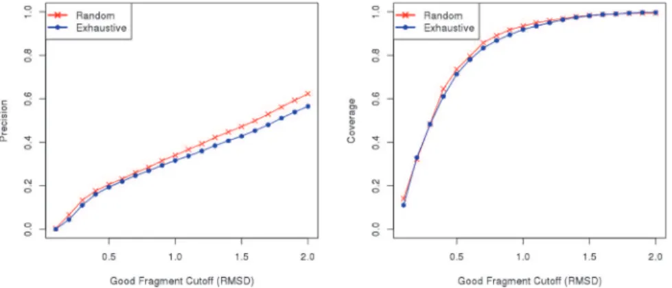
Fig 2. Comparison between Flib’s random extraction and exhaustive extraction methods.Analysis of the precision of fragment libraries generated by Flib using two different approaches for fragment extraction: random extraction (red), and exhaustive extraction (blue). We varied the RMSD to native structure cutoff to define a good fragment from 0.1 to 2.0 Angstroms (x-axis). The average precision on the 43 proteins in the test data set (left) and the average coverage (right) are shown for fragment libraries containing the top-1000 scoring fragments extracted exhaustively or at random. The precision indicates the proportion of good fragments in the generated libraries (y-axis).
fragments per position), when they are available, to LIB20 increases the precision of our meth-od (Fig 3).
Predominant Predicted Secondary Structure Determines Fragment
Quality
Secondary structures (α-helices andβ-strands) have restrictions by definition in the torsion an-gles of their residues, whilst loop regions are not so constrained and can assume a wider range of conformations [23]. Hence, secondary structure elements have a lower degree of
conformational variability.
Considering that fragments with a larger number of loop residues will present a higher vari-ability, we hypothesized that they would be harder to predict. In order to test our hypothesis, we have investigated the relationship between the RMSD to the native structure and fragments with different predominant predicted secondary structures.
Here a fragment is described as representing a target position N when it represents all the target residues between N and N+L (where L is the length of the fragment). We classify a target position as belonging to one of four distinct classes based on the predominant predicted sec-ondary structure of its residues. The four classes of secsec-ondary structure (SS classes) are: majori-tyα-helical,majorityβ-strand,majority loopandother(no predominant predicted secondary structure).
We analysed the RMSD to the native structure of fragments extracted at random. The frag-ments were grouped according to our predicted SS classes. The spread of fragment RMSD for the top 200 scoring fragments is shown for every position in the target 1E6K (Fig 4). THE RMSD spread of the top-200 fragments replicate the results obtained with LIB3000. This figure typifies our general observation that that there is a strong relationship between the RMSD to the native structure and our four SS classes. The RMSDs formajorityα-helicalfragments are significantly lower than the RMSDs for other SS classes.Majority loopfragments andother fragments show a wider variability and are much harder to predict accurately. The difference between SS classes is important in two ways. Firstly as current methods only offer coverage and precision across all SS classes, very poormajority loopandotherprecision may be hidden by highmajorityα-helicalprecision. Secondly, these results suggest that during fragment library Fig 3. Effect of protein threading hits on fragment library quality.Analysis of the impact of fragments extracted from protein threading hits. Precision and coverage are shown for the fragment libraries generated by LIB20, Protein Threading Hits and Flib (a combination of the other two approaches). We varied the RMSD to native structure cutoff to define a good fragment from 0.1 to 2.0 Angstroms (x-axis). The average precision and coverage on the 43 proteins in the test data set is shown for each approach. The precision indicates the proportion of good fragments in the generated libraries (y-axis). The coverage indicate the proportion of residues of the target represented by at least one good fragment.
generation, it may improve results if we treat fragments differently according to their predomi-nant predicted secondary structure. For that reason, Flib uses different cutoffs for accepting fragments based on SS class. Less stringent cutoffs for majority loop and other fragments are used as their variability is far higher. We have observed that adopting different cutoffs for each SS class improves the precision of the libraries generated by Flib (S3 Fig).
The usefulness of the fragments added at the protein threading step also differs between each SS classes. Adding the threading fragments to LIB20 increases the precision formajority
β-strand,majority loopandotherSS classes, but decreases the precision formajorityα-helical fragments (S4 Fig). Thus, no fragments from threading hits are added to themajorityα-helical target positions in our final library.
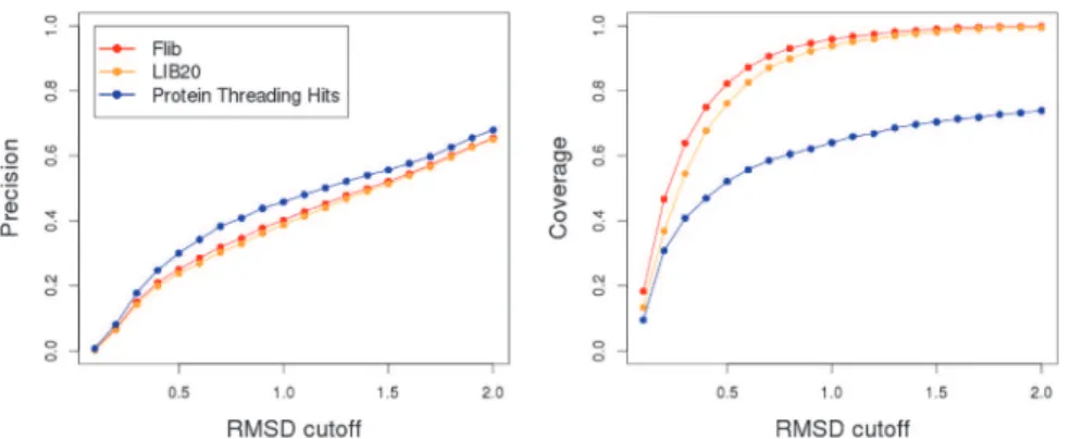
Cross-comparison between Flib and other software
We have compared Flib against NNMake and HHFrag (Figs5and6). A large scale comparison including SAFrag could not be performed because the software is only available as a web-serv-er. In order to perform a rigorous comparison, we have used two distinct validation sets: a set comprised of 275 protein domains from CASP9 and CASP10 (CASP set) and a set of 41 struc-turally diverse proteins (PDB-representative set). The second set was built to be representative of the PDB, both in terms of protein lengths and distribution of proteins amongst different SCOP classes.
In all analyses, fragments extracted from homologs were discarded (the impact of filtering out fragments from homologs is described in the next section). If we compare the overall preci-sion and coverage, Flib presents higher precipreci-sion compared to NNMake and higher coverage compared to HHFrag (Fig 5). This difference appears to be due to the increase in performance for the SS Classes majorityα-helical and majorityβ-strand (Fig 6). HHFrag coverage is signifi-cantly lower than that of the other two methods. At a 1.0Å RMSD cutoff, HHFrag's fragment li-braries describe slightly more than half of the target residues correctly (~55% coverage on Fig 4. Relationship between secondary structure class (SS-Class) and fragment quality.Boxplot of the RMSD to native structure (y-axis) of 200 fragments per target position (x-axis) for the protein 1E6K. The top-200 scoring fragments from its LIB3000 were selected and are displayed. This subset of LIB3000 was chosen to increase performance of data visualization. Four Different SS Classes are defined:majorityα-helical
(green),majorityβ-strand(red),majority loop(blue) andother(black). Positions for which fragments are
majorityα-helicalormajorityβ-strandpresent significantly lower RMSDs to the native structure and a smaller spread compared tomajority loopandotherpositions.
PDB-representative set, ~65% coverage on the CASP set). HHFrag failed to produce any frag-ments for ~13% of the positions. This can become a problem during structure prediction con-sidering that modelling routines generally require at least one fragment representing every target position.
Fig 5. Comparison between HHFrag, NNMake and Flib.Precision (left) and coverage (right) of fragment libraries generated using NNMake (red), HHFrag (green) and Flib (blue). The precision and coverage of the fragment libraries are averaged on a set of 41 structurally diverse proteins. We varied the RMSD cutoff to define a good fragment (x axis) and evaluated the precision (proportion of good Fragments in the libraries) and coverage (proportion of protein residues represented by a good fragment) for each method.
doi:10.1371/journal.pone.0123998.g005
When comparing the three programmes, Flib achieves the best balance between coverage and precision. Considering a good fragment cutoff of 1.0Å, HHFrag presents the highest aver-age overall precision, ~43%, compared to Flib, ~35%, and NNMake, ~29.1% (data shown for the PDB-Representative set). But HHFrag's precision is increased due to a reduced number of fragments output per position (see below). HHFrag also boosts its precision by not outputting any fragments for regions that are harder to predict (as stated above, on average, ~13% of the residues are not represented by any fragment in an HHFrag generated library). Not outputting fragments for low confidence regions will improve precision, but will also cause difficulties dur-ing protein structure prediction. Data for the CASP set can be found inS5 Fig.
On the PDB-representative validation set, Flib outputs, on average, 26 fragments per posi-tion, with an average length of ~7.4 residues. HHFrag outputs on average 10 fragments per po-sition, with an average length of 9.1 residues. Generating a smaller number of fragments can improve precision, but can represent a problem during modelling since less conformations will be sampled. NNMake always outputs 200 fragments per position with a constant length of nine residues.
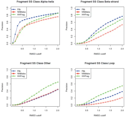
The three methods perform well at predicting fragments forα-helical segments, however, at 1.0Å RMSD cutoff, Flib's precision is 74.6%, which is higher than NNMake's 59% and
HHFrag's 64.7% (data shown for the PDB-representative set). Flib's precision forβ-strand frag-ments is also higher. At 1.0Å RMSD, Flib presents 41% precision against NNMake's 17.7% and HHFrag's 32.2% (data shown for the PDB-representative set). The precision of Flib for the other two SS classes are comparable to NNMake’s precision (less than 5% difference in preci-sion), but lower than HHFrag's. The coverage of Flib libraries slightly exceeds the coverage of NNMake for all SS classes. Results for the CASP set are included inS6 Fig.
To assess the statistical significance of our results, we have compared the distribution of RMSDs to the native structure of all fragments output by Flib and NNMake, for all targets in our PDB-representative validation set (S7 Fig). We performed a Kolmogorov-Smirnov test (al-ternative hypothesis that the cumulative distribution function of the RMSDs of Flib fragments is greater than the cumulative distribution function of the RMSDs of NNMake fragments) and we obtained a p-value of 2.2e-16. This indicates that Flib generate fragments with statistically significant lower RMSDs compared to NNMake.
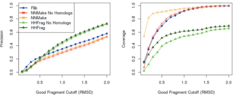
Effect of Homologs on Fragment Library Quality
We carried out an analysis to assess the impact of extracting fragments from sequence homo-logs of the target protein on fragment library quality. It has been shown that when a suitable template (a homolog) can be found for a specific target, template-based modelling is the most accurate way to model the structure of that target [21]. Hence,de novoprotein structure pre-diction tends to be used only in cases where no homologs can be found. For that reason, a frag-ment library that is representative of a realde novoprotein structure prediction case should not contain fragments extracted from homologs. Nonetheless, not all current methods for fragment library generation exclude such fragments from their outputs or from their published tests [e.g. 6, 10, 13, 19].
We have compared the precision and coverage of fragment libraries generated before and after homolog removal. If we consider the cutoff of 1.0Å to define a good fragment, homolog exclusion leads to a loss of precision from ~35% to ~29.1% for NNMake and from ~45% to 43.1% for HHFrag libraries. Homolog exclusion leads to a loss of coverage from ~61.6% to ~54% for HHFrag libraries and from ~96% to 92% for NNMake libraries. Fragments extracted from homologs increase the precision and coverage of fragment libraries.
Model Generation/Protein Structure Prediction
Our results indicate that Flib libraries present higher precision and coverage when compared to NNMake’s. However, in order to determine how well those results translate to protein struc-ture prediction, it is necessary to test the applicability of our libraries within a protein model-ling framework. Therefore, we have generated models using Flib libraries first to assess if accurate models could be generated using those libraries and second to compare to the models generated using NNMake’s libraries. We used our custom implementation of the fragment-basedde novostructure prediction software, SAINT2, to combine the fragments and to sample the conformational space (seeMethodsfor more details).
We generated 1,000 decoys for each of the proteins in our PDB-Representative set (S1 Table) using SAINT2 and Flib libraries. We compared our results to the results obtained by generating 1,000 decoys with NNMake libraries using SAINT2 (Fig 8). The Flib libraries gener-ated accurate models (TM-Score>0.5) for 12 of the 41 cases in our test set. The NNMake li-braries generated accurate models for 8 of the 41 cases. Of the 13 cases for which accurate models were generated by either method, Flib libraries performed better in 10. Flib failed to generate a correct model in only one case where NNMake libraries produced an accurate result, whereas NNMake libraries failed to generate a correct model in 5 cases where Flib libraries pro-duced good models.
Discussion
In this work, we have established that removal of homologs from any fragment library genera-tion pipeline is essential to ensure that the precision and coverage obtained are representative of a realisticde novostructure prediction scenario, otherwise overly promising results will be shown.
Fig 7. Effect of Homologs on fragment library quality.Precision (left) and coverage (right) of fragment libraries generated using three different methods: Rosetta’s NNMake (crosses), our method Flib (circles), and HHFrag (triangles). We varied the cutoff to define a good fragment (x axis) and evaluated the precision (proportion of good fragments in the libraries) and coverage (proportion of protein residues represented by a good fragment) for each of the methods when: homologs are included (red and orange) and when homologs are excluded (light and dark green). Homologs are always excluded from Flib (blue).
We tested different template databases (subsets of the PDB) in order to understand how da-tabase size and selectiveness can affect the quality of generated fragment libraries. Our analysis revealed that larger template databases give marginally better results. This implies that errors introduced by low quality structures are compensated for by the diversity introduced by using more proteins.
The correlation between sequence and secondary structure scores and fragment RMSD to the native structure was also investigated. We observed that, once homologs are excluded from template databases, sampling at random from fragments that satisfy a score cutoff produces better results than extracting fragments exhaustively. We opted to employ a combination of both methods (random sampling and exhaustive sampling) in Flib. Exhaustive extraction is useful for finding high scoring fragments that are likely to be good, whereas random methods increase the diversity of the final ensemble. We have observed that ranking fragments accord-ing to predicted torsion angles improved results. Previous results suggest that predicted torsion angles perform better than predicted secondary structure in assisting protein structure predic-tion [24]. Fragments extracted from protein threading hits were also added to our fragment li-braries. These fragments improved the accuracy of generated libraries and these fragment Fig 8. TM-Score of the best decoy as generated by Flib+SAINT2 and by NNMake +SAINT2.For each approach, 1,000 decoys were generated and the best decoy (highest TM-Score when superimposed to native structure) was chosen. Results are shown for the 41 proteins in our data set. We compared the TM-Score of best decoy generated by Flib + SAINT2 (x-axis) against NNMake + SAINT2. Each point represents a target. Point color represents the target's SCOP class and the point size is proportional to the protein length. The dotted lines indicate the cutoff for defining an accurate model (TM-Score>0.5). Flib libraries generated accurate models for 12 of the 41 cases in our PDB-representative set. NNMake libraries generated an accurate model for 8 of the 41 cases. On the 13 cases for which accurate models were generated, Flib libraries performed better in 10 cases. Flib outperforms NNMake in 31 of the 41 cases.
majorityα-helicalandmajorityβ-strandfragments. These results also suggest that it is harder to predict good fragments for positions that are represented bymajority looporother frag-ments. During model generation, it may be beneficial to concentrate sampling efforts into these harder to predict positions.
Flib presents a better balance between coverage and precision when compared against HHFrag and NNMake. Compared to NNMake, Flib can generate fragments with varying lengths. This has been previously shown to improve protein structure prediction. Flib frag-ments are, on average, 1 residue shorter than NNMake fragfrag-ments. Considering that RMSD is correlated with fragment length, we investigated whether Flib's higher precision could be ex-plained due to its shorter fragments. We built a new fragment library considering only the first eight residues of each of the fragments output by NNMake. We noticed a slight improvement in the precision of NNMake's libraries, but Flib libraries still presented higher precision and coverage. When compared to HHFrag, Flib presents a higher coverage. Flib also outputs frag-ments for every target position, which is necessary for structure prediction.
We have compared the improvement obtained by using Flib libraries against NNMake li-braries in a protein structure prediction framework. Flib lili-braries generated accurate models in 12 out of the 41 test cases. Further, our libraries outperform NNMake in 10 of the 13 cases where an accurate model was generated.
The number of decoys we have generated during our analysis is comparable to the number of decoys that were generated in previous works [10,19]. However, this number is relatively low and it is hard to assess the statistical significance of our results. For that reason, we com-pared the RMSD to the native structure of the best fragment for each target position obtained by each of Flib, NNMake and HHFrag (S8 Fig). In principle, if the fragment assembly is ex-haustive or has reached convergence, it is the best fragment within each window that ultimately determines the outcome ofde novostructure prediction. Therefore, this comparison describes how well a fragment library can be used to model a target independent of the number of decoys generated. The RMSDs of the best fragments for each target position between Flib and
NNMake are comparable and as Flib libraries are nearly 10 times smaller they are better suited for structure prediction. Furthermore, the modelling step in our analysis is computationally in-tensive. For that reason, we chose to work with a reduced number of targets (41 proteins). We believe that our data set is large enough to assess the impact of using better libraries in a struc-ture prediction context, despite probably not being large enough to be representative of the complete protein fold space.
Materials and Methods
Training Data Set
[26] protein classes: all alpha, all beta, alpha/beta, and alpha+beta. They are also evenly spread in terms of length, ranging from 50 to 500 residues. Each of the proteins in our dataset belongs to a different Pfam family [27]. Secondary structure for each protein was computed using the software DSSP [28] and predicted secondary structure was computed using PSIPRED [29]. Predicted torsion angles for each protein were computed using the software SPINE-X [24,25].
PDB-Representative Validation Data Set
Our fragment library generation method was validated using a set of 41 structurally diverse proteins extracted from the PDB [16]. A full list of these proteins is given inS1 Table. These proteins are all single chain, single domain proteins proportionally distributed into the four SCOP [26] protein classes: all alpha, all beta, alpha/beta, and alpha+beta. They are also evenly spread in terms of length, ranging from 50 to 500 residues.
CASP Validation Data Set
We have also validated Flib on a set of 275 domains that were used in CASP9 and CASP10. We have used all domains available from both experiments to compose this validation set.
Homolog Identification
Sequence homologue identification was performed using HHSearch [11]. We have used HHSearch with default parameters: database = PDB70_05Jun14, number of iterations = 2, E-value cutoff for inclusion in resulting alignment = 0.001. HHSearch hits with a probability of 99.5% or higher were considered to be homologs.
Template Databases
There are two main criteria used for culling protein structures from the PDB [16] when assem-bling template databases: pairwise sequence identity and resolution. NNMake accepts what it defines as non-identical sequences (50% identity cutoff) whereas HHFrag imposes a stricter cutoff of 25% pairwise sequence identity. NNmake only uses structure with a resolution better than 2.5Å, whereas HHFrag does not impose any resolution cutoff. We built three protein tem-plate databases by culling sequences from the PDB [16]: Database Flib, Database NNMake and Database HHFrag. For Database Flib, we removed any protein that presented a resolution worse than 5Å or that presented more than 90% sequence identity to another protein already in the database. For Database NNMake, we used the same selection criteria defined by NNMake (resolution cutoff of 2.5Å and 50% identity cutoff). Database HHFrag used the same criteria as HHFrag: the April, 2010 build of PDBselect25 [15].
These databases were further processed: we precomputed the secondary structure for every entry in each of the template databases using DSSP [28]. We classified each residue in all pro-tein sequences into seven distinct groups based on their backbone torsion angles. These seven groups are based on areas of the Ramachandran Plot as defined by Choiet al[30]. These areas define the environments for our environment-specific substitution matrices (seeFragment Scores). Therefore, each entry in a database is represented by three strings: sequence, secondary structure and Ramachandran region identifier.
Fragment Scores
Secondary Structure Score: this score is based on a pairwise comparison between the target fragments’predicted secondary structures as output by PSIPRED [29] to the database frag-ments’known secondary structure as output by DSSP. We used the following scoring scheme: Match = 2, Mismatch = -2.
Predicted Torsion Angle Score: the torsion angles (ϕ,ψ) for every database fragment was computed and compared to the predicted torsion angles for the target fragment as output by SPINE-X [24,25]. We define the predicted torsion angle score as the sum of the absolute differ-ences between predicted and realϕangles and between predicted and realψangles for each fragment residue.
Fragment Extraction with Flib
Fragments were generated for each of the proteins in our test data set using two extraction methods: random extraction and exhaustive extraction. In all cases, all fragments from homo-logs to the target were removed. We classified each target position according to its predicted predominant predicted secondary structure into four SS classes:majority alpha-helix,majority beta-strand,majority loopandother. For example, if more than half of the residues of a frag-ment are part of an alpha-helix, then the fragfrag-ment is classified as amajority alpha-helix frag-ment. If a fragment does not have a predominant SS type, we place it in theothercategory.
The random extraction method consisted of scoring 5,000 randomly selected fragments of varying length per target sequence position from the template databases. The length of each fragment was randomized to be between six to 20 residues. Each fragment was scored accord-ing to the Ramachandran score and the Secondary Structure score. Every fragment is accepted depending on whether its score satisfies an acceptance cutoff. We have selected the cutoffs that achieve the best precision whilst maximising the coverage (S3 Fig). Different cutoffs were de-termined and used within each fragment SS class. The resulting library presents, on average, 2,000 fragments per target position.
In exhaustive extraction all possible fragments from a template database were scored against every position in the target. Analogous to the random extraction, fragments were scored based on the Ramachandran-specific Sequence Score and the predicted secondary structure score. The top 1,000 scoring fragments are selected for each target position as we found that the preci-sion was not increased by the inclupreci-sion of more fragments.
The top 1,000 fragments per target position obtained by exhaustive extraction are merged with the 2,000 fragments per target position obtained by random extraction. The resulting frag-ment library presents approximately 3,000 fragfrag-ments per target position (LIB3000).
In the final step of our routine, we perform protein threading using the target sequence as input to HHSearch [17]. Default parameters for HHSearch were used to perform protein threading. Protein threading hits that originated from homologs, as described earlier, are moved from HHSearch's output. We extract every possible nine-residue fragment from the re-maining threading hits (Protein Threading Library). The fragments in the Protein Threading library are ranked according to hit score output by HHSearch. We select a maximum of 20 fragments per target position. Fragments belonging tomajority alpha-helicalpositions are re-moved from the Protein Threading Library in a post-processing step. All fragments in the Pro-tein Threading Library are added to LIB20 to generate the final output of Flib. This final library presents, on average, ~33 fragments per target position.
Validation
Two commonly used metrics to assess fragment library quality are global precision and cover-age. Precision is defined as the number of good fragments divided by the total number of frag-ments in a library (the proportion of good fragfrag-ments in the libraries). Coverage is defined as the number of residues represented by at least one good fragment divided by the number of residues of the target (the proportion of protein residues represented by a good fragment).
The quality of fragments was assessed by superimposing the fragment on to the target's known structure. We have varied the good fragment cutoff between 0.1 to 2.0 Å to compute a curve for precision and coverage. Fragments with an RMSD to the native structure below this varying cutoff are considered to be good fragments.
HHFrag
In order to generate fragment libraries using HHFrag, we have used HHFrag v2.1 with default parameters.
NNMake
We have used NNMake from the Build 3.5 of MiniROSETTA. In order to generate the frag-ment libraries, default parameters for NNMake were used.
Model Generation
We have generated 1,000 decoys for every protein in our Validation set using two different ap-proaches: Flib’s fragment libraries with SAINT2, NNMake's fragment libraries with SAINT2.
SAINT2
There is evidence that suggests that co-translational aspects of protein folding could assist pro-tein structure prediction [22,31,32,33,34]. SAINT2 is a co-translational protein structure pre-diction software programme [22]. It is a fragment-based approach that relies on sampling the conformational space in a sequential fashion. Unlike other fragment-based approaches, instead of starting with a fully elongated sequence, SAINT2 starts with a short peptide and moves from the heuristic routine are intercalated with an extrusion (a fragment replacement that happens at the end of the nascent chain and that elongates the peptide by one residue).
S2 Fig. Plot of fragment RMSD to native structure against three different scores: Rama-chandran sequence score (A), Predicted Secondary Structure Score (B), and Predicted Tor-sion Angle Score (C).Results are shown for 1,000 fragments extracted at random for each of the 43 proteins in our test data set.
(TIF)
S3 Fig. Example of Sequence Score cutoff selection.We have evaluated the average precision and coverage (y-axis) of fragment libraries generated by the random extraction method on our test set of 43 proteins. We have varied the Ramachandran-Specific Sequence Score cutoff (x-axis) for accepting fragments in the library and assessed the effect of the cutoff on the precision (bars) and the coverage (red line) of generated libraries. We select the cutoff that maximises precision while maintaining coverage as close as possible to 100% (illustrated by the blue line). (TIF)
S4 Fig. Analysis of the impact of fragments extracted from protein threading hits on the precision within each SS class.Precision is shown for the fragment libraries generated by LIB20, Protein Threading Hits and Flib (a combination of the two previous approaches). We varied the RMSD to native structure cutoff to define a good fragment from 0.1 to 2.0 Ang-stroms (x-axis). The average precision within each SS Class on the 43 proteins in the test data set are shown.
(TIF)
S5 Fig. Comparison between HHFrag, NNMake and Flib.Precision (left) and coverage (right) of fragment libraries generated using NNMake (red), HHFrag (green) and Flib (blue). The precision and coverage of the fragment libraries are averaged on a set of 275 protein do-mains that were used in CASP9 and CASP10. We varied the RMSD cutoff to define a good fragment (x axis) and evaluated the precision (proportion of good Fragments in the libraries) and coverage (proportion of protein residues represented by a good fragment) for
each method. (TIF)
S6 Fig. Precision of fragment libraries generated using NNMake (red), HHFrag (green), and Flib (blue) separated by SS Class.The precision of the fragment libraries were averaged on a set of 275 protein domains that were used in CASP9 and CASP10. We varied the cutoff to define a good fragment (x axis) and evaluated the precision (proportion of good fragments in the libraries) for each method within four different SS classes: majorityα-helical (top left), ma-jorityβ-strand (top right), majority loop (bottom right) and other (bottom left).
(TIF)
PDB-Representative validation set. (TIF)
S8 Fig. Distribution of RMSDs to the native structure of the best fragment per target posi-tion as generated by each of NNMake (red), HHFrag (green) and Flib (blue).Fragment li-braries were generated for the 41 proteins in our PDB-Representative validation set. Best fragments for each target position were selected using the RMSD to the native structure. (TIF)
S1 Table. The 41 proteins comprising our PDB-representative validation data set separated by SCOP classes.Proteins are single-domain, single chain, and belong to distinct
PFam families. (DOC)
S2 Table. The 43 proteins comprising our test data set separated by SCOP classes.Proteins are single-domain, single chain, and belong to distinct PFam families.
(DOC)
S3 Table. Accuracy of Contact Predictions as generated by PSICOV for our PDB-represen-tative validation data set of 41 proteins.The PDB IDs of each protein are described in the first column. The second column describes the accuracy (true positives/total predictions). The number of contacts predicted correctly can be observed on the third column and the total num-ber of predicted contacts can be observed in the fourth column.
(DOC)
Acknowledgments
The authors would like to thank Anthony Bradley for comments on the manuscript.
Author Contributions
Conceived and designed the experiments: SHPO JS CMD. Performed the experiments: SHPO. Analyzed the data: SHPO CMD. Wrote the paper: SHPO CMD. Contributed with ideas and with the analyses: JS.
References
1. Raman S, Vernon R, Thompson J, Tyka M, Sadreyev R, Pei J et al. Structure prediction for CASP8 with all-atom renement using Rosetta. Proteins 77 Suppl 9:89–99. (2009) doi:10.1002/prot.22540PMID: 19701941
2. Bradley P, Misura KM, Baker D. Toward high-resolution de novo structure prediction for small proteins. Science 309, 1868–71. (2005) PMID:16166519
3. Bonneau R, Strauss CE, Rohl CA, Chivian D, Bradley P, Malmstrom L et al. De novo prediction of three-dimensional structures for major protein families. J Mol Biol 322(1):65–78 (2002) PMID: 12215415
4. Bonneau R, Tsai J, Ruczinski I, Chivian D, Rohl C, Strauss CE et al. Rosetta in CASP4: progress in ab initio protein structure prediction. Proteins Suppl 5:119–26 (2001)
5. Holmes JB, Tsai J. Some fundamental aspects of building protein structures from fragment libraries. Protein Sci. 2004 Jun; 13(6):1636–50. PMID:15152094
6. Gront D, Kulp DW, Vernon RM, Strauss CE, Baker D. Generalized fragment picking in Rosetta: design, protocols and applications. PLoS One. 2011; 6(8):e23294. doi:10.1371/journal.pone.0023294Epub 2011 Aug 24. PMID:21887241
7. Kolodny R, Koehl P, Guibas L, Levitt M. Small libraries of protein fragments model native protein struc-tures accurately. J Mol Biol. 2002 Oct 18; 323(2):297–307. PMID:12381322
(13):i182–9. doi:10.1093/bioinformatics/btn165PMID:18586712
15. Griep S, Hobohm U. PDBselect 1992–2009 and PDBfilter-select. Nucleic Acids Res. 2010 Jan; 38(Da-tabase issue):D318–9. doi:10.1093/nar/gkp786Epub 2009 Sep 25. PMID:19783827
16. Bernstein FC, Koetzle TF, Williams GJ, Meyer EE Jr, Brice MD, Rodgers JR et al. "The Protein Data Bank: A Computer-based Archival File For Macromolecular Structures," J. of. Mol. Biol., 112 (1977): 535.
17. Söding J. Protein homology detection by HMM-HMM comparison. Bioinformatics. 2005 Apr 1; 21 (7):951–60. Epub 2004 Nov 5. PMID:15531603
18. Moult J, Fidelis K, Kryshtafovych A, Tramontano A. Critical assessment of methods of protein structure prediction (CASP)—round IX. Proteins. 2011; 79 Suppl 10:1–5. doi:10.1002/prot.23200Epub 2011 Oct 14. PMID:21997831
19. Shen Y, Picord G, Guyon F, Tuffery P. Detecting protein candidate fragments using a structural alpha-bet profile comparison approach. PLoS One. 2013 Nov 26; 8(11):e80493. doi:10.1371/journal.pone. 0080493PMID:24303019
20. CASP9 Proceedings. Proteins: Structure, Function, and Bioinformatics. Volume 79, Issue S10, Pages 1–207 (2011) doi:10.1002/prot.23200PMID:21997831
21. CASP10 Proceedings. Proteins: Structure, Function, and Bioinformatics. Volume 82, Issue Supple-ment S2, Pages 1–230 (2014)
22. Ellis JJ, Huard FP, Deane CM, Srivastava S, Wood GR. Directionality in protein fold prediction. BMC Bioinformatics. 2010 Apr 7; 11:172. doi:10.1186/1471-2105-11-172PMID:20374616
23. Choi Y, Agarwal S, Deane CM. How long is a piece of loop? PeerJ. 2013 Feb 12; 1:e1. doi:10.7717/ peerj.1Print 2013. PMID:23638343
24. Faraggi E, Yang Y, Zhang S, Zhou Y. Predicting continuous local structure and the effect of its substitu-tion for secondary structure in fragment-free protein structure predicsubstitu-tion. Structure. 2009 Nov 11; 17 (11):1515–27. doi:10.1016/j.str.2009.09.006PMID:19913486
25. Faraggi E, Zhang T, Yang Y, Kurgan L, Zhou Y. SPINE X: improving protein secondary structure pre-diction by multistep learning coupled with prepre-diction of solvent accessible surface area and backbone torsion angles. J Comput Chem. 2012 Jan 30; 33(3):259–67. doi:10.1002/jcc.21968Epub 2011 Nov 2. PMID:22045506
26. Murzin AG, Brenner SE, Hubbard T, Chothia C. SCOP: a structural classification of proteins database for the investigation of sequences and structures. J. Mol. Biol. 247, 536–540. 1995 PMID:7723011
27. Punta M, Coggill PC, Eberhardt RY, Mistry J, Tate J, Boursnell C et al. The Pfam protein families data-base. Nucleic Acids Research (2012) Database Issue 40:D290–D301 doi:10.1093/nar/gkr1065PMID: 22127870
28. Kabsch W, Sander C. Dictionary of protein secondary structure: pattern recognition of hydrogen-bond-ed and geometrical features. Biopolymers. 1983 22 2577–2637. PMID:6667333; UI: 84128824.
29. Jones DT. Protein secondary structure prediction based on position-specific scoring matrices. J. Mol. Biol. 1999 292: 195–202 PMID:10493868
30. Choi Y, Deane CM. FREAD Revisited: Accurate loop structure prediction using a database search algo-rithm. Proteins, 2009 78(6):1431–1440
31. Saunders R, Deane CM. Protein structure prediction begins well but ends badly. Proteins. 2010 Apr; 78 (5):1282–90. doi:10.1002/prot.22646PMID:20014025
32. Elcock AH. Molecular simulations of cotranslational protein folding: fragment stabilities, folding coop-erativity, and trapping in the ribosome. PLoS Comput Biol. 2006 Jul 8; 2(7):e98. Epub 2006 Jun 14. PMID:16789821
34. Jefferys BR, Kelley LA, Sternberg MJ. Protein folding requires crowd control in a simulated cell. J Mol Biol. 2010 Apr 16; 397(5):1329–38. Epub 2010 Feb 10. doi:10.1016/j.jmb.2010.01.074PMID: 20149797
35. Jones DT, Buchan DW, Cozzetto D, Pontil M. PSICOV: precise structural contact prediction using sparse inverse covariance estimation on large multiple sequence alignments. Bioinformatics. 2012 Jan 15; 28(2):184–90. doi:10.1093/bioinformatics/btr638Epub 2011 Nov 17. PMID:22101153