CASE REPORT
J of Evidence Based Med & Hlthcare, pISSN- 2349-2562, eISSN- 2349-2570/ Vol. 2/Issue 21/May 25, 2015 Page 3208
A RARE CASE OF BILATERAL MICROSPHEREPHAKIA
Pandu S1, Arunkumar B. Desai2, Sujatha V3, Srinivas B4, Muthukumaran R. S5HOW TO CITE THIS ARTICLE:
Pandu S, Arunkumar B. Desai, Sujatha V, Srinivas B, Muthukumaran R. S. ”A Rare Case of Bilateral
Microspherephakia”. Journal of Evidence based Medicine and Healthcare; Volume 2, Issue 21, May 25,
2015; Page: 3208-3211.
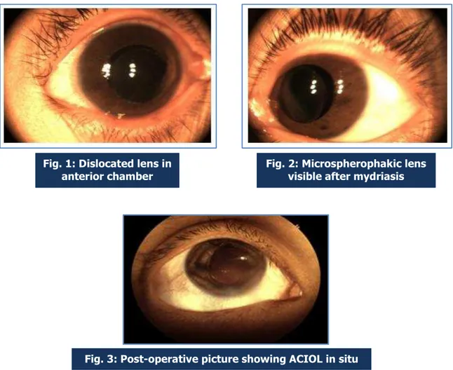
ABSTRACT: Microspherophakia is rare bilateral congenital anamoly of the crystalline lens. The condition may be isolated, familial or it may be associated with systemic affections like Marfan's syndrome, Weil-Marchesani syndrome, hyperlysinemia and congenital rubella. Microspherophakia results in lenticular myopia, lens dislocation, usually inferiorly and inverse glaucoma. We present a case in a 8 year old child who presented with bilateral microspherophakia and anterior dislocation of lens of right eye. visual acuity in right eye was counting fingers close to face and in left eye 6/60.IOP with perkins applanation tonometer was 30mmHg in right eye 22mmHg in left eye, cornea was hazy due to edema, anterior chamber was shallow in both eye patient was managed with emergency lens extraction of right eye and secondary ACIOL implantation. Left eye was managed by laser peripheral iridotomy. IOP was within normal limits postoperatively in both eyes without any antiglaucoma medications. Postoperatively best corrected visual acuity in right was 6/18 and 6/9 in left eye.
KEYWORDS: Microspherophakia, Anterior dislocated lens, Isolated microspherophakia.
INTRODUCTION: Microspherophakia is a rare congenital condition of crystalline lens where in the anteroposterior diameter is more than horizontal diameter and lens assumes a spherical shape,¹ Defective development of the zonules results in their deficiency, increased length, weakness and non-attachment of posterior zonules to the ciliary processes. This could be regarded as a simple arrest of lens development between the fifth and sixth month of intrauterine life.²-³ This leads to formation of a small spherical lens. Microspherophakia is associated with
some systemic affection like Weil Marchesani syndrome, Marfans syndrome or isolated microspherophakia.4
CASE REPORT
J of Evidence Based Med & Hlthcare, pISSN- 2349-2562, eISSN- 2349-2570/ Vol. 2/Issue 21/May 25, 2015 Page 3209 visual acuity in RE counting fingers close to face and in LE 6/36.She was put on I.V mannitol 20% and taken up for lens extraction of right eye under general anaesthesia and secondary ACIOL was implanted after 1 month. Post operatively VA in RE was 6/18.Laser iridotomy was done in LE. Post operatively IOP was within normal limits without any antiglaucoma medications.
DISCUSSION: Isolated microspherophakia is a rare condition. It is most often hereditary or familial or integrated into a general malformation syndrome.5 Investigators have hypothesized
that spherophakia occurs when an incompletely developed ciliary body and its loose elongated zonules do not exert sufficient pressure to flatten the developing lens. The lenses of patients with spherophakia therefore retain a fetal spherical conformation.²-³ It usually presents in first or
second decade of life with progressive myopia, angle closure glaucoma due to pupillary block by microspherophakic lens.Use of miotics causes forward displacement of lens iris diaphragm and cause inverse glaucoma in such patients.6-7
Familial microspherophakia, generally not associated with other systemic malformations, is inherited as an autosomal recessive trait and associated with ectopia lentis where the lens is most frequently displaced upwards.8 With our patient the manifestation of this condition is sporadic or
possibly familial as no previous case was identified in the immediate family. Fig. 1: Dislocated lens in
anterior chamber
Fig. 2: Microspherophakic lens visible after mydriasis
CASE REPORT
J of Evidence Based Med & Hlthcare, pISSN- 2349-2562, eISSN- 2349-2570/ Vol. 2/Issue 21/May 25, 2015 Page 3210
CONCLUSION: As there were no other signs or symptoms suggestive of Weil Marchesani, Marfans or other syndromes we concluded that this case is an isolated bilateral ectopic microspherophakia.
REFERENCES:
1. Duke-Elder Sir WS. System of ophthalmology vol. III. Normal and abnormal development Part 2 Congenital deformities. St Louis: CVMosby Co; 1963. pp. 694–696.
2. Johnson VP, Grayson M, Christian JC. Dominant microspherophakia. Arch Ophthalmol. 1971; 85: 534–7.
3. Farnsworth PA, Berke PA, Blanco J, Maltzman B. Ultra structural abnormalities in microspherical Ectopic lens. Exp Eye res. 1978; 27: 399–408.
4. Brown NP, Bron AJ. Lens disorders: A clinical manual of cataract diagnosis. London: Butterworth Heinemann Publishers; 1966. pp. 86–90.
5. Dietlein TS, Mietz H, Jacobi PC, Krieglstein GK. Spherophakia, nanophthalmia, hypoplastic ciliary body, and glaucoma in brachydactyly-associated syndromes. Graefes Arch Clin Exp Ophthalmol. 1996; 234: 187-192.
6. Kaushik S, Sachdev N, Pandav SS, Gupta A, Ram J. Bilateral acute angle closure glaucoma as a presentation of isolated microspherophakia in an adult: case report. BMC Ophthalmology. 2006; 6: 29.
7. Macken PL, Pavlin CJ, Tuli R, Trope GE. Ultrasound biomicroscopic features of spherophakia. Aust NZ J Ophthalomol. 1995; 23: 217–220.
CASE REPORT
J of Evidence Based Med & Hlthcare, pISSN- 2349-2562, eISSN- 2349-2570/ Vol. 2/Issue 21/May 25, 2015 Page 3211
AUTHORS:
1. Pandu S.
2. Arunkumar B. Desai
3. Sujatha V.
4. Srinivas B.
5. Muthukumaran R. S.
PARTICULARS OF CONTRIBUTORS:
1. Professor & HOD, Department of
Ophthalmology, M. V. J. Medical College of Research Institute.
2. Post Graduate, Department of
Ophthalmology, M. V. J. Medical College of Research Institute.
3. Associate Professor, Department of
Ophthalmology, M. V. J. Medical College of Research Institute.
4. Assistant Professor, Department of
Ophthalmology, M. V. J. Medical College of Research Institute.
5. Post Graduate, Department of
Ophthalmology, M. V. J. Medical College of Research Institute.
NAME ADDRESS EMAIL ID OF THE CORRESPONDING AUTHOR: Dr. Arunkumar B. Desai, Post Graduate,
Department of Ophthalmology,
M. V. J. Medical College & Research Institute, Bangalore.
E-mail: [email protected]
Date of Submission: 22/04/2015.