38
Eyelid lesion as a primary manifestation
of discoid lupus erythematosus
Lesão palpebral como manifestação
primária do lúpus eritematoso discóide
Juliana Grilo Teles¹, Suzana Matayoshi¹, Davi Araf¹, Juliana Mika Kato², Patricia Picciarelli de Lima³
¹ Sector of Ocular Plastic Surgery, Ophthalmic Clinic, Faculty of Medicine, University Hospital of the São Paulo University (USP), São Paulo (SP), Brazil.
² Medical Student, Faculty of Medicine, São Paulo University (USP), São Paulo (SP), Brazil.
³ Department of Pathology, Faculty of Medicine, University Hospital of the São Paulo University (USP), São Paulo (SP), Brazil.
Study conducted in the Ophthalmic Clinic of the University Hospital of the São Paulo State University.
The authors declare no conflicts of interest
A
BSTRACTThe discoid lupus erythematosus is an autoimmune disorder which generally affects the sun-exposed skin. Presentation of lesions on the eyelids in the absence of any other cutaneous abnormality is uncommon and the lower-eyelid involvement is seen in 6% of patients with cronic cutaneous lupus erythematosus. We have reported the case of a 40 year-old, woman who presented hyperemia, madarosis and ulceration on the lower eyelid of the left eye. She was treated for blepharitis without resolution. Before that, another similar lesion had been described on the upper eyelid of the right eye. Nevertheless, the incisional biopsies of that eyelid were inconclusive. Faced with a migratory lesion similar to the first one, the clinical hypothesis of discoid lupus erythematosus was suggested and diagnosis was confirmed by histopathological review. A high index of suspicion and early recognition may prevent misdiagnosis, clinical complications and inappropriate treatment, as described in the case of eyelid lesion as a primary manifestation of discoid lupus erythematosus.
Keywords: Lupus erythematosus, discoid/complications; Eyelids/pathology; Biopsy; Blepharitis/etiology; Case reports
R
ESUMOO lúpus eritematoso discóide é uma desordem autoimune que geralmente afeta áreas da pele expostas ao sol. A apresentação de lesões palpebrais na ausência de outras anormalidades cutâneas é incomum, sendo o envolvimento da pálpebra inferior prevalente em apenas 6% dos pacientes com lúpus eritematoso cutâneo crônico. Relatamos o caso de uma paciente do sexo feminino de 40 anos, com hiperemia, madarose e ulceração na pálpebra inferior do olho esquerdo refratária ao tratamento para blefarite. Inicial-mente, outra lesão semelhante havia sido descrita na pálpebra superior do olho direito. No entanto, as biópsias incisionais mostra-ram-se inconclusivas. Diante de uma lesão migratória palpebral de características semelhantes à primeira, a hipótese clínica de lúpus eritematoso discóide foi aventada e o diagnóstico confirmado por meio de revisão histopatológica. Uma forte suspeita clínica e o reconhecimento precoce podem evitar erros diagnósticos, complicações clínicas e tratamentos inapropriados, como descrito neste caso de lesão palpebral como manifestação primária do lúpus eritematoso discóide.
Descritores: Lupus eritematoso discóide/complicações; Pálpebras/patologia; Biópsia; Blefarite/etiologia; Relatos de casos
O
RIGINALA
RTICLEReceived for publication: 16/4/2012 - Accepted for publication: 11/6/2012
39
I
NTRODUCTIOND
iscoid Lupus Erythematosus (DLE) is a benign autoimmune disorder of the skin(1,2) and is consideredthe most common form of chronic cutaneous lupus erythematosus(2,3). The discoid lesions characteristic of the
condition are well-defined erythematous spots with thick adherent scales and follicular plugging. Old lesions show atrophic scars with hyperkeratosis(4). The condition usually affects women aged
between 20 and 40 years(2,5) and in many cases the distribution
of lesions is limited to the scalp and face(3). Involvement of the
lower eyelid occurs in 6% of patients, but eyelid lesions are rarely the only manifestation of DLE(4).
This paper presents an unusual case of eyelid discoid lupus erythematosus in which an erythematous lesion affected the eyelid of the contralateral eye.
Case report
Female, 40 year-old, mixed-race patient seen in June 2011 at the Ophthalmology Department of the University Hospital of the University of São Paulo. The patient complained of eye irritation and redness in the lower lid of the left eye, starting a year earlier and refractory to treatment for blepharitis.
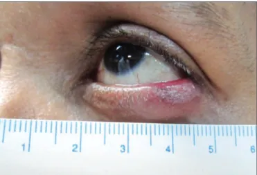
On examination, she had hyperaemia in the lower eyelid margin of the left eye, visual acuity of 20/20 without correction in both eyes and normal ocular motility. Biomicroscopy showed that the injury affected the lateral third of the lower lid of the left eye, with madarosis and ulceration with a friable, infiltrative appearance (Figure 1). The right eye showed an irregularity in the middle third of the upper eyelid margin, with dermatochalasis and mucocutaneous fistula in the lower eyelid.
The medical record showed that the patient had had a hypertrophic, erythematous, scaly lesion with madarosis in the upper eyelid of the right eye (Figures 2A and B). Incisional biopsies had shown chronic follicular reaction and lymphocytic infiltrates. The medical record also mentioned a worsening of symptoms and an excisional biopsy of the lesion followed by reconstruction of the upper eyelid by planes with auricular cartilage graft, orbicularis muscle flap and skin graft from the upper lid of the other eye. The frozen section procedure showed
lymphoid infiltration. The patient progressed with upper eyelid retraction and underwent reconstruction using the Cutler-Beard procedure, which consists of a flap from the lower to the upper eyelid. Additionally, trichiasis was corrected using the Van Milligan technique.
Family history revealed a sister with systemic lupus erythematosus, who died from the disease.
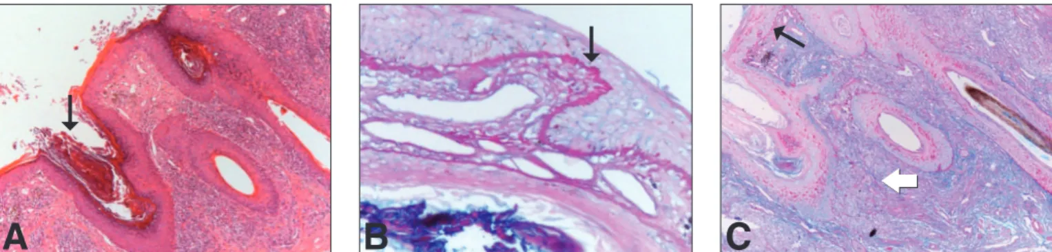
In view those findings, all slides and paraffin cubes from previous biopsies of the upper eyelid of the right eye were reviewed. The review indicated parakeratosis, follicular plugging, atrophy, vacuolisation of epithelial basal cells, thickening of the basement membrane and deposits of mucin in the dermis. In addition to these findings, subepidermal lymphocytic infiltrates were also seen around hair follicles, blood vessels and muscle fibers (Figures 3A, B and C). Histological findings were consistent with the hypothesis of discoid lupus erythematosus. Routine laboratory tests were normal, tests for autoantibodies (antinuclear antibody, extractable nuclear antigen, and anti-Ro/ SSA and anti-La/SSB) were negative and immunohistochemistry for CD 138 was positive.
Figure 2: Right eye. (A) Before surgical treatment, the original lesion (arrow) consisted of an erythematous plaque with localised loss of eyelashes in the middle and lateral thirds of the upper eyelid. (B) After surgical excision, the upper eyelid lacks the posterior lamella (white arrow), and a muco-cutaneous fistula (black arrow) can be seen in the lower eyelid due to the Cutler Beard procedure used to reconstruct the upper eyelid
Figure 1: Before treatment, the left eye had a loss of eyelashes and a hypertrophic erythematous lesion limited to the lateral third of the lower eyelid
A
B
Eyelid lesion as a primary manifestation of discoid lupus erythematosus
40
It was therefore concluded that the patient had discoid lupus erythematosus in the lower lid of the left eye and had had a similar lesion in the upper eyelid of the right eye. The palpebral discoid lesions improved within four months with oral hydroxychloroquine sulfate (400 mg/day for 12 weeks and 200 mg/day for another 8 weeks), topical steroids and sunscreen.
D
ISCUSSIONThis case highlights the difficulty in diagnosing eyelid discoid lupus erythematosus. Although initially considered an unclear chronic inflammatory process, successive biopsies were performed in an attempt to find evidence of malignancy, which would have been an important diagnosis if a sclerodermiform basal cell carcinoma had been detected (macroscopic aspect). In view of a migratory eyelid lesion with features similar to the first, the possibility of DLE was suggested and the diagnosis was confirmed through histopathology. The patient had no other skin lesions.
Eyelid discoid lupus erythematosus was first described in 1875 by Kaposy and Hebra(6), who stated that the eyelids could
be a primary site of typical lesions. According to Koga, DLE presenting as chronic blepharitis was first reported in an ophthalmology journal in 1930 by Aubaret et al.(7) In 1989, Burge
et al.(8) reported that eyelid lesions were present in 4 (6%) out
of 69 patients with cutaneous lupus erythematosus. Cyran et al.(9)
later reviewed the literature in English and reported 21 cases of DLE involving the eyelids, in some cases exclusively. Exclusive involvement of the eyelid is uncommon(2,3,10), and the lower eyelid
is affected in around 6% of patients with chronic cutaneous lupus erythematosus(4,5,10,11).
DLE lesions commonly occur in sun-exposed areas(2,3,10)
and the ocular form of the disease typically presents with symmetrical erythema and oedema of the outer third of the lower eyelids, progressing with permanent scarring and loss of eyelashes(4,10). Localised DLE occurs when only the head and
neck are affected, while the generalised form occurs when other areas are affected, regardless of head and neck involvement(1,4,11).
Patients with generalised involvement often have blood and serum abnormalities, are more likely to develop systemic lupus erythematosus (SLE), and are more difficult to treat(11,12). DLE
is typically a benign, chronic skin condition, even though 2-20% of these patients develop SLE.(3)
The diagnosis is difficult when DLE affects primarily the eyelids. The correct diagnosis is made on average 2 to 3 years after the onset of symptoms(3). The uncommon location can lead
to diagnostic errors and inadequate treatment(5). Patients with
this presentation will likely be treated as chronic blepharitis or eczema(7). The differential diagnosis of eyelid DLE includes
chronic blepharitis, allergic dermatitis, sarcoidosis, tinea, vitiligo, acne rosacea, lichen planus, psoriasis, cicatricial pemphigoid(1,3,4,11),
basal cell carcinoma, sebaceous gland carcinoma, and lymphoma(4,13). Early diagnosis and treatment can prevent
complications such as permanent scarring, entropion, ectropion, adhesion, trichiasis, and visual dysfunction(2,3). The
histopathological findings of eyelid DLE are no different from those of lesions involving other skin sites(7), with irregular
epidermal thickness, vascularisation of the basal layer, dermis with superficial and deep perivascular infiltration with vasodilation, oedema and dissociation of collagen(13). In our case,
the histopathology of eyelid lesions was typical of DLE. Skin biopsy and a careful dermatological examination for similar lesions is important because patients with eyelid DLE may show erythema and/or lesions elsewhere, and some may develop SLE. Although approximately 50% of patients with DLE have abnormal levels of antinuclear antibodies, only 5% develop SLE(4).
Thus, DLE should be considered as a differential diagnosis, especially in cases of chronic blepharitis or persistent madarosis(4,10,11). Treatment includes protection against
ultraviolet rays, which can cause and exacerbate skin lesions by inducing apoptosis of epidermal cells, exposing antigenic material on the surface of keratinocytes, inducing inflammation, promoting the expression of antigens Ro/SSA, and facilitating the formation of immune complexes in situ(14). Pharmacotherapy
includes intralesional or topical steroids(2,10), antimalarial drugs
and immunosuppressive agents for resistant cases(2, 3,10). It is
important to note that chronic DLE lesions, if left untreated, can lead to permanent scarring and deformities on the eyelid margin(10).
A strong clinical suspicion, early recognition and communication between the pathologist and the surgeon can help avoid misdiagnosis, clinical complications and inappropriate treatment(1,3), as described in this case of eyelid lesions as a
primary manifestation of DLE. Patients should be periodically examined due to the risk of systemic involvement, especially those with positive serological findings or widespread skin involvement(3).
Figure 3: A sample of eyelid skin. (A) The epidermis shows parakeratosis with follicular plugs (arrow) and atrophy. A lymphocytic infiltrate can be seen in the dermis around hair follicles and blood vessels, and telangiectasias are visible in the upper dermis. Haematoxylin and eosin (H/E) 200X. (B) Detail of the thickened basement membrane with deposition of PAS positive material (arrow). PAS/Alcian Blue, 400X. (C) Mucin deposits throughout the dermis between collagen strands (blue colour – white arrow) with a thickened basement membrane and deposition of PAS positive material (pink colour, black arrow). PAS/Alcian Blue, 200X
A
B
C
Teles JG, Matayoshi S, Araf D, Kato JM, Lima PP
41
Corresponding author:
Juliana Grilo Teles
Rua Dr. Eduardo Amaro, nº 99 - apto 714
CEP 04104-080 – - São Paulo (SP), Brasil
Tel: (011) 7037-1844
E-mail: [email protected]
R
EFERENCES1. Uy HS, Pineda R 2nd, Shore JW, Polcharoen W, Jakobiec FA, Foster CS. Hypertrophic discoid lupus erythematosus of the conjunctiva. Am J Ophthalmol. 1999;127(5):604-5.
2. Acharya N, Pineda R 2 nd, Uy HS, Foster CS. Discoid lupus erythema-tosus masquerading as chronic blepharoconjunctivitis. Ophthalmol-ogy. 2005;112(5):e19-23.
3. Giménez-García R, Sánchez-Ramón S, De Andrés A. Discoid lupus erythematosus involving the eyelids. J Eur Acad Dermatol Venereol. 2005;19(1):138-9.
4. Ena P, Pinna A, Carta F. Discoid lupus erythematosus of the eyelids associated with staphylococcal blepharitis and Meibomian gland dys-function. Clin Exp Dermatol. 2006;31(1):77-9.
5. Pianigiani E, Andreassi A, De Aloe G, Rubegni P, Rufa A, Motolese E. Chronic erythematous desquamative plaques of the eyelids: discoid lupus erythematosus (DLE) solely localized to the eyelids. Arch Dermatol. 2002;138(4):527-32.
6. Kaposi M, Hebra F. On diseases of the skin, including the exanthe-mata. London: New Sydenham Society; 1875.
7. Koga M, Kubota Y, Kiryu H, Nakayama J. A case of discoid lupus erythematosus of the eyelid. J Dermatol. 2006;33(5):368-71. 8. Burge SM, Frith PA, Juniper RP, Wojnarowska F. Mucosal
involve-ment in systemic and chronic cutaneous lupus erythematosus. Br J Dermatol. 1989;121(6):727-41.
9. Cyran S, Douglass MC, Silverstein JL. Chronic cutaneous lupus erythe-matosus presenting as periorbital edema and erythema. J Am Acad Dermatol. 1992;26(2 Pt 2):334-8.
10. Selva D, Chen CS, James CL, Huilgol SC. Discoid lupus erythematosus presenting as madarosis. Am J Ophthalmol. 2003;136(3):545-6. 11. Yaghoobi R, Feily A, Behrooz B, Yaghoobi E, Mokhtarzadeh S.
Palpe-bral involvement as a presenting and sole manifestation of discoid lupus erythematosus. TheScientificWorldJournal. 2010;10:2130-1. 12. Frith P, Burge SM, Millard PR, Wojnarowska F. External ocular
find-ings in lupus erythematosus: a clinical and immunopathological study. Br J Ophthalmol. 1990;74(3):163-7.
13. Kormann RB, Cunha E, Silva AT, Beckhauser AP, Werner B. Lúpus discóide em pálpebras: relato de caso. Arq Bras Oftalmol. 2009;72(4):549-51.
14. Martins MA, Carrilho FJ, Alves VAF, Castilho EA, Cerri GG, Wen CL, editores. Clínica médica. Barueri: Manole; 2009.
Eyelid lesion as a primary manifestation of discoid lupus erythematosus