251
C
ASER
EPORTGranuloma macular por tuberculose
sem manifestação pulmonar
Macular granuloma due to tuberculosis
without pulmonary symptoms
Albert Costa Rebello
1, João Helio Leonardo de Sousa
2, José Gilberto de Sá
3, Karime Kalif de Sousa Rebello
41,4 Centro de Oftalmologia Rio – Rio de Janeiro, RJ, Brazil; 2,3 Hospital Municipal Jesus – Rio de Janeiro, RJ, Brazil.
Received for publication 10/07/2012 - Accepted for publication 26/03/2013
The authors declare no conflicts of interest
R
ESUMOTuberculose é uma doença infecciosa causada pelo Mycobacteriumtuberculosis, também conhecido como bacilo de Koch. O principal
sítio de acometimento é o pulmonar, porém o bacilo pode disseminar-se por via linfo-hematogênica para outros órgãos, dentre eles: o olho. A incidência de tuberculose ocular é de 1 a 2% dos casos extra pulmonares. Os autores apresentam um caso clínico de um paciente do sexo feminino de 28 anos que procura atendimento médico devido a redução da acuidade visual em olho esquerdo há 7 dias. Apresentava a melhor acuidade visual corrigida no olho acometido de 20/200, e no olho contralateral de 20/20. Na fundoscopia era evidenciado um granuloma em área macular do olho esquerdo, com edema e hemorragia intrarretiniana adjacente. Após investigação diagnóstica, a paciente foi tratada com esquema antibiótico para tuberculose durante 6 meses, obtendo regressão do granuloma e melhora da acuidade visual deste olho para 20/50.
Descritores: Tuberculose ocular/diagnóstico; Tuberculose ocular/tratamento; Macula lutea; Uveíte/diagnóstico; Uveíte/trata-mento; Relatos de casos
Study conducted at the Municipal Hospital Jesus and the Ophthalmology Center Rio – Rio de Janeiro (RJ), Brazil.
Rev Bras Oftalmol. 2015; 74 (4): 251-3
A
BSTRACTTuberculosis is an infectious disease caused by Mycobacterium tuberculosis. The main site of involvement is the lung, but the bacillus may spread by hematogenous/lymph systems to other organs, including the eye. The incidence of ocular TB is 1-2% of extra-pulmonary cases. The authors present a case of a 28 years old female patient seeking medical care due to reduction of visual acuity in the left eye for 8 days. She had the best corrected visual acuity in the affected eye of 20/200, and the opposite eye was 20/20. At fundoscopy was shown a granuloma in the macular area of the left eye, with retinal edema and hemorrhage. After diagnostic investigation the patient was treated with antibiotic therapy for tuberculosis during 6 months, obtaining lesion regression and visual acuity improvement to 20/50.
Keywords: Tuberculosis, ocular/diagnosis; Tuberculosis, ocular/drug therapy; Macula lútea; Uveitis/diagnosis; Uveitis/therapy; Case reports
252 RebelloAC, SousaJHL, SáJG, RebelloKKS
Rev Bras Oftalmol. 2015; 74 (4): 251-3
I
NTRODUCTIONB
razil is one of 22 countries prioritized by the World HealthOrganization (WHO) covering 80% of the global cases
of tuberculosis(1). Currently the country is in the 19th
place, and was formerly the 14th in 2004. In 2009, there were 71,700 new cases of the disease, with an incidence rate of 37 per each group of 100 thousand inhabitants. In Rio de Janeiro the incidence rate is 73.99 per 100,000 inhabitants, giving the state a
sad first place when compared to other States(2). According to
data collected by the Ministry of Health and the Department of Health Surveillance, in the case of extra pulmonary tuberculosis the incidence rate drops to 10.06 per 100,000 inhabitants in Rio de Janeiro. This number includes the cases of pleural, lymph node, osteoarticular, genitourinary, intestinal, peritoneal,
pericardial, central nervous system, eye and skin tuberculosis(3,4).
The authors present a case of ocular manifestation of tuberculosis that was satisfactorily responsive to the treatment against tuberculosis for 6 months.
C
ASER
EPORTFemale patient, 28, white, sought medical care due to decreased visual acuity in the left eye for 7 days, with sudden and spontaneous onset. Past medical history without relevant data, but reporting contact with a tuberculosis patient two years ago in her own home.
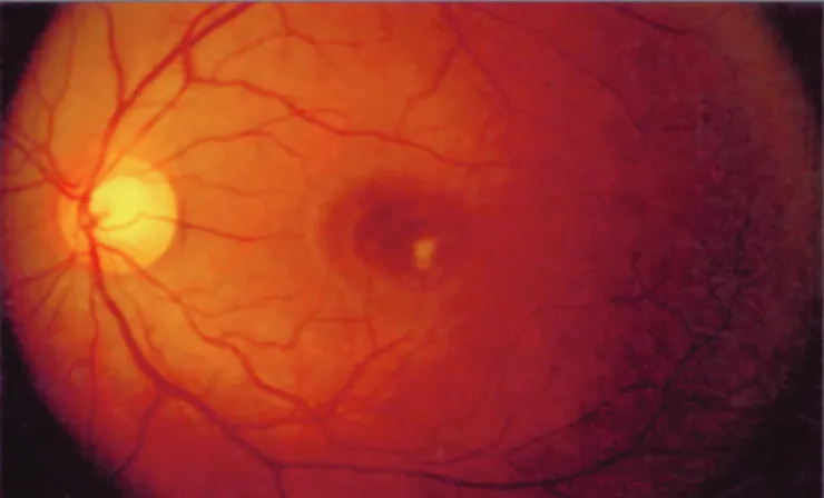
The eye exam showed better corrected visual acuity for farsight on the right eye: 20/20, and on the left eye: 20/200; Ocular motility without change; Normal biomicroscopy; Tonometry RE: 12 mmHg and LE: 13 mmHg; Fundoscopy: stained buds, physiological excavations, preserved arteriovenous ratio, presence of a yellowish granulomatous lesion with adjacent retinal edema and hemorrhage in the macula of the left eye (Figure 1).
Initial diagnostic research showed no changes in blood count, microbiology, biochemistry and immunochemistry. It is important to emphasize the negative serology for toxoplasmosis, both IgM as IgG. Normal chest radiograph. Tuberculin skin test equal to 20mm. Antituberculosis oral therapy was initiated with follow-up in specialized unit of the public health service. To improve the retinal edema, she received topical treatment with dexamethasone eyedrops for 30 days, with gradual dose reduction and monitoring of the intraocular pressure.
After follow-up for 6 months, with the respective end of the oral treatment, the patient improved the visual acuity of the left eye to 20/50. In the fundoscopy, the lesion improved and the retinal edema disappeared, the hemorrhage was absorbed and the retinal pigment epithelium in the macular area was atrophied (Figure 2).
D
ISCUSSIONGiven the high prevalence of the disease in our country, it is always advisable to investigate tuberculosis in cases of chorioretinitis. It is a especially important fact, considering that some treatments directed to other etiologies may worsen the tuberculosis condition (as an example we can mention the use of systemic corticosteroids, used in some cases of toxoplasmosis chorioretinitis).
The standard test for the diagnosis finding Mycobacterium
tuberculosis in ocular specimens, but due to the paucibacillary lesions and the noble condition of the visual apparatus significantly limit the histological study, the diagnosis of ocular tuberculosis is supported by epidemiological data, clinical examination, positive tuberculin skin test and laboratory exclusion
of other conditions(5), among them toxoplasmosis.
The tuberculin skin test is still the exam which guides us
about possible infections with Mycobacterium tuberculosis,
although a positive result does not mean active disease. Therefore, other criteria are needed to confirm the diagnosis of ocular tuberculosis. Among them are: observing if there is improvement with the basic scheme in the first two months, if there is no recurrence of the disease during treatment, or if there is no recurrence in the first two years after interrupting the medication. Otherwise, probably the etiology of tuberculosis is not
considered, forcing the search for other causes(6).
We must also take into account the differential diagnosis with syphilis, sarcoidosis, brucellosis, histoplasmosis and toxocariasis.
Any tissue in the eye may be affected, though the most common site is the uvea. As illustrated in the present case, the disease may be present even without systemic disease. The changes can be caused by bacterial invasion or be immunological, by hypersensitivity reaction type IV. Some of the possible ocular manifestations are anterior, posterior, intermediate uveitis, panuveitis, serpiginous-like choroiditis, subretinal abscesses, Figure 1: Granulomatous lesion with hemorrhage and retinal edema.
253
serous retinal detachment, retinitis, choroiditis, retinochoroiditis,
retinal vasculitis and endophthalmitis(6).
In this case, the improved healing of the macular injury after the use of anti tuberculous medication is an important evidence to establish the diagnosis, along with the positive history of contact. But also, there was healing and regression of the lesion after the beggining of the treatment without reincidence, and apparently it continues healed even after the end of 6-month therapy. As the prognosis is influenced by the location of the lesion and the adherence to treatment, in this case the significant improvement in vision draws attention, despite the proximity between the granuloma and the macula.
R
EFERENCES1. Arakaki D, Oliveira G, Barreira D, Moherdaui F, Codenotti S, Bartholomay P. Novo sistema de tratamento da tuberculose para adultos e adolescentes no Brasil. Informe Técnico de Tuberculose. Edição nº 5, julho de 2010. Disponível em: http://portal.saude.gov.br/ portal/arquivos/pdf/informe_tb_julho10_certo_22_07_2010.pdf 2. Brasil. Ministério da Saúde. Departamento de Informatica do
SUS. DATASUS. Disponível em http://tabnet.datasus.gov.br/cgi/ e http://www2.datasus.gov.br/DATASUS
Macular granuloma due to tuberculosis without pulmonary symptoms
Rev Bras Oftalmol. 2015; 74 (4): 251-3 Corresponding author:
Albert Costa Rebello
Av. Passos, 101 / 1609 - Centro ZIP Code: 20051-040 - Rio de Janeiro (RJ), Brazil
Fax No.: (21) 2263-4838
E-mail: [email protected]
3. Brasil. Ministério da Saúde. Secretaria de Políticas de Saúde. Departamento de Atenção Básica. Manual técnico para controle da tuberculose. (Cadernos de Atenção Básica, 6). Brasília: Ministério da Saúde; 2002.
4. Brasil. Ministério da Saúde. Secretaria de Vigilância em Saúde. Departamento de Vigilância Epidemiológica. Doenças infecciosas e parasitárias: guia de bolso. 8a ed. rev. Brasília: Ministério da Saúde; 2010.
5. Lopes AJ, Capone D, Mogami R, Tessarollo B, Cunha DL, Capone RB, et al. Tuberculose extrapulmonar: aspectos clínicos e de imagem. Pulmão RJ. 2006;15(4):253-61.