MYC
cis
-Elements in
PsMPT
Promoter Is
Involved in Chilling Response of
Paeonia
suffruticosa
Yuxi Zhang☯, Tingzhao Sun☯, Shaoqing Liu, Lei Dong, Chunying Liu, Wenwen Song,
Jingjing Liu, Shupeng Gai*
College of Life Sciences, Qingdao Agricultural University, Key Lab of Plant Biotechnology in Universities of Shandong Province, Changcheng Road 700, Qingdao, China
☯These authors contributed equally to this work.
Abstract
The MPT transports Pi to synthesize ATP.PsMPT, a chilling-induced gene, was previously reported to promote energy metabolism during bud dormancy release in tree peony. In this study, the regulatory elements ofPsMPTpromoter involved in chilling response were further analyzed. ThePsMPTtranscript was detected in different tree peony tissues and was highly expressed in the flower organs, including petal, stigma and stamen. An 1174 bp of the PsMPTpromoter was isolated by TAIL-PCR, and thePsMPTpromoter::GUStransgenic Arabidopsiswas generated and analyzed. GUS staining and qPCR showed that the pro-moter was active in mainly the flower stigma and stamen. Moreover, it was found that the promoter activity was enhanced by chilling, NaCl, GA, ACC and NAA, but inhibited by ABA, mannitol and PEG. In transgenic plants harboring 421 bp of thePsMPTpromoter, the GUS gene expression and the activity were significantly increased by chilling treatment. When the fragment from -421 to -408 containing a MYCcis-element was deleted, the chilling response could not be observed. Further mutation analysis confirmed that the MYC element was one of the key motifs responding to chilling in thePsMPTpromoter. The present study provides useful information for further investigation of the regulatory mechanism ofPsMPT during the endo-dormancy release.
Introduction
The mitochondrial phosphate transporter (MPT) shuttles inorganic phosphate (Pi) into the mitochondrial matrix, where Pi is utilized for oxidative phosphorylation to synthesize ATP from ADP.MPTencoding genes have been cloned from mammals [1–3], yeast [4], and wood frogs [5] with most studies focusing mainly on the structure and catalytic function of the transporters.
Recently, cloning and characterization ofMPTwere reported in several plants [6–12]. Plant MPTgenes were identified to be involved in abiotic stress responses, and their expression
a11111
OPEN ACCESS
Citation:Zhang Y, Sun T, Liu S, Dong L, Liu C, Song W, et al. (2016) MYCcis-Elements inPsMPT Promoter Is Involved in Chilling Response ofPaeonia suffruticosa. PLoS ONE 11(5): e0155780.
doi:10.1371/journal.pone.0155780
Editor:Keqiang Wu, National Taiwan University, TAIWAN
Received:January 14, 2016
Accepted:May 4, 2016
Published:May 26, 2016
Copyright:© 2016 Zhang et al. This is an open access article distributed under the terms of the
Creative Commons Attribution License, which permits unrestricted use, distribution, and reproduction in any medium, provided the original author and source are credited.
Data Availability Statement:All relevant data are within the paper and its Supporting Information files.
Funding:This work was supported by National Natural Science Foundation of China (31071828, 31372104 and 31471908).
patterns showed tissue preferences. BirchMpt1was ozone-inducible and highly expressed in the tissue of dividing cells, such as root tips, shoot apices and developing root nodules [6]. AtMPTsplay an important role in response to salt stress inArabidopsis. Furthermore, with dif-ferent expression profiles in various tissues and conditions, transcription ofAtMPTshas been detected in all tissue except siliques [12]. The sequences and structures of 26 potential PT fam-ily genes in rice were analyzed, and sixMPTs also showed tissue preferential expression pro-files, among whichOsPT17andOsPT19were differently regulated under hormone treatment conditions. In addition, six putativecis-elements were found in all of theOsPTgenes including ARR1AT, CAATBOX1, CACTFTPPCAL, GATABOX, GT1CONSENSUS and GTGANTG10. Specifically, GATABOX and GT1CONSENSUS are light-responsivecis-elements, and CACTFTPPCAL is necessary for carbon metabolism [11]. Current knowledge ofMPT regula-tion and the molecular mechanisms mediating its biological funcregula-tions in plants is still incomplete.
Tree peony (Paeonia suffruticosaAndrews) is one of the most well-known horticultural and medicinal plants in the world. One of the main production mechanisms in the tree peony industry, especially for the Spring Festival flower market in China, is forcing culture. Dor-mancy is a major obstacle for the forced culture of tree peony in winter, and sufficient chilling is an efficient way to break dormancy. Therefore, it is important to determine how chilling induces dormancy release in tree peony.PsMPTwas previously isolated from the tree peony subtractive cDNA library of burst buds and strongly induced by chilling treatment to promote ATP production during the release of bud dormancy. In addition, ectopic-expression of PsMPTinArabidopsisshowed thatPsMPTenhanced ATP synthesis and affected plant growth and development [10]. These results suggested thatPsMPTplays an important role in energy production during bud dormancy release in tree peony [13]. However, the expression charac-teristics ofPsMPTand its regulatory mechanisms are unclear.
In this study, we isolated the promoter ofPsMPTand constructedPsMPTpromoter::GUS engineeredArabidopsis. We investigated: 1) the temporal and spatial characteristics of the PsMPTpromoter inArabidopsisandPsMPTexpression in tree peony; 2) how plant hormones and abiotic stresses, including chilling, affects the activity of thePsMPTpromoter; 3) which one ofcis-elements among thePsMPTpromoter is involved in the chilling response.
Materials and Methods
Plant materials
Four-year-old tree peonies (Paeonia suffruticosa‘Luhehong’) were obtained from the Tree Peony Research Center of Heze (Shandong, China). According to the method of Huang et al. [10], plants were treated in cold conditions (0–4°C) for 21 days to break bud dormancy, as the daily mean temperature was under 10°C in Qingdao, Shandong, China. The plants were then transferred to a greenhouse (18–22°C, 8-h-light/16-h-dark cycle) to resume growth. Tissues (root, stem, leaf, calyx, petal, stamen and carpel at the early stage of flowering) were collected and stored at -80°C until use. One hundredμmolL-1ABA and 50μmolL-1GA3were applied to non-chilling buds with double-distilled water as the control, and buds were collected after 0, 1, 6, 12, 24 and 48 h. Three replicates (3 plants/replicate) were performed for all treatments.
Isolation of the
PsMPT
promoter
Genomic DNA was extracted from tree peony buds using the cetyltrimethylammonium bro-mide (CTAB) extraction method as previously described [14]. DNA samples were qualified photometrically, then checked on agarose gel, and stored at -20°C for use. Based on the cDNA sequence ofPsMPT(Genbank accession No.: EU072922), three gene-specific primers, SP1, SP2
and SP3, in nested positions were designed with primer premier 5.0 and synthesized (Table 1). ThePsMPTpromoter was amplified using gene-specific primers and four short arbitrary prim-ers (AP) with the Genome Walking Kit (TaKaRa). The PCR products were ligated into the pMD18-T vector (TaKaRa), and sequenced at BGI-Beijing, China.
Bioinformatics analysis of the promoter sequence
Regulatory elements in the promoter were analyzed using the online program PLACE (http:// www.dna.affrc.go.jp/PLACE/) [15,16] and Plantcare (http://bioinformatics.psb.ugent.be/ webtools/plantcare/html) [17].
Construction of the promoter-reporter plasmids
To construct the binary vector consisting of theβ-glucuronidase (GUS) coding sequence driven by thePsMPTpromoter, a fragment from -1117 to -1 relative to the translation initiation codon was obtained using the high fidelity DNA polymerase and promoter specific primers (FP6 and RP,Table 1). The serial deletions of thePsMPTpromoter (-909, -621, -574, -421, and -282 to -1) were amplified by PCR with the corresponding forward (FP1, FP2, FP3, FP4, FP5) and reverse primers (RP) that contained aHind III andBamH I site at the 5' end of each primer, respectively. For chilling response element identification, the MYC element -413 rela-tive to the translation initiation site was subsequently deleted from the P2 construct with the FDP primer, and mutated to form the MP construct by recombination PCR with the FM1 and RM1 primers (Table 1). PCR products were retrieved and cloned into a pMD18-T simple vec-tor (TaKaRa) followed by sequencing conformation at BGI Beijing, China. TheHind III/BamH I digested DNA fragments were inserted into the corresponding sites of pBI121, in place of the deleted CaMV35S promoter upstream of theGUScoding sequence. The pBI121 vector with theCaMV35S::GUSwas used as the positive control. The resulting constructs were named P1 Table 1. The primers used in this paper.
Primers Sequences (5’-3’) purpose
SP1 CTGATGTTAGGGTTCTACTTTCCTCTTTCTCTC’ Promoter isolation
SP2 ATGTCTGCGTTACCCAAGGTCGTCCC Promoter isolation
SP3 ATAGGGCATTCCCAGAAACGATTGTCC Promoter isolation
FP1 GGGAAGCTTGGGACCCAGTGTGT (Hind III) Promoter analysis
FP2 GGGAAGCTTTGGGGACTCAATTGT (Hind III) Promoter analysis
FP3 GCGAAGCTTGATACAATGGGAGAGGAG (Hind III) Promoter analysis
FP4 GGGAAGCTTGGTCGCATTCGTCG (Hind III) Promoter analysis
FP5 GGGAAGCTTAGAACAAGAATCGTGGAG (Hind III) Promoter analysis
FP6 GGGAAGCTTAGCTCGGCATTCAGTG (Hind III) Promoter analysis
RP CGAGGATCCCATATCTGATGTTAGG (BamH I) Promoter analysis
FDP GGGAAGCTTCATATTATGTCAAATTGG (Hind III) Motif identification
RM1 TCCAATTTGACATAATATGTTACAAGGT Motif identification
FM1 TTATGTCAAATTGGAGACTTGATT Motif identification
GUSFW AGTGGCAGTGAAGGGCGAACAGT qPCR ofGUS
GUSRV TCAGCGTAAGGGTAATGCGAGGT qPCR ofGUS
ActinFW GAGAGATTCCGTTGCCCTGA qPCR ofβ-actin
ActinRV CTCAGGAGGAGCAACCACC qPCR ofβ-actin
MPTFW GCTGGAGGAATAATGAGTTGTGG qPCR ofPsMPT
MPTRV GCACCTTGAGCACTGTAACCA qPCR ofPsMPT
(-282), P2 (-421), P3 (-574), P4 (-621), P5 (-909), P6 (-1117), DP (-404), and MP (-574), respectively.
Arabidopsis
transformation
Arabidopsis thalianawild-type (Col-0) plants were transformed using theAgrobacterium tumefaciensstrain EHA105 carrying the above constructs according to the floral dip method [18]. The transformants were screened on MS medium containing 50 mg L-1kanamycin, and positive plants were identified by PCR amplification using GUS specific primers (GUSFW and GUSRV,Table 1). The corresponding T1transgenic seedlings that segregated at a ratio of 3:1 (resistant: sensitive) were selected to propagate T2individuals for further analysis. Five to ten transgenic lines were obtained for every construct.
Hormone and abiotic stress treatments
The transformedArabidopsisplants grew at 18°C in a controlled growth chamber (16-h-light/ 8-h-dark cycle), and 5-week-oldArabidopsisplants were used to analyze the response of the PsMPTpromoter to hormone and abiotic stresses. For chilling treatment, plants were cultured under 4°C for 24 h, and those at 18°C were used as controls. Moreover, the inflorescence of 5-week-oldArabidopsisplants were sprayed by GA3(50μmol L-1), NAA (100μmol L-1), ABA (100μmol L-1), ACC (250 mmol L-1), NaCl (200 mmol L-1), mannitol (40μmol L-1) and PEG (100 mmol L-1) treatments for 24 h, respectively. Double-distilled water was used as a control. Samples were collected after 0, 1, 3, 6, 12 and 24 h treatments and stored at -80°C.
GUS histochemical assay and quantitative analysis of GUS activity
GUS activity was determined in tissue extracts using a standard protocol [19]. GUS fluores-cence was measured with a Microplate Spectrofluorometer, the data were obtained by subtract-ing the background 4-methyiumbelliferyl glucuronide of thePPsMPT::GUStransgenic plants. The average GUS activity was obtained from at least five independent transformants, and each assay was repeated three times. GUS histochemical staining was performed using identified homozygous transgenic plants by a modified Jefferson’s method [19]. In brief, plant tissues were incubated in a 100 mmol L-1sodium phosphate buffer (pH 7.0) containing 0.1% Triton X-100, 10 mmol L-1EDTA, 1 mmol L-1X-gluc and 0.5 mmol L-1potassium ferricyanide at 37°C overnight. The stained tissues were then washed several times with 70% ethanol to bleach the chlorophyll.Quantitative real-time PCR (qPCR)
Results
Expression characteristics of
PsMPT
in tree peony
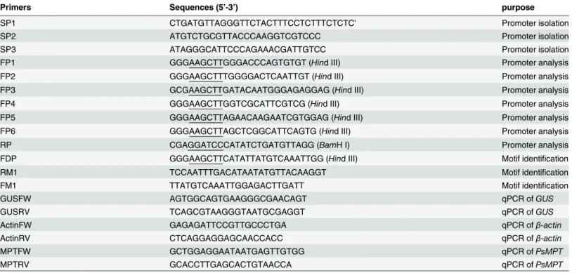
The expression ofPsMPTwas previously reported during dormancy release in tree peony [13]. In this study, the temporal and spatial expression ofPsMPTwas further detected at the early stage of flowering in tree peony. The results of qPCR indicated that the transcription ofPsMPT was detected in all tree peony tissues; however, thePsMPTtranscript was very low in root, stem, leaf and calyx, but high in flower organs, including petal, stamen and stigma (Fig 1A). ThePsMPTtranscripts in the stamen were expressed 6-fold as compared to that of the root. The results indicated thatPsMPTwas expressed preferentially in flower organs of tree peony.
The response ofPsMPTin dormant buds to Gibberellic Acid (GA) and Abscisic Acid (ABA) were analyzed by qPCR. When GA was applied, thePsMPTtranscript was quickly pro-moted and peaked at 24 h, then declined slightly. Conversely, ABA application dramatically decreased the expression ofPsMPT(Fig 1B).
Isolation of the
PsMPT
promoter and the putative
cis
-acting element
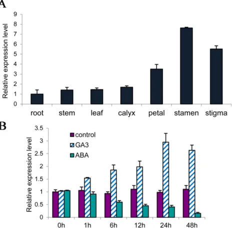
In this study, the 1174 bp upstream genomic DNA sequence ofPsMPTwas isolated by TAIL-PCR (Fig 2). The adenosine of the translation initiation codon (ATG) of thePsMPT gene was defined as +1 (Fig 2). A motif search was carried out using PLACEandPlantCareto
Fig 1. Tissue-specific expression ofPsMPTin germinated buds (A) and transcriptional levels of
PsMPTin response of GA3and ABA of tree peony by qPCR (B).100μmol L-1ABA and 50μmol L-1GA3
was sprayed to the dormant buds in green house (18–22°C, 8-h-light/16-h-dark cycle). Values are means±SD
of three replicates.
analyze the putativecis-elements. As shown inFig 2, the putative TATA box was found at posi-tion -189/-185 and the CAAT box at posiposi-tion -210/-207. In addiposi-tion, a number of regulatory motifs potentially related to environmental signals were found, which included auxin-, GA-and dehydration-responsive GA-and tissue-specific elements. Among thecis-elements, one puta-tive pyrimidine box and two GA-responsive elements (GAREs) were located at -991/-986, -913/-907 and -780/-771, respectively, which were related to GA response and sugar repression [22,23]. Four putative Myelocytomatosis viral oncogene homolog (MYC) (5’-CANNTG -3’) motifs and four Myeloblastosis viral oncogene homolog (MYB) (5’-WAACCA -3’, 5’ -YAACKG -3’or 5’-GGATA-3’) motifs were located at positions -797/-792, -737/-732, -589/-584, -412/-408 and -1127/-1122, -1056/-1051, -427/-422, -40/-35, respectively. These motifs had previously been identified in response to dehydration inArabidopsis[24–26]. Sev-eral MYB and MYC motifs were thought to respond to chilling or freezing inArabidopsis[24–
26]. There were two putative sulfur-responsive element (SURE) motifs at positions -647/-643 and -387/-382, and one putative TGA element (auxin responsive element) at position -764/-759. SURE contains the auxin response factor binding sequence [27]. Therefore,PsMPTmight be regulated by chilling and auxin. Interestingly, eight putative GATA boxes were present in the sequence, which were thought to be involved in tissue-specific expression and light response [28,29]. Moreover, three putative pollen1lelat52 elements, related to the pollen spe-cific expression [2,30], were found at -775/-771, -782/-777, and -1005/-1001, respectively. Fig 2.PsMPTpromoter sequence of the 5’region upstream from the start codon (ATG) and putative
cis-elements predicted in the promoter region.Numbers indicate the positions relative to the translation start codon starting from the adenosine (+1). The putativeTATAbox at position -189, CAAT box at position -210, and the ATG start codon is framed in box and denoted with blue color. The other important putative cis-elements are framed and labeled below.
Temporal and spatial expression of the
PsMPT
promoter in
Arabidopsis
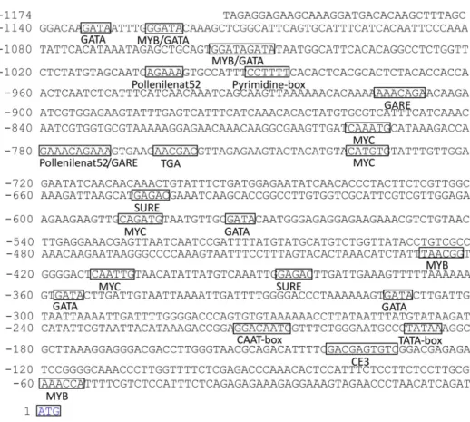
To identify the expression patterns of thePsMPTpromoter, the promoter::GUSchimeric con-struct (PPsMPT::GUS) was transformed intoArabidopsis, and histochemical GUS staining was carried out in various organs throughout plant development (Fig 3I). These results showed that GUS activity was not detected in the seedling tissues (Fig 3IA and 3IB) but were observed in the flower organs (Fig 3IC, 3IE–3IG). GUS activity in transgenic plants was more pronounced in the stigma and stamen compared with the sepals (Fig 3II–3IL). GUS staining was notFig 3. Histochemical localization (I) and tissue-specific expression ofGUS(II) in transgenicArabidopsis thaliana
carrying thePsMPTpromoter::GUSconstruct.(I)Histochemical localization of GUS expression by staining with X-gluc in transgenicArabidopsis thalianacarrying thePsMPTpromoter::GUSconstruct. Arrow bar shows GUS staining in flower. A7-day-plants from seeding;B21-day-plants from seeding;C28-day-plants from seeding;DPositive control (Ca35S promoter driven);Eroots;F: stem;Gleaf;H: mature silique;Iflower;Jstigma;Kstamen;Lpetal. (II)Total RNA was isolated from roots (R), rosette leaf (RL), cauline leaf (CL), flower bud (F), bloomed flower (BF), stamen (S), stigma (St), petal (P) and siliques (Si) of 35-day-old transgenic plants from seeding. The transcriptional levels were analyzed by qPCR using GUS gene-specific PCR primers, which were normalized with beta-actin. Values are means±SD of three replicates.
observed in the silique and seeds (Fig 3IH). These results suggested thatPsMPTis preferentially expressed in the flower tissues.
qPCR was performed to evaluate the spatial expression of thePsMPTpromoter. The results showed that theGUStranscript was only detected in the flower (Fig 3II). No transcript ofGUS was detected in the roots, rosette leaves, cauline leaves and silique.GUStranscript levels in bloomed flowers were approximately 10-fold higher compared with flower buds. In bloomed flowers, the most abundant expression was found in stigma, followed by in stamen, with the lowest amount in the petals. In summary, the GUS staining results are in accordance with that of qPCR. The GUS reporter did not appear in the immature flower buds possibly due to the low abundance of the transcripts. The flower-specific expression characteristics of the pro-moter implied thatPsMPTmight participate in plant anthesis and gametophyte development.
Responses of the
PsMPT
promoter to hormones and abiotic stresses
The transgenic plants carrying thePPsMPT::GUScassette were treated with hormones and abi-otic stresses, and the transcription ofGUSwas evaluated by qPCR, respectively. Overall,GUS activity changed rapidly and fluctuated during the entire period for all of the treatments (Fig 4). Chilling increased theGUStranscript during 3 h to 6 h after treatment, with a peak approxi-mately 2.5-fold higher at 6 h (Fig 4A), which was also verified by GUS staining (Fig 4B). The results of qPCR showed that GA3and NAA treatments enhancedGUSexpression at 1 h, then decreased, followed by another peak at 24 h. ACC accelerated the transcript ofGUSduring the entire process. Conversely, ABA inhibitedGUSexpression throughout the process. In addition, theGUStranscript was continuously decreased by mannitol and PEG until 3 h, followed by a slight increase at 12 h and 24 h; however, theGUStranscript levels were lower than that of con-trol. Notably, NaCl dramatically enhanced GUS activity, and it reached a peak at 3 h with an approximate 25-fold increase (Fig 4C). The results of GUS activity were consistent withGUS expressions of qPCR (Fig 4D).
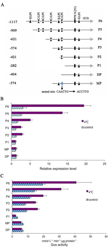
Identification of chilling response elements
As reported previously,PsMPTwas chilling inducible. Furthermore,PsMPTwas involved in chilling induced dormancy release in tree peony. To confirm thePsMPTpromoter region involved in the chilling response, a number of truncated promoter fragments (P1 (-282), P2 (-421), P3 (-574), P4 (-621), P5 (-909) and P6 (-1117)) were isolated and fused to theGUS reporter gene into pBI121 vector (Fig 5A). Transgenic Arabidopsis plants with each promoter-GUS construct were generated. The transcription levels of promoter-GUS were detected by qPCR (Fig 5B). In the transgenic plants, the highest level ofGUSexpression was detected in the engi-neered Arabidopsis with the P6 construct, which contained the full-lengthPsMPTpromoter (−1117/−1). GUS transcript decreased in order from the P6 to P1 construct, and P2 and P3 had similar abundance. These results suggested that the transcription enhancer might exist in the upstream of thePsMPTpromoter.
We also detectedGUSexpression of the successive deletions at 4°C for 6 h compared to 18°C (Fig 5B). After exposure to 4°C, theGUStranscripts were up-regulated in the P2 to P6 constructs. The largest increase inGUSabundance was observed in plants with the P6 con-struct (6.34-fold). The next-largest increase was observed in the P5 concon-struct (-909/-1, 5.40-fold). Similar enhancement was observed in the P2 and P3 constructs with 2.45 and 2.41-fold, respectively. However, the P1 construct (-282/-1), which only contained basic tran-scriptional elements, could not induce GUS expression at 4°C. These results indicated that the
Fig 4. Relative expression levels ofPsMPTpromoter in response to hormone and various abiotic stresses treatments in transgenic Arabidopsis plants.(A)GUSexpressions when exposed to 4°C temperature. Plants were transferred to a cold chamber maintained at 4°C, and the control grew at 18°C. Error bars represent±SD. (B) GUS staining of transgenicArabidopsisgrew at 18°C (left) and at 4°C for 6 h (right). (C)GUSexpressions were measured by qPCR using 35-day-old plants from seeding. 50μmol L-1GA
3,
100μmol L-1NAA, 100μmol L-1ABA, 250 mmol L-1ACC, 200 mmol L-1NaCl, 40μmol L-1mannitol and 100 mmol L-1PEG was sprayed to the inflorescence at 18°C, and double-distilled water treatment was used as
control. (D)GUSfluorescence (nmol L-1min-1μg protein-1) were measured by a Microplate Spectrofluorometer
using 35-day-old plants from seeding. 50μmol L-1GA
3, 100μmol L-1NAA, 100μmol L-1ABA, 250 mmol L-1
ACC, 200 mmol L-1NaCl, 40μmol L-1mannitol and 100 mmol L-1PEG was sprayed to the inflorescence at 18°C, and double-distilled water treatment was used as control.
found within the promoter. We speculated thus that the MYC elements in the promoter might be involved in the chilling response.
To verify our hypothesis, another deletion construct (DP) (-404) and one mutated construct (MP) (-574) were constructed (Fig 5A). In the DP construct, the MYC element located at -412 in the promoter was deleted. When exposed to 4°C, no significant difference was observed as compared to that of 18°C treatment, which indicated that there was a loss of function for chill-ing induction with the MYC element deleted in thePsMPTpromoter (Fig 5B). In the MP con-struct where the MYC element (-412) was mutated from CAATTG to ACCTTG, a complete elimination of the chilling response was observed (Fig 5B).
To confirm the results obtained by qPCR, a quantitative measurement of GUS activity was performed using the series constructs, P1-P6, DP and MP. Consistent with the results obtained by qPCR, the GUS activity was observed to increase from the P2 to P6 after exposed to 4°C, and no difference was observed in the P1, DP and MP. In summary, we concluded that the MYC element involved in chilling treatment responses (Fig 5C).
Discussion
MPT can shuttle inorganic phosphate (Pi) into the mitochondrial matrix, where ATP is syn-thesized. Thus, MPT plays a key role in cellular ATP regeneration. ATP is essential for almost all biological processes in the cell, andMPTshave been reported to be involved in abiotic responses [6,12], bud dormancy release, growth and development [13]. Although several MPTs have been cloned and functionally annotated, their characteristics and regulatory mech-anisms are poorly understood.
We previously clonedPsMPT, a chilling induced gene, in tree peony, which accelerated ATP synthesis and dormancy release [13]. In this study, we isolated thePsMPTpromoter and detected its activity using GUS as the reporter in transgenicArabidopsis. GUS-staining and qPCR of the transgenic plants revealed that thePsMPTpromoter was preferentially expressed in the flower, mainly in stamen and stigma. Therefore, the tissue-specific expression may be related to the putative GATA boxes and pollen1lelat52 elements founded in the promoter [30,
31]. The results suggested thatPsMPTmight play an important role during gametophyte devel-opment, pollination and fertilization, and Pi might be transported to reproductive organs dur-ing the reproductive development stage. Differdur-ing from ectopic expression of thePsMPT promoter,PsMPTmRNA was detected in all tissues of tree peony by qPCR. This discrepancy has also been reported in mice [32,33] and is believed to be related to different biological spe-cies or incomplete isolation of promoter sequences.
Fig 5. Assays for GUS expression driven by the seriesPsMPTpromoter.(A) Schematic diagram of the PsMPTpromoter deletions and mutation that were used to analyze the activity of different fragments of the PsMPTpromoter. All fragmented promoter were fused to a GUS reporter gene. (B) Quantitative analyses of GUS expression in transgenic plants driven by deletion or muted constructs ofPsMPTpromoter in response to chilling. (C) GUS activity in transgenic Arabidopsis plants. The inflorescence of 5-week-old Arabidopsis plants was used as material, and five independent lines for every treatment.
Therefore, we speculated that the discrepancy might be due to species-specific independent evolution of MPTs and their promoters.
The response of thePsMPTpromoter to abiotic stress and hormones was analyzed by qPCR.GUStranscript increased during chilling treatment in the transgenic plants driven by thePsMPTpromoter. In addition, thePsMPTpromoter was also induced by salt, GA3, ACC and NAA, while ABA, mannitol and PEG suppressed its activity. In rice,OsPT19was sup-pressed by five hormone treatments including ABA, 2, 4-D, GA3, KT, and NAA, whereas OsPT17was induced by the hormone treatments with NAA or GA3[11]. These responses might be due to thecis-elements in the promoter, for instance, the putative pyrimidine box and two GARE motifs to GA response, putative TGA and SURE elements to NAA response, and MYB and MYC to abiotic stress response, such as chilling. Interestingly, NaCl treatment could significantly increase the expression of the reporter gene with a maximum of approximately 25-fold. Similar results were reported forArabidopsis[12].
Among all of the factors influencing promoter activity, chilling and GA3treatments are of interest because they effectively accelerate the dormancy release in winter [35–38]. Huang et al. found thatPsMPTwas induced by chilling [13]. Interestingly, it was reported that the chilling-induced expression ofPsMPTwas not maintained after being transferred to a greenhouse (18– 22°C) when less than 21 days of chilling were applied. On the other hand, the levels ofPsMPT transcripts remained high with a 21 d or longer chilling duration after returning to growth tem-perature [13]. In this study, ectopic expression analyses provided more evidence that the PsMPTpromoter could be induced by chilling. We speculated that the early increase of the PsMPTtranscript might be induced by chilling, and GA production might be a downstream effect of chilling, as proposed for dormant seeds [39,40]. Meanwhile, chilling temperature was reported to enhance the accumulation of endogenous GA, and exogenous GA could partially replace chilling to accelerate endo-dormancy release [41]. In this study, we found that exoge-nous GA could activate the expression of thePsMPTpromoter. Therefore, the reactivation of thePsMPTtranscripts might be due to the high endogenous GA induced by sufficient chilling when transferred to growth condition. Buds chilled for less than 21 days had relatively low GA levels that could not activate thePsMPTexpression required for the recovery of plant growth ability.
Considering the central role ofPsMPTin energy metabolism during dormancy release, it is important to elucidate how chilling acceleratesPsMPTexpression. It is well-known that the transcription of mRNA is mainly regulated through the cooperation of transcript factors and correspondingcis-elements. Severalcis-elements have been identified to be involved in chilling or cold responses, such as ABRE (ABA responsive element), DRE/CRT (dehydration-respon-sive element/C-repeat element, A/GCCGAC), MYB, MYC, and the E-box [24,26,42,43,44,
45]. Bioinformatics analysis of the isolatedPsMPTpromoter showed that four MYB and four MYC elements were present upstream of the promoter, which might be responsible for the chilling response. Based on the location of the MYC and MYB elements, deletion experiments were conducted to identify the candidate chilling response elements in the promoter.GUS expression and activity revealed that the P2 construct containing one MYC element (-412/-408, CAATTG) effectively responds to chilling, and the addition of a MYB element (P3 con-struct) did not improve the chilling response ability. When MYC was deleted or mutated, the chilling response character abated. Alternatively, increase of MYC elements in the P4, P5 and P6 constructs enhanced the chilling response activity, indicating there was an additive effect of MYC elements in the chilling response. This study demonstrates that the MYC elements in the PsMPTpromoter play a crucial role in the chilling responses.
mannitol, auxin and GA. Deletion and mutation analyses demonstrated that the MYCcis -ele-ment functioned in the chilling response. This work provides useful information for further investigation of the regulatory mechanisms ofPsMPTpromoter during endo-dormancy release.
Supporting Information
S1 Fig. Organization of the PsMPT and AtMPTs promoters. (DOCX)
S1 Table. The identity (%) between PsMPT protein and theArabidopsisand rice MPTs. (DOC)
Acknowledgments
We would like to thank Dr. Chunhai Dong for constructive suggestions in writing and revision of this manuscript.
Author Contributions
Conceived and designed the experiments: SG. Performed the experiments: TS SL LD YZ CL WS JL. Analyzed the data: SG YZ TS. Contributed reagents/materials/analysis tools: YZ SG TS. Wrote the paper: YZ SG TS.
References
1. Runswick MJ, Powell SJ, Nyren P, Walker JE. Sequence of the bovine mitochondrial phosphate carrier protein: structural relationship to ADP/ATP translocase and the brown fat mitochondria uncoupling pro-tein. EMBO J. 1987 May; 6 (5):1367–1373. PMID:3038521
2. Ferreira GC, Pratt RD, Pedersen PL. Energy-linked anion transport. Cloning, sequencing, and charac-terization of a full length cDNA encoding the rat liver mitochondrial proton/phosphate symporter. J Biol Chem. 1989; 264 (26):15628–15633. PMID:2670944
3. Dolce V, Fiermonte G, Messina A, Palmieri F. Nucleotide sequence of a human heart cDNA encoding the mitochondrial phosphate carrier. DNA Seq. 1991; 2 (2):133–135. PMID:1777677
4. Phelps A, Schobert CT, Wohlrab H. Cloning and characterization of the mitochondrial phosphate trans-port protein gene from the yeastSaccharomyces cerevisiae. Biochem. 1991 Jan; 30 (1):248–252. 5. De Croos JA, McNally JD, Palmieri F, Storey KB. Upregulation of the mitochondrial phosphate carrier
during freezing in the wood frog Rana sylvatica: potential roles of transporters in freeze tolerance. J Bioenerg Biomembr. 2004 Jun; 36 (3):229–239. PMID:15337853
6. Kiiskinen M, Korhonen M, Kangasjärvi J. Isolation and characterization of cDNA for a plant mitochon-drial phosphate translo2 cator (Mpt1): ozone stress induces Mpt1 mRNA accumulation in birch (Betula pendula Roth). Plant Mol Biol. 1997 Oct; 35(3):271–279. PMID:9349251
7. Takabatake R, Hata S, Taniguchi M, Kouchi H, Sugiyama T, Izui K. Isolation and characterization of cDNAs encoding mitochondrial phosphate transporters in soybean, maize, rice, and Arabidopsis. Plant Mol Biol. 1999 Jun; 40 (3): 479–486. PMID:10437831
8. Nakamori K, Takabatake R, Umehara Y, Kouchi H, Izui K, Hata S. Cloning, functional expression, and mutational analysis of a cDNA for Lotus japonicus mitochondrial phosphate transporter. Plant Cell Phy-siol. 2002 Jul 25; 43 (10):1250–1253. PMID:12407206
9. Hamel P, Saint Georges Y, De Pinto B, Lachacinski N, Altamura N, Dujardin G. Redundancy in the function of mitochondrial phosphate transport inSaccharomyces cerevisiaeandArabidopsis thaliana. Mol Microbiol. 2004 Jan; 51 (2):307–317. PMID:14756774
10. Huang X, Xue T, Dai S, Gai S, Zheng C, Zheng G. Genes associated with the release of dormant buds in tree peonies (Paeonia suffruticosa). Acta Physiol Plant. 2008a Nov; 30:797–806.
12. Zhu W, Miao Q, Sun D, Yang G, Wu C, Huang J, et al. The mitochondrial phosphate transporters modu-late plant responses to salt stress via affecting ATP and gibberellin metabolism in Arabidopsis thaliana. PLoS ONE. 2012 Aug; 7:e43530. doi:10.1371/journal.pone.0043530PMID:22937061
13. Huang X, Zhu W, Dai S, Gai S, Zheng G, Zheng C. The involvement of mitochondrial phosphate trans-porter in accelerating bud dormancy release during chilling treatment of tree peony (Paeonia suffruti-cosa). Planta. 2008b Sep; 228:545–552.
14. Gai S, Zhang Y, Mu P, Liu C, Liu S, Dong L, et al. Transcriptome analysis of tree peony during chilling requirement fulfillment: assembling, annotation and markers discovering. Gene. 2012 Apr 15; 497 (2):256–262. doi:10.1016/j.gene.2011.12.013PMID:22197659
15. Prestridge DS. SIGNAL SCAN: a computer program that scans DNA sequences for eukaryotic tran-scriptional elements. Comput Appl Biosci. 1991 Apr; 7 (2): 203–206. PMID:2059845
16. Higo K, Ugawa Y, Iwamoto M, Korenaga T. Plant cis-acting regulatory DNA elements (PLACE) data-base: 1999. Nucleic Acids Res. 1999 Jan 1; 27 (1):297–300. PMID:9847208
17. Lescot M, Déhais P, Thijs G, Marchal K, Moreau Y, Van de Peer Y, et al. PlantCARE, a database of plant cis-acting regulatory elements and a portal to tools for in silico analysis of promoter sequences. Nucleic Acids Res. 2002 Jan 1; 30 (1):325–327. PMID:11752327
18. Clough SJ, Bent AF. Floral dip: a simplified method for Agrobacterium-mediated transformation of Ara-bidopsis thaliana. Plant J. 1998 Dec; 16 (6):735–743. PMID:10069079
19. Jefferson RA. Assaying chimeric genes in plants: the GUS gene fusion system. Plant Mol Biol Rep. 1987 Dec; 5 (4):387–405.
20. Gai S, Zhang Y, Liu C, Zhang Y, Zheng G. Transcript Profiling of Paoenia ostii during Artificial Chilling Induced Dormancy Release Identifies Activation of GA Pathway and Carbohydrate Metabolism. PLoS ONE. 2013 Feb 6; 8:e55297. doi:10.1371/journal.pone.0055297PMID:23405132
21. Livak KJ, Schmittgen TD. Analysis of Relative Gene Expression Data Using Real-Time Quantitative PCR and the 2−ΔΔCTMethod. Methods. 2001 Dec; 25 (4):402–408. PMID:11846609
22. Morita A, Umemura T, Kuroyanagi M, Futsuhara Y, Perata P, Yamaguchi J. Functional dissection of a sugar-repressed alpha-amylase gene (RAmy1A) promoter in rice embryos. FEBS Lett. 1998; 423 (1):81–85. PMID:9506846
23. Mena M, Cejudo FJ, Isabel-Lamoneda I, Carbonero P. A role for the DOF transcription factor BPBF in the regulation of gibberellin-responsive genes in barley Aleurone. Plant Physiol. 2002 Aug 8; 130: 111–119. PMID:12226491
24. Abe H, Urao T, Ito T, Seki M, Shinozaki K, Yamaguchi-Shinozaki K. ArabidopsisAtMYC2(bHLH) and AtMYB2(MYB) function as transcriptional activators in abscisic acid signaling. Plant Cell. 2003 Jan; 15:63–78. PMID:12509522
25. Chinnusamy V, Ohta M, Kanrar S, Lee B, Hong X, Agarwal M, et al. ICE1: a regulator of cold-induced transcriptome and freezing tolerance in Arabidopsis. Gene Dev. 2003 Apr 2; 17 (8):1043–1054. PMID: 12672693
26. Agarwal M, Hao Y, Kapoor A, Dong C, Fujii H, Zheng X, et al. A R2R3 typeMYBtranscription factor is involved in the cold regulation of CBF genes and in acquired freezing tolerance. J Biol Chem. 2006 Oct 2; 281 (49):37636–37645. PMID:17015446
27. Maruyama Nakashita A, Nakamura Y, Watanabe Takahashi A, Inoue E, Yamaya T, Takahashi H. Iden-tification of a novel cis-acting element conferring sulfur deficiency response in Arabidopsis roots. Plant J. 2005 Feb 17; 42 (3):305–314. PMID:15842617
28. Lam E, Chua N. ASF-2: a factor that binds to the cauliflower mosaic virus 35S promoter and a con-served GATA motif in Cab promoters. Plant Cell. 1989 Dec; 1 (12):1147–1156. PMID:2535536 29. Vieweg MF, Frühling M, Quandt H, Heim U, Bäumlein H, Pühler A, et al. The promoter of theVicia faba
L. leghemoglobin geneVfLb29is specifically activated in the infected cells of root nodules and in the arbuscule-containing cells of mycorrhizal roots from different legume and nonlegume plants. Mol Plant Microbe In. 2004 Jan; 17 (1): 62–69.
30. Bate N, Twell D. Functional architecture of a late pollen promoter: pollen-specific transcription is devel-opmentally regulated by multiple stage-specific and co-dependent activator elements. Plant Mol Biol. 1998 Jul; 37 (5):859–869. PMID:9678581
31. Filichkin SA, Leonard JM, Monteros A, Liu PP, Nonogaki H. A novel endo-beta-mannanase gene in tomatoLeMan5is associated with anther and pollen development. Plant Physiol. 2004 Mar; 134 (3):1080–1087. PMID:14976239
33. Kristiansen MT, Rasmussen LM, Olsen N, Asa SL, Jørgensen JO. Ectopic ACTH Syndrome:
Discrep-ancy between Somatostatin Receptor Status in vivo and ex vivo, and between Immunostaining and Gene Transcription for POMC and CRH. Horm Res. 2002; 57 (5–6): 200–204. PMID:12053094 34. Taylor MS, Kai C, Kawai J, Carninci P, Hayashizaki Y, Semple CAM. Heterotachy in mammalian
pro-moter evolution. PLoS Genet. 2006 Apr; 2(4): e30. PMID:16683025
35. Horvath DP, Anderson JV, Chao WS, Foley ME. Knowing when to grow: signals regulating bud dor-mancy. Trend Plant Sci. 2003 Nov; 8 (11):534–540.
36. Schrader J, Moyle R, Bhalerao R, Hertzberg M, Lundeberg J, Nilsson P, et al. Cambial meristem dor-mancy in trees involves extensive remodelling of the transcriptome. Plant J. 2004 Oct; 40 (2):173–187. PMID:15447645
37. Rohde A, Ruttink T, Hostyn V, Sterck L, Van Driessche K, Boerjan W. Gene expression during the induction, maintenance, and release of dormancy in apical buds of poplar. J Exp Bot. 2007 Sep 28; 58 (15–16): 4047–4060. PMID:18039739
38. Rentzsch S, Podzimska D, Voegele A, Imbeck M, Müller K, Linkies A, et al. Dose-and tissue-specific interaction of monoterpenes with the gibberellin-mediated release of potato tuber bud dormancy, sprout growth and induction ofα-amylases andβ-amylases. Planta. 2012 Jan; 235 (1): 137–151. doi:10. 1007/s00425-011-1501-1PMID:21858448
39. Hazebroek JP, Metzger JD, Mansager ER. Thermoinductive regulation of gibberellin metabolism in Thlaspi arvenseL. (II. Cold induction of enzymes in gibberellin biosynthesis). Plant Physiol. 1993 Jun; 102 (2):547–552. PMID:12231843
40. Yamauchi Y, Ogawa M, Kuwahara A, Hanada A, Kamiya Y, Yamaguchi S. Activation of gibberellin bio-synthesis and response pathways by low temperature during imbibition of Arabidopsis thaliana seeds. Plant Cell. 2004 Feb; 16 (2): 367–378. PMID:14729916
41. Zheng GS, Gai SP, Gai WL. Changes of endogenous hormones during dormancy release by chilling in tree peony. Scientia Silvae Sinicae. 2009; 45:48–52. (Chinese in English abstract)
42. Chinnusamy V, Schumaker K, Zhu JK. Molecular genetic perspectives on cross-talk and specificity in abiotic stress signalling in plants. J Exp Bot. 2004 Dec 12; 55(395):225–236. PMID:14673035 43. Kim JC, Lee SH, Cheong YH, Yoo CM, Lee SI, Chun HJ, et al. A novel cold-inducible zinc finger protein
from soybean, SCOF-1, enhances cold tolerance in transgenic plants. Plant J. 2001 Feb; 25(3): 247–
259. PMID:11208017
44. Feller A, Machemer K, Braun EL, Grotewold E. Evolutionary and comparative analysis of MYB and bHLH plant transcription factors. Plant J. 2011 Apr; 66(1): 94–116. doi:10.1111/j.1365-313X.2010. 04459.xPMID:21443626