Recebido em 07.01.2002. / Received in January, 07thof 2002.
Aprovado pelo Conselho Consultivo e aceito para publicação em 05.10.2002. / Approved by the Consultive Council and accepted for publication in October, 05thof 2002. * Trabalho realizado nos serviços de Dermatologia e Anatomia Patológica da Universidade Federal Fluminense-UFF. / Work done at "the services of Dermatology and Pathologic Anatomy,
Fluminense Federal University – UFF"
1Sócio Efetivo SBD/ SBCD; Mestre em Dermatologia - UFF; Doutor em Dermatologia - UFRJ; Responsável pela Disciplina de Dermatologia Cirúrgica e Oncologia Cutânea - PGRJ/IPGMCC. / Effective Member SBD/SBCD; M.Sc. in Dermatology – UFF; Ph.D. in Dermatology – UFRJ; Responsible for the Department of Surgical Dermatology and Cutaneous Oncology – PGRJ/IPGMCC.
2Especialista em Dermatologia pela UFF / Dermatology Specialist – UFF
3Sócia Colaboradora SBD; Professora Adjunta do Departamento de Patologia da UFF; Doutora em Anatomia Patológica - UFF; Pós-Doutorado em Dermatologia - A. B. Ackermann - Institute for Dermatopathology - Thomas Jefferson University / Collaborating Member of the SBD; Adjunct Professor, Department of Pathology – UFF; Ph.D. in Pathologic Anatomy – UFF; Post-Doctorate in Dermatology – A. B. Ackermann – Institute of Dermatopathology – Thomas Jefferson University
©2004by Anais Brasileiros de Dermatologia
Halo Nevo Spilus
*Halo Nevus Spilus
*Flávio Barbosa Luz
1Beatriz França da Mata
2Mayra Carrijo Rochael
3Resumo: Os autores descrevem o caso de paciente de 15 anos que apresenta um componente jun-cional de seu nevo spilus envolto por halo acrômico na coxa direita. O mecanismo imunológico con-stante no fenômeno halo e a resposta imune ao melanoma estão intimamente relacionados. Aparentemente, o fenômeno halo representa uma reação imunológica mediada por células contra um antígeno desconhecido presente nas lesões melanocíticas. Há também presença de anticorpos nessa reação.
Palavras-chave: imunidade; melanoma; nevo pigmentado.
Summary:A patient with a junctional component from a nevus spilus surrounded by an achromic halo on the right thigh is reported. The link between the immune response to melanoma and the mechanism responsible for halo nevus is closely related. There is evidence that the halo phenome-non results from a mediated cell immunologic reaction against an unknown antigen present in melanocytic lesions. Antibodies are also found in these lesions.
Key words: immunity; melanoma; nevus, pigmented.
Caso Clínico / Case Report
INTRODUÇÃO
O halo nevo é fenômeno relativamente comum, que ocorre sobretudo em adolescentes, em especial no dorso, e pode ser múltiplo. Sua evolução é característica: um nevo melanocítico preexistente é circundado por halo despig-mentado que gradualmente desaparece com o nevo. Na tabela 1 observa-se essa progressão do ponto de vista his-topatológico. Frank e Cohen1 relataram que pelo menos 50% dos halos nevos desaparecem espontaneamente. As áreas de despigmentação podem permanecer por meses ou anos, bem como repigmentar totalmente.2,3
A malignização das lesões do nevo spilusé
conhe-cida. Foram encontrados descritos na literatura 15 casos de melanoma oriundos de lesões dessa natureza.4-18
O nevo spilusé um tipo especial de nevo
tico congênito. Alguns autores crêem que nevos melanocí-ticos pequenos apresentam risco aumentado de evolução
INTRODUCTION
Halo nevus is a relatively common phenomenon that occurs mainly in adolescents, especially in the back and can be multiple. Its clinical course is characteristic: a preexisting melanocytic nevus is surrounded by a depig-mented halo that gradually disappears with the nevus. Table 1 shows this progression from the histopathological standpoint. Frank and Cohen1
reported that at least 50% of the halo nevi disappear spontaneously. However, areas of depigmentation can persist for months and even years or become totally repigmented.2,3
The malignant potential of nevus spiluslesions has been recognized. Fifteen cases were found in the literature of melanoma originating from this type of lesion.4-18
para melanoma. Tal risco de malignização aumenta após a puberdade. Estima-se entre 0,8 e 4,9% a probabilidade cumulativa em pacientes com mais de 60 anos.19,20
RELATO DE CASO
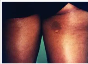
Adolescente de 15 anos, parda, estudante, solteira apresentando desde a infância uma mácula hipercrômica de limites precisos, porém irregulares, localizada na face pos-terior da coxa direita (Figura 1). Um exame mais minucio-so evidenciou máculas puntiformes enegrecidas na superfí-cie da lesão. Há seis meses notou o surgimento de uma mácula acrômica numular de limites bem definidos no inte-rior da lesão. O exame histopatológico mostrou tratar-se de uma reação tipo nevo halo de Sutton sobre um nevo juncio-nal envolto por lentigo simples.
DISCUSSÃO
A reação tipo halo nevo é caracterizada clinicamen-te pelo surgimento de mácula acrômica em torno de lesão melanocítica tumoral, assemelhando-se a uma mancha de vitiligo. Do ponto de vista histopatológico, essa reação é formada por infiltrado mononuclear ao redor de células névicas, as quais vão sendo progressivamente destruídas.
Em l952, Ito e Hamada19descreveram o nevo
spi-luscomo uma mácula castanho-clara de limites nítidos e
irregulares com pontos enegrecidos em seu interior. Histopatologicamente, a área castanho-clara corresponde a um lentigo simples, e os pontos mais escuros a nevos juncionais ou compostos. O nevo spilusé uma lesão
mela-nocítica congênita. A lesão costuma surgir no primeiro ano de vida, e os pontos mais escuros entre os seis e os 39 anos de idade.
Nessa reação vitiligóide tipo halo nevo de Sutton sobre uma lesão de nevo spilusnão há sinais histológicos
de atipia. Tal achado pode representar tanto uma resposta eficaz contra um melanoma bem inicial quanto um forte argumento contra a hipótese de esse tipo de resposta
signi-The risk of becoming malignant increases after puberty, the cumulative probability in patients aged over 60 years is considered to be between 0.8 and 4.9%.19,20
CASE REPORT
Adolescent 15 years old, mixed race, student and single, presenting from infancy a hyperchromic stain with precise though irregular margins, located in the subsequent face of the right thigh (Figure 1). A more meticulous exam revealed punctiform and blackened stains in the surface of the lesion. Six months previously she had noticed the appea-rance of an achromic nummular stain with very well defi-ned edges within the lesion. Histopathological exam demonstrated that this was a Sutton’s halo nevus type lesion in a junction nevus surrounded by lentigo simplex.
DISCUSSION
Halo nevus type lesion is clinically characterized by the appearance of a nonpigmented patch bordering a mela-nocytic tumoral lesion, resembling a vitiligo stain. From the histopathological point of view, this lesion is formed by a mononuclear infiltrate around nevoid cells, which are pro-gressively destroyed.
In l952, Ito and Hamada19
described nevus spilusas a light-chestnut patch with precise but irregular edges and with blackened spots inside. Histopathologic exam shows that the light-chestnut area corresponds to a lentigo simplex and the darker points to junctional or compound nevi. Nevus spilusis a congenital melanocytic lesion. The lesion tends to appear within the first year of life and the darker points between six and 39 years of age.
In such a Sutton’s nevus type vitiliginous lesion within a nevus spiluslesion there are no histological signs of atypia. Such a finding could represent both an effective response against a very initial melanoma and a strong argument against the hypothesis that this type of response means a defense mechanism against a malignant process. Estágio I ou pré-regressão o nevo é envolto por número moderado de células T, B e macrófagos.
Stage I or pre-regression the nevus is surrounded by a moderate quantity of T and B cells and macrophages.
Estágio II ou regressão precoce grande número de linfócitos T estão em contato com sítio de ligação de células névicas.
Stage II or precocious regression a great number of T lymphocytes are in contact with the site at which the nevoid cells are binded.
Estágio III ou regressão tardia aumento do número de linfócitos T, macrófagos e células de Langerhans.
Stage III or tardive regression an increase in the number of T lymphocytes, macrophages and Langerhan’s cells.
Estágio IV ou regressão completa ausência de células névicas e poucos linfócitos T. Macrófagos contendo células névicas.
Stage IV or complete regression absence of nevoid cells and few T lymphocytes. Macrophages containing nevoid cells.
Tabela 1: Estágios para regressão de um halo nevo / Table 1: Stages in the regression of a halo nevus
Figura 1: Fenômeno halo ocorrendo sobre o componente juncional
de um nevo spilus
Figure 1: Halo phenomenon occurring on the junctional component of a nevus spilus
The presence of immu-nological mechanisms in the pathogenesis of halo nevus can be demonstrated by two factors: mononuclear cell infiltrate progressively invol-ving the nevoid cells in dege-neration and the presence of antibodies directed to the anti-gens that react in vitrowith nevoid and melanocytic cells.21
The immunity mediated by T cells in the develop-ment of halo nevus can be demonstrated by immunohisto-chemical studies, which reveal that the majority of the lymphocytes of the infiltrate are derived from the T lymphoid lineage and it is estimated that the largest part of which, are CD8+ T cells.22
Natural killer cells were found in small amounts and the lymphokines they activate have little influence in the cytotoxicity of the melanocytes in vitiligo,23
enabling the supposition that the same are not involved in the immune reactions that lead to the depigmentation.
Although no direct demonstration of death of the melanocytes due to the effector cells present in the halo has been observed, the abundance of antigen presenter cells within the nevus in regression and the presence of T lymphocytes in the site of the depigmentation suggest the participation of these cells in the halo phenomenon. Within the latter population of cells, evidence points to the involve-ment of CD8+ T cells as important destructive agents of melanocytic nevi. Possible factors unchaining the migration and activation of lymphocytes in nevi apparently without alterations have yet to be clarified.21
Mooney et al.,24
stud-ying 142 halo nevi, observed that the depigmentation pheno-menon occurs in a variety of types of histologically atypical nevi. This study demonstrates that there can be a broad spectrum of atypia among the halo nevi and in those with intense atypia it can be difficult to differentiate them from the rare phenomenon of malignant halo melanoma.
Research into the halo phenomenon in melanoma is of fundamental importance for the understanding of its regression, as well as that of halo nevus. Copeman and Eliot25
demonstrated the presence of antibodies against cytoplasmatic components of melanoma cells in patients with halo nevus in involution. They concluded that circula-ting antibodies contribute to the occurrence of halo nevus and that this fact suggests the vitiligous reaction could sig-nify a rejection of the organism to an initial melanoma that is growing in a nevoid lesion. Grispan et al.26
have also
sug-ficar um mecanismo de defe-sa contra um processo de malignização.
A presença de meca-nismos imunológicos na patogênese do halo nevo pode ser evidenciada por dois
fatores: infiltrado celular mononuclear envolvendo progres-sivamente as células névicas em degeneração e a presença de anticorpos direcionados aos antígenos que reagem com células névicas e melanócitos in vitro.21
A imunidade mediada por células T no desenvolvi-mento do halo nevo pode ser comprovada por estudos imuno-histoquímicos, os quais revelam que a maioria dos linfócitos do infiltrado é derivada da linhagem linfóide T, estimando que, em sua maior parte, sejam células T CD8+.22 As células natural killer foram encontradas em
pequenas quantidades, e linfocinas por elas ativadas não têm muita influência na citotoxicidade dos melanócitos no vitili-go,23permitindo supor que as mesmas não estejam envolvi-das em reações imunes que levam à despigmentação.
Embora nenhuma demonstração direta de morte dos melanócitos pelas células efetoras presentes no halo tenha sido observada, a abundância de células apresentadoras de antígeno no nevo em regressão e a presença de linfócitos T no local de despigmentação sugerem a participação dessas células no fenômeno halo. Dentro desta última população de células, as evidências apontam para envolvimento de células T CD8+ como importantes agentes destruidores de nevos melanocíticos. Possíveis fatores deflagradores da migração e ativação de linfócitos em nevos aparentemente sem altera-ções permanecem inexplicados.21Mooney
et al.,24estudando 142 halos nevos, observaram que o fenômeno de despig-mentação ocorre numa variedade de tipos de nevos histolo-gicamente atípicos. Esse estudo demostra que pode haver um amplo espectro de atipia entre os halos nevos, sendo que naqueles com intensa atipia pode ser difícil sua diferencia-ção do raro fenômeno halo do melanoma maligno.
desenvolvendo sobre uma lesão névica. Grispan et al.26 tam-bém sugerem que esse fenômeno ocorra sobre lesões displá-sicas de nevos spilus.
A tirosinase é um auto-antígeno em potencial envol-vido na resposta contra melanócitos no vitiligo auto-imune27 e células malignas do melanoma.28Outro estudo comprova que em ambas as doenças ocorre produção de anticorpos devida aos antígenos comuns aos melanócitos e melano-ma.29Como bem conhecido na prática clínica, anticorpos direcionados contra o melanoma são citotóxicos também para melanócitos normais, podendo desencadear lesões de vitiligo em pacientes com melanoma.
Apesar de existir relação entre a despigmentação do halo nevo e a produção de anticorpos contra células névi-cas, a regressão do nevo não ocorre simultaneamente com o surgimento de anticorpos circulantes reagentes contra o citoplasma das células do melanoma. Krebs et al.30 sugeri-ram que a citotoxicidade imunologicamente mediada des-truiria células névicas que, por sua vez, produziriam anti-corpos. A lesão da célula alvo por linfócitos T citotóxicos parece ser o evento primário na formação do halo nevo.
Após estudar 46 casos de nevos halo e melanomas em regressão, baseando-se em aspectos imuno e histopato-lógicos, Campos31 concluiu que o fenômeno halo aparenta ser determinado por antígenos que não especificam o tipo nem o caráter benigno ou maligno das lesões, uma vez que o halo nevo, o melanoma e os melanócitos normais compar-tilham alguns dos alvos moleculares responsáveis pelo fenômeno de regressão.
Os autores relatam o caso de uma paciente com nevo spilus no qual foi observado fenômeno halo clínica e histo-patologicamente. A possibilidade de reação auto-imune contra células atípicas ou mesmo típicas presentes nessas lesões, à semelhança do que ocorre no melanoma, tem sido apresentada por alguns autores, embora sua causa permane-ça inexplicada. q
gested that this phenomenon occurs against dysplastic lesions of nevus spilus.
Tyrosinase is an auto-antigen in potential involved in the response against melanocytes in the auto-immune vitiligo27
and malignant melanoma cells.28
Another study has demonstrated that in both diseases there is a production of antibodies due to the antigens common to both the mela-nocytes and melanoma.29
As is well known in clinical prac-tice, antibodies directed against the melanoma are also cytotoxic to normal melanocytes and can unchain vitiligo lesions in patients with melanoma.
Although there is a relationship between depigmen-tation of the halo nevus and production of antibodies against nevoid cells, regression of the nevus does not occur simultaneously with the appearance of circulating reactive antibodies against the cytoplasm of the melanoma cells. Krebs et al.30
have suggested that the immunologically mediated cytotoxicity could destroy nevoid cells that, in turn, would produce antibodies. Lesion of the cell targeted by cytotoxic T lymphocytes appears to be the primary event in the formation of the halo nevus.
After studying 46 cases of halo nevus and melano-mas in regression, based on immune and histopathological aspects, Campos31
concluded that the halo phenomenon appears to be determined by antigens that do not specify the type nor benign or malignant character of the lesions, since the halo nevus, melanoma and normal melanocytes share some of the molecular targets responsible for the regression phenomenon.
The authors report the case of a patient with nevus
19. Ito M, Hamaada Y. Nevus spilusen nappe. Tohuku J Exp Med l952;55:44-8.
20. Cohen LM, Bennion SD, Johnson TW, Golitz LE. Hypermelanotic nevus: clinical, histopathologic, and ultrastruc-tural features in 316 cases. Am J dermatol 1997;19(1):23-30. 21. Zeff RA, Freitag A, GrincM, Grant-Kels JM. The immune response in halo nevi. J Am acad Dermatol 1997;37(4):620-4. 22. Akasu R, Fron L, Kahn HJ. Characterization of the mononu-clear infiltrate involved in regression of halo nevi. J Cutan Patholl 1994;21:302-11.
23. Durham-Pierre DG, Walters CS, Halder RM et al. Natural killer cell and lymphokine-activated killer cell activity against melanocytes in vitiligo. J Am Acad Dermatol l995;33:26-30. 24. Mooney MA, Barr RJ, Buxton MG. Halo nevus or halo phe-nomenon? A study of 142 cases. J Cutan Pathol 1995;22:342-8. 25. Copeman PWM, Elliot PG. Melanoma cytoplasmic humoral antibody test. Br J Dermatol l976:94:565.
26. Grispan D et al. Melanoma on dysplastic nevus spilus. Int J Dermatol l997;36:499-502.
27. Song YH, Connor E, Li Y et al. The role of tyrosinase in autoimmune vitiligo. Lancet l994;344:1049-52.
28. Merimsky O, Baharav E, Schoenfeld Y et al. Anti-tyrosinase antibodies in malignant melanoma. Cancer Immunol Immunother l996; 42:297-302.
29. Cui J, Bystryn J-C. Melanoma and vitiligo are associated with antibody responses to similar antigens on pigment cells. Árch Dermatol l995;121:314-8.
30. Krebs JÁ, Roenigk HH, Deodhar SD et al. Halo nevus: Competent surveillance of potential melanoma? Cleve Clin Q l976;43:11-5.
31. Campos MGSC. Nevo halo e melanoma em regressão – uma avaliação da imunorreatividade com os marcadores HMB-45, Melan A e tirosinase. Tese de Doutorado. Rio de Janeiro: Rio de Janeiro, 2000.
REFERÊNCIAS / REFERENCES
1. Frank SB, Cohen HJ. The halo nevus. Arch Dermatol l964;89:367. 2. Rhodes AR. Neoplasms: benign neoplasias, hyperlasias and dysplasias of melanocytes. In: Fitzpatrick TB, Eisen az, Wolf K, Freedberg IM, Austen KF, EDS. Dermatology in general medi-cine: textbook and atlas. New York: McGraw-Hil Book Company, l987:915.
3. Wayte DM, Heelwig EB. Halo nevi. Cancer l968;22:69 4. Rhodes AR, Mihm MC Jr. Origin of cutaneous melanoma in a congenital dysplastic nevus spilus. Arch Dermatol 1990;126:500-5. 5. Casanova D. Bardot J, Aubert JP. Andrac L, Magalon G. Management of nevi Spilus. Pediatr Dermatol l996;13:233-8. 6. Vion B, Belaich S, Grossin M et al. Les aspects evolutifs du naevus sur naevus: revue de la literature a propos de 7 observa-tions. Ann Derm Venereol 1985;112:813-9.
7. Kopf AW, Levine LL, Rigel DS et al. Congenital nevus-like nevi, nevi spili, and café-au-lait spots in patients with malignant melanoma. J Dermatol Surg Oncol 1985;275-80.
8. Brufau C, Moran M, Armijo M. Naevus sur naevus. A propos de 7 observations, trois associees a d’autres dysplasies et une a un melanome malin invasif. Ann Derm Venereol 1986;113:409-18. 9. Wagner RF, Cottel WI. In situ malignant melanoma arising in a speckled lentiginous nevus. J Am Acad Dermatol 1989;20:125-6. 10. Stern JB, Haupt HM, Aaronson CM. Malignant melanoma in a speckled zosteriform lentiginous nevus. Int J Dermatol 1990;29: 583-4.
11. Rutten A, Goos M. Nevus spilus with malignant melanoma in a patient with neurofibromatosis. Arch Dermatol 1990;126:539-40. 12. Bolognia JL. Fatal melanoma arising in a zosteriform speck-led lentiginous veus. Arch Dermatol 1991;127:1240-1.
13. Breuillard F, Duthoit DP. Melanome malin in situsur naevus spilus. Ver Eur Dermatol 1991;3:476-8.
14. Guillot B, Bessis D, Barneon G et al. Malignant melanoma occurring on a “naevus on naevus”. Br J Dermatol 1991;124:610-11. 15. Kurban RS, Preffer FL, Sober AJ et al. Occurrence of melanoma in “dysplastic” nevus spilus: report of case and analy-sis by flow cytometry. J Cutan Pathol 1992;19:423-8.
16. Vazquez-Doval J, Sola MA, Contreras-Mejuto F et al. Malignant melanoma developing in a speckled lentiginous nevus. Int J Dermatol 1985;34:637-8.
17. Breitkopf C, Ernst K, Hundeiker M. Neoplasms in nevus spilus. Hautarzt 1996;47:759-62.
18. Krahn G, Thoma E, Peter RU. Two superficially spreading malignant melanomas on nevus spilus. Hautarzt 1992;43:32-4.
ENDEREÇO PARA CORRESPONDÊNCIA: / MAILINGADDRESS: Flávio Barbosa Luz
Rua Desembargador Izidro, 28 / 606 - Tijuca 20521-160 Rio de Janeiro RJ