MORPHOMETRIC EVALUATION, BY ULTRASONOGRAPHIC EXAM, OF THE PORTAL VEIN, CAUDAL VENA CAVA AND ABDOMINAL AORTA IN HEALTHY
DOGS OF DIFFERENT BODY WEIGHTS
Raquel Sartor1, Maria Jaqueline Maprim1, Regina Kiomi Takahira1
1
Universidade Estadual Paulista (UNESP) – Campus de Botucatu - raquelsartor@yahoo.com.br
ABSTRACT: The diameters and areas of portal vein, caudal vena cava and abdominal aorta are
useful measurements in dogs. These values can be easily measured by ultrasonographic exam, and variations of normality can be an important indicator of hepatic or extra-hepatic alterations. This study aimed to measure the diameter and areas of portal vein, caudal vena cava and abdominal aorta in healthy dogs, with normal corporal score, divided in groups according to the body weight, and assess whether the data are influenced by animal weight. Thirty dogs were examined and divided into three groups (Group A: ≤ 10 kg Group B: from 10.1 to 20.0 kg; Group C: ≥ 20.1 kg). To measure the diameters and areas of portal vein, caudal vena cava and abdominal aorta, the animal was kept in left lateral decubitus position and the transducer was placed on the right lateral abdominal wall, at approximately the 10th or 11th intercostal space, in the porta hepatis region. The diameters and areas
of the portal vein, caudal vena cava and abdominal aorta were significantly lower for dogs in Group A with respect to other groups and the dogs from Groups B and C had similar results with each other. The diameters and areas of the portal vein, caudal vena cava and abdominal aorta may vary with the animal size, and reference values must be specific for small, medium and large dogs.
Key words: abdominal vessels; area; diameter; measurement; ultrasonographic exam
AVALIAÇÃO MORFOMÉTRICA, REALIZADA PELO EXAME
ULTRASSONOGRÁFICO, DA VEIA PORTA, VEIA CAVA CAUDAL E AORTA ABDOMINAL EM CÃES SAUDÁVEIS DE DIFERENTES PESOS CORPÓREOS
RESUMO: Os diâmetros e áreas da veia porta, veia cava caudal e aorta abdominal são
mensurações importantes a serem realizadas durante o exame ultrassonográfico abdominal no cão. Tais valores podem ser facilmente mensurados, e a detecção de valores fora do padrão de normalidade pode ser considerada um importante indicador de alterações hepáticas, ou extra-hepáticas. O objetivo deste estudo é mensurar os diâmetros e as áreas da veia porta, veia cava caudal e da aorta abdominal em cães hígidos, divididos em grupos, de acordo com o peso corporal, para determinar valores de normalidade e avaliar a influência do peso corpóreo nestes valores. Foram examinados 30 cães divididos em três grupos (Grupo A: ≤ 10 kg; Grupo B: de 10,1 a 20,0 kg; Group C: ≥ 20,1 kg). Para medir os diâmetros e áreas da veia porta, veia cava caudal, e aorta abdominal, os animais foram mantidos em decúbito lateral esquerdo e o transdutor foi posicionado na parede abdominal lateral direita, aproximadamente no 10º ou 11º espaço intercostal, na região porta hepatis. Os diâmetros e áreas da veia porta, veia cava caudal e aorta abdominal, foram menores nos cães do Grupo A com relação aos outros Grupos (P<0,01), enquanto os cães dos Grupos B e C tiveram resultados semelhantes entre eles (P> 0,05). Concluiu-se que tanto o diâmetro quanto a área da veia porta, veia cava caudal e aorta abdominal variam com o tamanho do animal, e os valores de referência devem ser específicos de acordo com o porte do cão.
INTRODUCTION
B-mode ultrasound is a
noninvasive and safe method for the assessment of the vascular architecture, it is possible to observe vascular wall thickening, some intraluminal thrombus and to measure the caliber of the vessels (Szatmári et al., 2004).
The diameters and areas of portal vein, caudal vena cava and abdominal aorta are useful measurements in dogs. These values can be easily measured
by ultrasonographic exam, and
variations of normality can be an important indicator of hepatic or extra-hepatic alterations (D’ Anjou, 2007).
Portosystemic shunts are frequent in small dogs and may lead to either an increase or a decrease of the caliber of these vessels. Right-sided heart failure has also been described as a common cause of caudal vena cava dilatation (D’ Anjou, 2007). In humans, an increase in portal vein caliber is one of the main signs of portal hypertension (Moriyasu et al., 1986).
The measurements of these areas and the knowledge of normal values, in dogs, are also required to make and to interpret the Doppler evaluation of the hepatic vessels, such as to calculate the flow volume and the congestion index (Nyland and Fisher, 1990).
In literature, there are only reports about the determination of these normal values in healthy dogs of weight range restricted to medium-sized animals. No study measuring, and comparing, these variables in animals of different body weights is reported (Nyland and Fisher,
1990; Evans, 1993; Lamb and
Mahoney,1994).
The values reported in the literature for the diameters of these vessels were 1.20cm, for the portal vein, in dogs of indeterminate weight (Evans,1993), and 1.00cm, for the caudal vena cava, in large-sized dogs (Finn and Hudson, 1998). For the area
the values reported to the portal vein were 0.66 ± 0.14cm2, in sixteen Beagles (Lamb and Mahoney, 1994), and 0.65 ± 0.15cm2, in mixed breed dogs, weighing between 16 and 26kg (Nyland and Fisher, 1990).
Due to the lack of standardization of the portal vein, caudal vena cava and abdominal aorta diameters in dogs, the relationship among the diameters of these vessels is widely used to evaluate those measures. The relationship among the diameters of the portal vein and abdominal aorta is considered the most reliable to be assessed, since the diameter of caudal vena cava may vary easily, and its normal range was between 0.70cm and 1.25cm (D’ Anjou, 2007).
The hypothesis is that such values are not similar among dogs of different sizes that cover the range encountered in clinical veterinary practice. The aim of this study was to measure using B-mode ultrasound images, the diameters and their relationships, and the areas of portal vein, caudal vena cava and abdominal aorta; in order to detect variations among groups with different sizes and body weights.
MATERIAL AND METHODS
Experimental design was
completely randomized, with three groups and ten replicates per group. Thirty healthy dogs, males and females, of several breeds were evaluated. They had different weights and were from the Veterinary Hospital, School of Veterinary Medicine and Animal Science-FMVZ, São Paulo State University-UNESP, Botucatu, São Paulo State, Brazil.
Animals were divided into three groups, with 10 dogs each, according to
weight range: Group A, dogs weighed ≤
kg. These animals had corporal score considered normal, in other words, this study did not include animals with weight range considered above or below, in order to exclude the possibility of any interference of this factor in the study. The animals in Group A, B and C were called small, medium and large dogs, respectively.
The animals were considered
healthy based on physical,
hematological and biochemical assays,
as well as on abdominal
ultrasonography. A triplex scan
ultrasonographic device (GE, Logic 3 model) was used. It had two multi-frequency transducers, convex from 3.5 to 5 MHz and linear from 6 to 10 MHz.
It is known that several factors may change the caliber of these vessels, such as respiratory and postural variations, and postprandial state. Thus, measurements should be undertaken with dogs in fasting, in the same position and with a quiet breathing (Machado et al., 2004).
Dogs were deprived of food for 12 hours before the test and received dimethicone, (Dimeticolin® 75mg/ml, Hipolabor Farmacêutica LTDA, Brazil), orally, at a dose of 4 drops/kg, three times from the beginning of the fasting period to 20 minutes before the
ultrasonographic evaluation.
Dimethicone is an antiflatulent and was used to avoid intestinal gas formation. No sedatives were used.
To measure the diameters and areas of portal vein, caudal vena cava and abdominal aorta, the animal was kept in left lateral decubitus position and the transducer was placed on the right lateral abdominal wall, at approximately the 10th or 11th intercostal space, in the
porta hepatis region, or as far as an
image of the liver could be obtained, in which the right kidney was not observed, as previously proposed by other authors (D’ Anjou, 2007). For caudal vena cava, when only an ovoid format of the vessel
was obtained through transversal section, measures were done in both directions and the diameter was estimated based on a mean. The relationship among the portal vein and caudal vena diameters (PV/CVC), and the relationship among the portal vein and abdominal aorta diameters (PV/AA) were calculated.
After diameters were determined, vessel areas were calculated using the following formula (Nyland and Fisher,
1990):
4 ) ( 2×π
= D
A
Data were analyzed through F-test and means compared by Tukey’s test, at 5% significance.
RESULTS AND DISCUSSION
According to the statistical analysis, the diameters and areas of portal vein, caudal vena cava and abdominal aorta were smaller in Group A (P<0.01) and similar between Groups B and C (P> 0.05). As regards the relationships among the diameters of such vessels, values were similar among the three groups (Table 1).
The diameters and areas of portal vein, caudal vena cava and abdominal aorta in dogs of different weights are important measurements to establish a standard for these animals, since
portosystemic shunts, arterioportal
fistula, right-sided heart failure, and portal hypertension may affected dogs and may lead to either an increase or a decrease in the portal vein, caudal vena cava and abdominal aorta calibers (Moriyasu et al., 1986; Lamb, 1998; D’ Anjou, 2007).
Table 1- Means (± SD) diameter (D) and area (A), obtained from evaluation through B mode ultrasonography, of portal Vein (PV), caudal Vena Cava (CVC) and abdominal aorta (AA), according to groups
Group
(kg) PVD (cm) CVCD (cm) AAD (cm) (cm/cm) PV/CVC (cm/cm) PV/AA (cm²) PVA CVCA (cm²) (cm²) AAA
G A ≤ 10.0 (6.3)*
0.65 ±
0.11b 0.65 ± 0.12 b 0.61 ± 0.11 b 1.01 ± 0.08 a 1.07 ± 0.09 a 0.34 ± 0.12 b 0.34 ± 0.14 b 0.30 ± 0.12 b G B
10.1 –
20.0 (16.4)*
0.90 ±
0.08 a 0.98 ± 0.12 a 0.90 ± 0.07 a 0.93 ± 0.18 a 1.01 ± 0.08 a 0.64 ± 0.12 a 0.77 ± 0.19 a 0.64 ± 0.09 a G C
≥ 20.1
(27.8)* 0.97 ± 0.12 a 1.04 ± 0.08 a 0.95 ± 0.07 a 0.11 a 0.93 ± 1.02 ± 0.09 a 0.76 ± 0.18 a 0.88 ± 0.22 a 0.72 ± 0.11 a
a, b – in each column, means followed by the same letter did not differ by Tukey test (P> 0.05). * average weight of each group
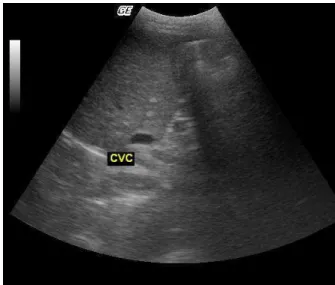
Figure 1- Transverse ultrasound image of the AA (abdominal aorta), CVC (caudal vena cava) and PV (portal vein). Transducer (5.0MHz) on the right lateral abdominal wall, at the 10th intercostal
space, in the porta hepatis region.
ved in the same image for all vessels. Thus, the transducer was angled to obtain the ideal image of each vessel to be measured (Figure 2).
It was also noted that while the portal vein and abdominal aorta were visualized in the circular cross section, the caudal vena cava appeared in oval shape, suffering lateral compression, as cited in the literature (Szatmári et al., 2004, Kamikawa, 2008). The oval shape of the caudal vena cava, sometimes, was due to pressure exerted to the transducer in the abdominal wall as cited
before (Kamikawa,2008) but in some
animals, even exerting minimum
pressure, it was not possible to obtain the circular shape of the vessel (Figure 3a, 3b).
It was observed that the areas and diameters of the vessels measured were influenced by the weight of the animal, so this is a significant factor to be considered for a correct interpretation of the measurements.
Figure 2- Transverse ultrasound image of the PV (portal vein), measuring the vessel diameter. Transducer (7.0MHz) on the right lateral abdominal wall, at the 11th intercostal space, in
the porta hepatis region.
Figure 3a- Transverse ultrasound image of the CVC (Caudal vena cava), showing the oval shape of the vessel, due to the compression of the transducer in the abdominal wall. Transducer (5.0MHz) on the right lateral abdominal wall, at the 10th intercostal space, in the porta hepatis
region.
Figure 3b- Transverse ultrasound image of the CVC (Caudal vena cava), showing the circular shape of the vessel, ideal to measure the diameter. Transducer (5.0MHz) on the right lateral abdominal wall, at the 10th intercostal space, in the porta hepatis region.
clinician to the misinterpretation of the results. It was observed that the values of diameter and area measured were similar to those cited in the literature (Nyland and Fisher, 1990; Evans,1993; Lamb and Mahoney, 1994; Finn and Hudson, 1998), only when compared with dogs of similar body weight.
In humans, an increase in portal vein caliber is one of the main signs of portal hypertension (Moriyasu et al., 1986). However Nyland and Fisher (1990) assessed the caliber of the portal vein in dogs before and after the induction of cirrhosis, and they observed no alterations in the area of the vessel with the establishment of cirrhosis.
The relationships of these vessels are also considered important measures since some alterations can be excluded or presumed by these values. The value of the relationships PV/AA and PV/CVC did not change with the weight of the
animal, since the variations in
measurements were proportional. These values were similar to those found in the literature reviewed (Lamb and Mahoney, 1994; Kamikawa, 2008).
In the literature reports of variations in the caliber of those vessels are based on changes in their relationship. D’Anjou
(2007) observed that dogs with PV/AA ≤
0.65 may have extra hepatic shunt or non-cirrhotic idiopathic hypertension, while dogs with this ratio ≥ 0.80 the
existence of extra hepatic portosystemic shunt can be completely excluded. The values found in healthy dogs, of any size, in this study, are consistent with those reported previously by the author, for dogs in which alterations such as extra hepatic portosystemic shunt were completely excluded.
CONCLUSION
REFERENCES
D’ ANJOU, M. A. Thesonographic search for
portosystemic shunts. Clinical Techiniques in
Small Animal Practice, v.22, n.3, p.104-114,
2007.
EVANS, H. E. Miller´s anatomy of the dog. 3.ed. Philadelphia: Saunders; 1993.1113p. FINN-BODNER, S. T.; HUDSON, J. A. Abdominal vascular sonography. Veterinary Clinics of North America: Small Animal
Practice, v.28, n.4, p.920-930, 1998.
KAMIKAWA, L. Avaliação morfométrica e
hemodinâmica comparativa dos vasos envolvidos no desvio portossistêmico em cães. 2008. São Paulo. Tese (Doutorado em
Medicina Veterinária) - Curso de Pós-graduação em Medicina Veterinária, Universidade de São Paulo.
LAMB, C. R. Ultrasonography of Portosystemic Shunts in Dogs and Cats. The Veterinary Clinics of North America – Small Animal
Practice, v.28, n.4, p.725-796, 1998.
LAMB, C. R.; MAHONEY, P. N. Comparison of three methods for calculating portal blood flow velocity in dogs using duplex-Doppler
ultrasonography. Veterinary Radiology and
Ultrasound, v.35, n.3, p.190-194, 1994.
MACHADO, M. M; ROSA, A. C. F.; NESTOR, B.; MARTINS, L.; ROSA, J.B.F; CERRI, L. M. O.; CHAMMAS, M. C; DAHER, T.; DAHER, T. R.; SAAD, W. A.; CERRI, G. G. Estudo doppler na hipertensão portal. Radiologia Brasileira,
v.37, n.1, p.1-10, 2004.
MORIYASU, F.; NISHIDA, O.; BAN, N.; NAKAMURA, T.; SAKAI, M.; MIYAKE, T.; UCHINO, H. “Congestion index” of the portal
vein. American Journal of Roentgenology,
v.46, n.2, p.735-739, 1986.
NYLAND, T. G.; FISHER, P. E. Evaluation of experimentally induced canine hepatic cirrhosis using duplex Doppler ultrasound.Veterinary
Radiology, v.31, n.4, p.189-194, 1990.
SZATMÁRI, V.; ROTHUIZEN, J.; VOORHOUT, G.; Standard planes for ultrasonographic examination of the portal system in dogs.
Journal of American Veterinary Medicine