348
Pediatric Heart Surgery Department – Hospital de Base – Medical School of São José do Rio Preto
Correspondence adress: Ulisses Alexandre Croti
Hospital de Base – FAMERP – Av. Brigadeiro Faria Lima, 5416 CEP 15090-000 – São José do Rio Preto - São Paulo
E-mail: [email protected]
Ulisses Alexandre CROTI, Domingo Marcolino BRAILE, Sírio HASSEM SOBRINHO, Carlos Henrique DE MARCHI
Braz J Cardiovasc Surg 2005; 20(3): 348-349 CLINICAL-SURGICAL CORRELATION
RBCCV 44205-771
Cavopulmonar total extracardíaco com homoenxerto criopreservado
Extracardiac total cavopulmonary connection with
cryopreserved homograft
CLINICAL DATA
We report on a 23-year-old male patient who had suffered from cyanosis since birth. In the first year of life he was submitted to a hemodynamics examination and complex heart disease was diagnosed which was clinically accompanied without the use of medications. The patient suffered no limitations in respect to routine physical activities and was in functional class I (NYHA). In the six months previous to arriving in our department, the patient evidenced sudden episodes of dizziness together with dyspnea. He was in a good general state, eupneic at rest but cyanotic 4+/6. The thorax was symmetrical the heart rhythm regular with a single second click, hyperphonic at a pulmonary focus (4+/6) and ejective at a high sternal border. The pulmonary auscultation was symmetrical without adventitious sounds. The abdomen presented with no changes. The pulses were palpable for the four limbs without pressure differences or presence of digital clubbing.
ELECTROCARDIOGRAM
An electrocardiogram demonstrated sinus rhythm and a heartbeat of 72 bpm. The P wave axis was +90º, the QRS complex axis was also +90º, and the PR and QRS interval within normal limits.
Article received in May, 2005 Article accepted in June, 2005
Case 6/2005
349 CROTI, UA ET AL - Extracardiac total cavopulmonary connection
with cryopreserved homograft
Braz J Cardiovasc Surg 2005; 20(3): 348-349
RADIOGRAM
By radiography, a cardiothoracic index of 0.40 was calculated. Bulging was evidenced by the left medial arch of the heart associated with a deformation at the right cardiac border suggestive of juxtaposition of the atrial appendixes to the left. Slight accentuation of the pulmonary vascular network was also evidenced. The aorta and cardiac apex were to the right (Figure 1).
ECHOCARDIOGRAM
An echocardiogram identified situs solitus. The venoatrial connections were normal, the atrioventricular connections were univentricular without a right connection and the
ventriculo-arterial connection exhibited a double outflow tract from one main ventricle. Significant pulmonary valvar and subvalvar stenosis was diagnosed along with a significant interatrial shunt. A Doppler examination demonstrated a maximum instantaneous pressure gradient of 77 mmHg caused by the pulmonary stenosis.
DIAGNOSIS
Coronary cineangiography confirmed the echocardiographic findings of double outflow tract from a single right-type main ventricle and determined a mean pressure in the pulmonary trunk of 14 mmHg, RVP 0.3 w/m2, RVS 21 w/m2, RVP/RVS 0.01.
The results of an ergospirometric test gave maximum consumed VO2 of 23 mL/kg/min (high fractions of expired O2 and low fractions of CO2). The peripheral saturation of O2 was 74% and after exercise 62%. There were frequent, uniform, isolated triple extra-systoles, as well as two short periods of non-sustained ventricular tachycardia.
OPERATION
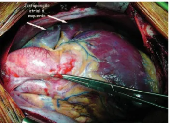
A median transsternal thoracotomy was made. Cardiopulmonary bypass was established at a temperature of 28ºC without aortic clamping. The diagnosis was confirmed as situs solitus in dextrocardia, atrial appendixes to the left (Figure 2), the aorta anterior, pulmonary trunk posterior and the presence of a right-type main ventricle. The superior vena cava was sectioned near the right atrium and anastomosed end-to-side to the right pulmonary artery (Glenn bidirectional). The pulmonary trunk was sectioned near to the pulmonary valve and positioned to the side of the heart in the direction of the inferior vena cava [1]. The inferior vena cava was sectioned from the right atrium and anastomosed to a cryopreserved descending aorta homograft tube [2], which was also anastomosed to the pulmonary trunk (Figure 3). Thus, the total cavopulmonary operation was completed with an extracardiac tube. The cardiopulmonary bypass time was 85 minutes. For all the sutures, 4-0 polypropylene thread was utilized. During the evolution, the patient stayed for two days in the intensive care unit taking intravenous diuretic and inotropic agents. On the 5th day of hospitalization, he was
discharged but continued to take warfarin, diuretic agents, amiodarone and angiotensin II receptor antagonists.
BIBLIOGRAPHIC REFERENCES
1. Van Son JA, Reddy M, Hanley FL. Extracardiac modification of the Fontan operation without use of prosthetic material. J Thorac Cardiovasc Surg. 1995;110(6):1766-8.
2. Mavroudis C, Backer CL, Deal BJ, Johnsrude CL. Fontan conversion to cavopulmonary connection and arrhythmia circuit cryoablation. J Thorac Cardiovasc Surg. 1998;115(3):547-56. Fig. 2 - Juxtaposition of the left and right atrial appendixes to
the left.