CASE REPORT
Agenesis of inferior vena cava associated with deep venous
thrombosis
Agenesia de veia cava inferior associada à trombose venosa profunda
Clovis Luis Konopka1, Marcelo Salame2, Geórgia Andrade Padulla2,Raquel Rodrigues Muradás3, Julio César Batistella4
Introduction
Deep venous thrombosis (DVT) is relevant because of its high frequency and morbidity/mortality rates. Its preva-lence in the western population is estimated to be 1:1,000 individuals per year1-3. he prevalence varies according to
age, being ten times lower in 20 to 40 year-old individuals than in older age groups2,3.
In young patients, its etiology is frequently associ-ated with risk factors such as congenital and acquired thrombophilia, autoimmune diseases, pregnancy and pu-erperium, use of oral contraceptives, neoplasms, surgical procedures, prolonged immobilization and trauma. he most common types of thrombophilia are: protein C and S deiciencies, resistance to C-reactive protein, prothrom-bin G20210A gene mutation, hyperhomocysteinemia, and antiphospholipid syndrome. Such thrombophilias may be
found in 5 to 10% of the DVT cases1, and one or more risk
factors may be identiied in over 80% of these patients1,4,5.
The increasing use of CT angiography and mag-netic resonance imaging (MRI) has allowed physicians to identify more frequently the presence of inferior vena cava (IVC) malformations associated with DVT of the lower limbs1,4,5. Some studies report the presence of a
type of IVC anomaly in approximately 5% of young pa-tients, which suggests that this condition is a new risk factor for DVT1,6.
he prevalence of IVC anomalies in the general popu-lation is estimated in 0.07 to 8.7%1,7.Such conditions may be
associated with vague and unspeciic symptoms or, in many cases, may be completely asymptomatic.
In most studies, the most common IVC malforma-tions are hypoplasia of the prerenal and renal segments, followed by hypoplasia of the postrenal segment and IVC
Abstract
he agenesis of the inferior vena cava is a rare congenital anomaly, which was recently identiied as an important risk factor for the development and recurrence of deep venous thrombosis especially in young people. he goal of this work was to report the case of a patient who presented deep venous thrombosis approximately two months after varicose vein surgery. he CT angiotomography demonstrated a complex venous anomaly with absence of the inferior vena cava.
Keywords: cardiovascular abnormalities ; vena cava, inferior; venous thrombosis.
Resumo
A agenesia da veia cava inferior é uma anomalia congênita rara, que foi recentemente identiicada como um importante fator de risco para o desenvolvimento e a recorrência de trombose venosa profunda de membros inferiores em jovens. O objetivo deste trabalho foi relatar o caso de uma paciente que apresentou trombose venosa profunda dois meses após a realização de cirurgia de varizes. A angiotomograia computadorizada demonstrou a presença de anomalia venosa complexa com ausência da veia cava inferior.
Palavras-chave: anormalidades cardiovasculares; veia cava inferior; trombose venosa.
Study carried out at the University Hospital Santa Maria of UFSM, Santa Maria (RS), Brazil.
1 Assistant Professor of Vascular Surgery at the Service of Endovascular Surgery of Hospital Universitário de Santa Maria (UFSM), Santa Maria (RS), Brazil. 2 Medical student at UFSM; Instructor at the Service of Vascular Surgery of University Hospital Santa Maria of UFSM, Santa Maria (RS), Brazil. 3 Medical student at UFSM, Santa Maria (RS), Brazil.
4 Radiologist at DIX – Diagnóstico por Imagem, Santa Maria (RS), Brazil. No conlict of interest was declared concerning the publication of this article. Received on: Mar 19, 2010. Accepted on: Jun 1, 2010
Deep venous thrombosis associated with agenesis of IVC - Konopka CL et al. J Vasc Bras 2010, Vol. 9, Nº 3 197
duplicity5. In such cases, the irst episode of DVT generally
occurs before the age of 30, with similar incidence in men and women5.
his paper describes a case of complex congenital mal-formation with absent IVC in association with DVT.
Case report
A 43-year-old Caucasian saleswoman presented with superficial, dilated and tortuous symptomatic veins (clinical classification: CEAP II) affecting the lower limbs. She had a history of two pregnancies with cesar-ean delivery at term and family history of varicose veins of the lower limbs. The surgical treatment of varicose veins was indicated after Color Doppler ultrasonogra-phy. The patient underwent bilateral great saphenous vein stripping under epidural anesthesia and discharged 24 hours later.
Two months ater the procedure, the patient presented sudden pain and edema on the right inferior limb. Color Doppler ultrasonography conirmed the suspected acute iliofemoral DVT on the right side and showed absence of IVC in its usual topography and reduced diameter of the right common iliac vein.
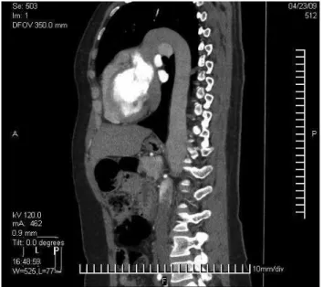
horacic and abdominal CT angiography conirmed the absence of the IVC. (Figure 1) and stenosis on the tho-racoabdominal transition of a large vein located on the let side of the aorta (Figure 2). In the lower abdomen, reduction of the right common iliac vein diameter was ob-served in comparison to its let counterpart. he common iliac veins and both renal veins drained into the anoma-lous vein on the let side of the aorta (Figure 3), which ascended to the thorax, with conluence of let upper limb and let cervical region vessels. he veins of the right up-per limb, cranium and right cervical region formed the superior vena cava.
he patient was treated with anticoagulant therapy with unfractionated heparin by endovenous infusion (18 UI/kg/h) adjusted by activated partial thromboplas-tin time (aPTT) (2.5 times the initial value) for 7 days, followed by oral anticoagulation with antivitamin K (Warfarin sodium®). he International Normalized Ratio
(INR) was maintained between 2 and 3 during 6 months. At the end of treatment, the patient still had mild edema of the right ankle and almost complete venous recanali-zation, conirmed by color Doppler ultrasonography. An investigation for thrombophilia was performed by dosing antithrombin, homocysteine and C and S proteins, Factor
V of Leiden, prothrombin mutation,anticardiolipin
anti-bodies(IgM and IgG), and lupus anticoagulant antibody.
he results were negative.
Discussion
Embryologic abnormalities of the IVC and its tribu-taries are rare1,2,4,8. To date, 15 diferent types have been
reported, and the most common ones are let IVC, double
Figure 1 – Coronal reconstruction of contrast CT angiography showing
absence of the inferior vena cava in its habitual topography, right com-mon iliac vein with reduced diameter, venous drainage by a vessel on the left side of the aorta
Figure 2 – Sagittal reconstruction of CT angiography showing
Deep venous thrombosis associated with agenesis of IVC - Konopka CL et al. J Vasc Bras 2010, Vol. 9, Nº 3
198
Figure 3 – Axial cut of contrasted CT angiography showing that the
right renal vein drains into the vessel that corresponds to the inferior vena cava, to be found at the left side of the aorta
IVC, azygos continuation, retroaortic let renal vein and complete agenesis of the IVC. hese anatomical variations occur between the sixth and eighth weeks of embryonic development1,4,7,9.
The embryogenesis of the IVC is a complex event that involves formation, regression and fusion of three pairs of embryonic veins1,3,7,8,10. The infrahepatic segment
of the IVC may be divided in three parts: suprarenal, re-nal and infrarere-nal; the right subcardire-nal vein becomes the suprarenal segment; the supracardinal anastomoses with the subcardinal vein originates the renal segment, and the infrarenal segment emerges from the right su-pracardinal vein1,5,8,9. Agenesis of the IVC suggests the
si-multaneous occurrence of a defect in all three embryonic segments1,4,6,8.
The etiology of the IVC agenesis is controversial in literature. Some authors suggest that thrombosis on the IVC during the perinatal period is the origin of its disappearance, hence no embryologic abnormalities are seen1,3,9. IVC agenesis or hypoplasia may be accompanied
by other congenital abnormalities such as splenic anom-alies, disorders of intestinal rotation, pulmonary dysgen-esis, renal agendysgen-esis, dextrocardia and other congenital heart diseases1,3,4,9,11. These anomalies are identified in
over 1% of the patients, but the incidence may reach 2% of the patients with congenital heart disease1,6,12.
In total absence of IVC, the venous drainage through thoracolumbar, pelvic and abdominal veins may re-sult in symptoms on thorax, hypogastrium, lumbar and
genital regions before the occurrence of lower limbs’ DVT6.
Although these symptoms are rare and unspeciic, their early detection in young patients may indicate the presence of an IVC malformation5.
Patients with IVC anomalies are prone to DVT due to the venous stasis of the lower limbs1,4. In the case described,
the patient had iliofemoral DVT of the right lower limb, which could be related to the markedly decreased right common iliac vein caliber observed in the imaging exams (Figure 1).
he improvement in imaging exams and their increas-ing use have led to frequent detections of anatomical varia-tions and IVC anomalies4. he best imaging methods for
the diagnosis of IVC anomalies are CT angiography and MRI and the diagnosis is considered to be diicult when ultrasonography is used alone3,4,6,13,14.
he presence of spontaneous, recurrent and sometimes bilateral proximal lower limb DVT in young patients must call the assistant physician’s attention to the possibility of an IVC anomaly. MRI and contrast CT angiography are espe-cially useful in such cases5,11,15. However, DVT as a
paraneo-plasic manifestation or as a result of a hypercoagulable state must also be excluded11.
he most appropriate treatment in such cases is anti-coagulation for at least six months. he possibility of recur-rence is high when the patient has the anticoagulation treat-ment discontinued before this period5.
Conclusion
Patients with congenital IVC anomalies associated with DVT are signiicantly younger in comparison to patients with isolated DVT of the lower limbs. he DVT is related to venous stasis of the lower limbs, which may be bilateral in over 50% of the cases. Besides, there are elevated indexes of thrombosis recurrence due to inadequate venous return and consequent venous stasis.
Surgical intervention is rarely indicated; only an-ticoagulation is recommended as the ideal treatment. There is no consensus in literature about the duration of the anticoagulant treatment. Some authors1,3,7
recom-mend life-long anticoagulation even when thrombo-philia investigation is negative in order to reduce the recurrence risk.
Deep venous thrombosis associated with agenesis of IVC - Konopka CL et al. J Vasc Bras 2010, Vol. 9, Nº 3 199
References
1. Cho BC, Choi HJ, Kang SM, et al. Congenital absence of inferior vena cava as a rare cause of pulmonary thromboembolism. Yonsei Med J. 2004;45:947-51.
2. Milani C, Constantinou M, Berz D, et al. Left sided inferior vena cava duplication and venous thromboembolism: case report and review of literature. J Hematol Oncol. 2008;1:24-7.
3. Iqbal J, Nagaraju E. Congenital absence of inferior vena cava and thrombosis: a case report. J Med Case Reports. 2008;2:46-9.
4. Suh HJ, Kim WT, Kim MY, et al. Combined anomaly of the right hepatic lobe agenesis and absence of the inferior vena cava: a case report. Koren J Radiol. 2008;9:s61-4.
5. García-Fuster MJ, Forner MJ, Flor-Lorente B, et al. Inferior vena cava malformation and deep venous thrombosis. Rev Esp Cardiol. 2006;59:171-5.
6. Gayer G, Luboshitz J, Hertz M, et al. Congenital anomalies of the inferior vena cava revealed on CT in patients with deep vein thrombosis. AJR Am J Roentgenol. 2003;180:729-32.
7. Obernosterer A, Aschauer M, Schnedl W, Lipp RW. Anomalies of the inferior vena cava in patients with iliac venous thrombosis. Ann Intern Med. 2002;136:37-41.
8. Yigit H, Yagmurlu, Yigit N, et al. Low back pain as the initial symptom of inferior vena cava agenesis. AJNR Am J Neuroradiol. 2006;27:593-96.
9. Bass JE, Redwine MD, Kramer LA, et al. Absence of the infrarenal infe-rior vena cava with preservation of the suprarenal segment as revealed by CT and MR Venography. AJR Am J Roentgenol. 1999;172:1610-2.
10. Mano A, Tatsumi T, Sakai H, et al. A case of deep venous thrombo-sis with a double inferior vena cava efectively treated by suprare-nal ilter implantation. Jpn Heart J. 2004;45:1063-9.
11. Atmatzidis K, Papaziogas B, Pavlidis T, et al. Surgical images: soft tissue. Recurrent deep vein thrombosis caused by hipoplasia of the inferior vena cava. Can J Surg. 2006;49:285.
12. Felício ML, Martins AS, Andrade RR, et al. Ausência parcial de veia cava inferior associada à malformação intestinal. Rev Bras de Cirurgia Cardiovascular. 2007;22:362-4.
13. Gay SB, Armistead JP, Weber ME, et al. Left infrarenal region: ana-tomic variants pathologic conditions, and diagnostic pitfalls. Radiographics. 1991;11:549-70.
14. Onzi RR, Costa LF, Angnes RF. Malformação de veia cava inferior e trombose venosa profunda: fator de risco de trombose venosa em jovens. J Vasc Bras. 2007;6:186-9.
15. Viana SL, Mendonça JLF, Freitas FMO, et al. Hipoplasia da veia cava inferior: relato de caso e revisão da literatura. Rev Imagem. 2006;28:203-7.
Correspondence:
Clovis Luis Konopka Rua Duque de Caxias, 1.668/803 – Centro CEP: 97015-190 – Santa Maria (RS), Brasil E-mail: [email protected]
Authors’ contributions