Structural and Ultrastructural Analysis of the
Cervical Discs of Young and Elderly Humans
Ricardo Braganca de Vasconcellos Fontes1,2*, Josemberg Silva Baptista3, Said Rahnamaye Rabbani4, Vincent C. Traynelis2, Edson Aparecido Liberti1
1Department of Anatomy, Instituto de Ciencias Biomedicas, Universidade de Sao Paulo, Sao Paulo, SP, Brazil,2Department of Neurosurgery, Rush University Medical Center, Chicago, IL, United States of America,3Department of Morphology, Universidade Federal do Espirito Santo, Vitoria, ES, Brazil,
4Department of General Physics, Instituto de Fisica, Universidade de Sao Paulo, Sao Paulo, SP, Brazil
Abstract
Several studies describing the ultrastructure and extracellular matrix (ECM) of intervertebral discs (IVDs) involve animal models and specimens obtained from symptomatic individuals during surgery for degenerative disease or scoliosis, which may not necessarily correlate to changes secondary to normal aging in humans. These changes may also be segment-spe-cific based on different load patterns throughout life. Our objective was to describe the ECM and collagen profile of cervical IVDs in young (G1 -<35 years) and elderly (G2 ->65 years)
presumably-asymptomatic individuals. Thirty cervical discs per group were obtained during autopsies of presumably-asymptomatic individuals. IVDs were analyzed with MRI, a morphological grading scale, light microscopy, scanning electron microscopy (SEM) and immunohistochemistry (IHC) for collagen types I, II, III, IV, V, VI, IX and X. Macroscopic degenerative features such as loss of annulus-nucleus distinction and fissures were found in both groups and significantly more severe in G2 as expected. MRI could not detect all morphological changes when compared even with simple morphological inspection. The loose fibrocartilaginous G1 matrix was replaced by a denser ECM in G2 with predominantly cartilaginous characteristics, chondrocyte clusters and absent elastic fibers. SEM demon-strated persistence of an identifiable nucleus and Sharpey-type insertion of cervical annulus fibers even in highly-degenerated G2 specimens. All collagen types were detected in every disc sector except for collagen X, with the largest area stained by collagens II and IV. Colla-gen detection was significantly decreased in G2: although significant intradiscal differences were rare, changes may occur faster or earlier in the posterior annulus. These results dem-onstrate an extensive modification of the ECM with maintenance of basic ultrastructural fea-tures despite severe macroscopic degeneration. Collagen analysis supports there is not a “pathologic”collagen type and changes are generally similar throughout the disc. Under-standing the collagen and ultrastructural substrate of degenerative changes in the human disc is an essential step in planning restorative therapies.
OPEN ACCESS
Citation:Fontes RBdV, Baptista JS, Rabbani SR, Traynelis VC, Liberti EA (2015) Structural and Ultrastructural Analysis of the Cervical Discs of Young and Elderly Humans. PLoS ONE 10(10): e0139283. doi:10.1371/journal.pone.0139283
Editor:Xing-Ming Shi, Georgia Regents University, UNITED STATES
Received:April 13, 2015
Accepted:September 9, 2015
Published:October 1, 2015
Copyright:© 2015 Fontes et al. This is an open access article distributed under the terms of the Creative Commons Attribution License, which permits unrestricted use, distribution, and reproduction in any medium, provided the original author and source are credited.
Data Availability Statement:Data have been deposited to Figshare: (http://dx.doi.org/10.6084/m9. figshare.1510907).
Introduction
The basic structure of the human intervertebral disc (IVD) has been known since at least 1858, while the first studies concerning the morphological changes secondary to aging (i.e., disc degeneration) date from the 1920s [1,2]. Macroscopic modifications of human IVDs related to aging, such as disappearance of vascular channels, annular fissures, osteophyte formation and ingrowth of blood vessels into the annulus fibrosus (AF) had been described by 1950, as well as an expected sequence of degenerative events, all thought to be precipitated by the largely avas-cular nature of the human IVD. Lumbar discs have been the main object of these studies, with only a small fraction involving cervical discs.
Countless subsequent studies have analyzed different microscopic and molecular aspects of disc degeneration but a relatively small number focused on the primary constituent of the IVD, i.e., the extracellular matrix and its collagen content. Several concerns exist over the direct application of these results to cervical discs—extrapolation of lumbar results, utilization of sur-rogates for normal human discs (e.g., adjacent discs obtained during surgery in symptomatic individuals or for deformity indications), age heterogeneity, undisclosed disc region (e.g., ante-rior or posteante-rior AF) and analytical problems resulting from the use of“semi-quantitative”
methods are just some of them [3–6]. Therefore, in this study we describe and compare the morphology, ultrastructure and collagen content of cervical discs from presumably asymptom-atic young (under 35 years) and elderly (over 65 years) individuals. Our hypotheses are: 1) disc ultrastructure and collagen content are significantly modified during normal aging and 2) these modifications impact anterior and posterior disc regions differently.
Material and Methods
Thirty C4-6 vertebral blocks were collected from unselected autopsies of recently-deceased (<6
hours) cadavers at the SVOC-USP. This study was reviewed and approved by the ICB-USP IRB (811/2007). Next of kin provided consent and were interviewed to exclude cadavers with known history of neck or back pain, neoplasms or rheumatological conditions as previously described [7]. In order to allow for degenerative changes to accumulate in the elderly group, a relevant time interval should separate both groups—ten years is the minimal amount demon-strated to cause a significant accumulation of these changes [8]. Here we arbitrarily defined 30 years as the interval: therefore, Group 1 (G1) included 15 cadavers younger than 35 years old
and Group 2G2), 15 cadavers aged 65 or older (Table 1). Throughout the study, C4-5 and
C5-6 discs were analyzed jointly, thus resulting in 30 discs/age group. Specimens were assigned random identifiers and masked to researchers.
MR imaging
The IFUSP 1.5T MR scanner (Philips S15/ACSII, Netherlands) was employed to obtain T2 mid-sagittal and 2-mm axial images through the level of the C4-5 and C5-6 discs of five
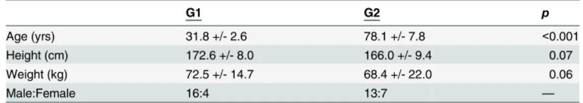
Table 1. Cadaver data: average +/- standard deviation.
G1 G2 p
Age (yrs) 31.8 +/- 2.6 78.1 +/- 7.8 <0.001
Height (cm) 172.6 +/- 8.0 166.0 +/- 9.4 0.07
Weight (kg) 72.5 +/- 14.7 68.4 +/- 22.0 0.06
Male:Female 16:4 13:7 —
p, Student’s T analysis ofG1versusG2.
doi:10.1371/journal.pone.0139283.t001
cadavers (ten discs) fromG1andG2each. MR parameters were adapted to our specimens to
replicate a T2 sequence (matrix = 512x225, TR/TE = 5000/130ms and FOV = 140x140mm). Specimens were placed in a tray, surrounded by air and scanned at room temperature (20–23 degrees Celsius). Discs were analyzed semi-quantitatively with a modified Okada grading sys-tem: individual scores (0, 1 or 2) were added and resulted in a final grade 0 (least) to 6 (most degenerated)[9].G1andG2results were compared with the Mann-Whitney test (GraphPad
Prism 6, San Diego, CA). A significance level of .05 was utilized throughout the study.
Morphological grading
Following fixation in 4% formaldehyde for six months, all specimens were sectioned in the mid-sagittal plane and graded semi-qualitatively with the Thompson scale [10]. Degeneration was graded 1 to 5 and aG1versusG2comparison made with the Mann-Whitney rank-order
test.
Light microscopy
Discs and their intact endplates were decalcified in 0.25M EDTA for 30 days, followed by 1M EDTA for 5 days immediately before processing. Cervical discs were divided in the axial plane at their mid-point in two sectors,anteriorandposterior. The NP was not visible to the naked eye in most cases. Fragments were frozen-sectioned on a sagittal (20 discs/group) or coronal (10 discs/group) orientation. Semi-serial, 8μm sections stained according to hematoxylin-eosin (HE), Sirius Red (SR), Verhoeff’s iron-hematoxylin (mature elastic fibers) and Weigert’s resorcin-fuchsin (elastic and elaunin fibers) techniques [11]. Photomicrographs were acquired under normal and polarized (Sirius Red) light.
Scanning electron microscopy (SEM)
Six discs fromG1andG2each were randomly selected for SEM. Following decalcification and
sectioning as described, the clean-cut mid-point surface of each specimen was attached, face-up, to an SEM stub [11]. Anterior and posterior cervical blocks from each disc were dehydrated (45°C for 12 hours), gold-coated and analyzed in a scanning electron microscope (Leo 435 VP, Cambridge, England).
Collagen immunohistochemistry (IHC)
Six discs fromG1andG2each were randomly selected for this experiment. A
commercially-available ABC kit was utilized (ImmunoCruz ABC, SantaCruz Biotechnology, California). Eightμm-thick sections of each disc sector (anteriorandposterior) were sequentially prepared according to manufacturer’s instructions—protease-based antigen unmasking (Table 2- 30
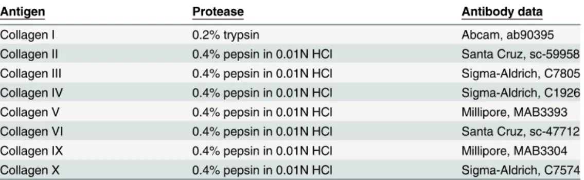
Table 2. Antibodies and proteases utilized in the study.
Antigen Protease Antibody data
Collagen I 0.2% trypsin Abcam, ab90395
Collagen II 0.4% pepsin in 0.01N HCl Santa Cruz, sc-59958
Collagen III 0.4% pepsin in 0.01N HCl Sigma-Aldrich, C7805
Collagen IV 0.4% pepsin in 0.01N HCl Sigma-Aldrich, C1926
Collagen V 0.4% pepsin in 0.01N HCl Millipore, MAB3393
Collagen VI 0.4% pepsin in 0.01N HCl Santa Cruz, sc-47712
Collagen IX 0.4% pepsin in 0.01N HCl Millipore, MAB3304
Collagen X 0.4% pepsin in 0.01N HCl Sigma-Aldrich, C7574
minutes, 37°C), neutralization of endogenous peroxidase (1% H2O2in PBS, 5 minutes), block-age of non-specific sites (1.5% blocking serum, 30 minutes), incubation with primary (12 hours, 4°C) and secondary antibodies (30 minutes, 37°C) and avidin-peroxidase conjugate (30 minutes, 25°C). DAB chromogen was allowed to react for 3 minutes and slides assembled in usual manner [6]. Each step was preceded by two PBS washes.
Primary antibodies against human collagen types I, II, III, IV, V, VI, IX and X were utilized. Single- (no primary antibody) and double-negative (no antibody) controls were included. Quantification was performed with an area-based method as previously described and
expressed as a percentage of total area [12]. Ten random microscopy fields at 1000x magnifica-tion from each disc sector were quantified, resulting in a total of 60 data entries per disc sector, per group. Expression of each collagen type was analyzed against the negative controls with one-way ANOVA andpost-hocTukey tests to determine if it was significantly different than background or non-specific binding. Comparisons between disc regions (anteriorversus poste-rior) and groups (G1versusG2) were performed with T test. Staining unable to be
differenti-ated from artifact was only included in theG1versusG2comparison and excluded from sector
and segment analyses. No adjustments were made for multiple comparisons as all modifica-tions in collagen staining were considered part of the same pathophysiological process and ana-lytical priority was given to the general picture over any individual comparison [13].
Results
Magnetic resonance imaging
Cervical discs fromG1were not devoid of degenerative findings, though these were generally
incipient (Figs1and2). The most common finding inG1was simply the loss of AF-NP
dis-tinction; when more advanced findings were seen, these were mostly horizontal tears. These tears were usually confined to only one of the two discs in each segment, occasionally leading to widely different Okada scores within the same cadaver. InG2, advanced degeneration was
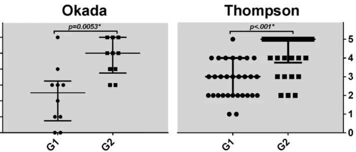
usually manifested as complete disc collapse (Fig 1), which typically impacted both graded discs in a similar manner. Semi-qualitative grading confirmed progression of degenerative fea-tures fromG1toG2as expected (p= .005 -Fig 2).
Morphological grading
Although Thompson grades 1 through 5 (Th1–Th5) were represented in our samples, Th1 discs were rare inG1(2/30) and non-existent inG2(Fig 2). Since identification of a distinct
nucleus is necessary for grades Th1 and Th2, even Th2 discs were rare inG2(3/30). Advanced
degeneration inG1was usually due to small osteophytes and fissures but not complete
ankylo-sis—accordingly, 7 of 30 discs were graded Th4 but only 1 received a Th5 grade. Tears could also affect the two graded discs of the same cadaver differently as seen inFig 1C and 1Dbut the difference in degeneration was always restricted to one Thompson grade. Accumulation of degenerative features fromG1toG2was also confirmed through semi-quantitative grading (p
<.001 -Fig 2).
Light microscopy
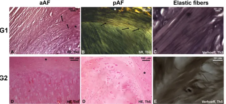
A morphologically“young”fibrocartilaginous phenotype consisted of an anterior AF (aAF) with dense alternating lamellae, predominantly longitudinal in more superficial areas and obli-que in deeper sectors, and a thinner posterior AF (pAF) with predominantly longitudinal fibers (Fig 3). Even inG1, a central area of discernible loose connective tissue was seen in only 2/30
clusters in its interior. Under polarized light, the AF lamellae can be seen to firmly insert them-selves into the adjacent endplates in the manner of Sharpey’s fibers—regardless of how degen-erated the disc was, the central area never exhibited these insertions. Elastic fibers were found aligned with the connective tissue lamellae in the AF and staining was concordant between the Verhoeff and Weigert techniques, suggesting these fibers are of the mature elastic type. Elastic Fig 1. MR and corresponding mid-sagittal views of cervical vertebral blocks. G1specimen with only incipient degenerative findings is demonstrated inAandB. Advanced degeneration was manifested inG1
primarily through horizontal tears such as seen in the C4-5 disc of this anotherG1specimen (CandD). Complete disc collapse was present in over 50% ofG2specimens as seen here (EandF). The C3-4 disc was included in this last specimen but not graded.
doi:10.1371/journal.pone.0139283.g001
Fig 2. Distribution of Okada (n = 10) and Thompson (n = 30) scores per group.Bars represent median and interquartile range. Comparison performed with Mann-Whitney test.
fibers were found in smaller quantities in more degeneratedG1specimens but were still
present.
A morphologically“elderly”phenotype seen in mostG2discs included marked disruption
of the lamellar structure, endplate hypertrophy and more numerous and larger chondrocyte clusters within the disc, while SR staining revealed a substitution of green-refringent fibers for opaque material. With the exception of a single cervical disc,G2specimens uniformly lacked
elastic fibers regardless of Thompson score.
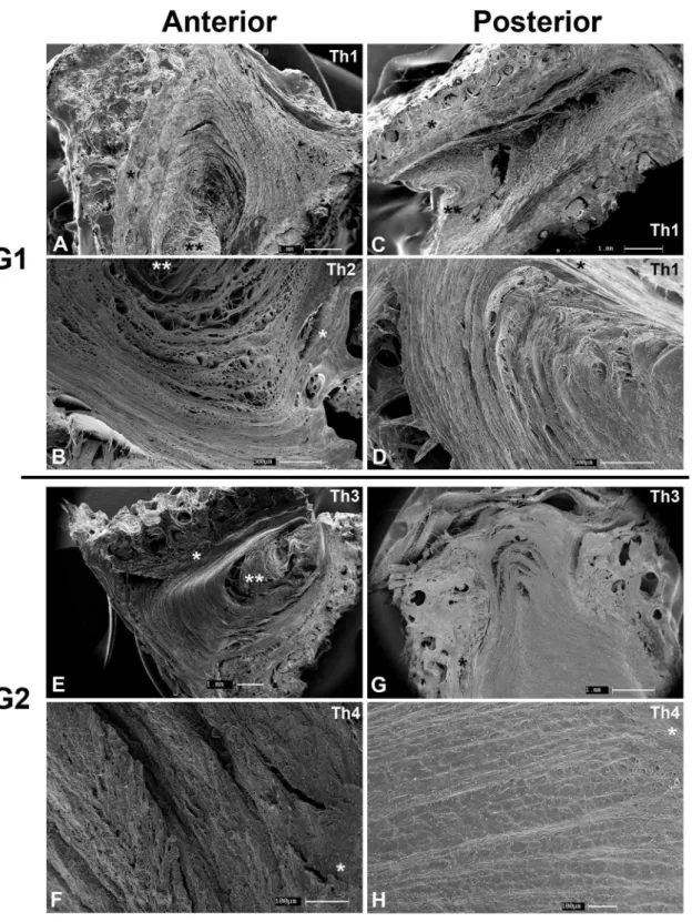
SEM
The superficial layers of the anterior and posterior AF were indistinct from the anterior (ALL) or posterior (PLL) longitudinal ligaments, while the pattern of intertwined lamellae predomi-nates in deeper areas (Fig 4). A fine diagonal mesh can be seen betweenG1lamellae. Endplate
insertion was perpendicular in the anterior AF and obtuse in the posterior AF. SEM was the only method that could visualize the NP as a separate structure in all specimens due to its lack of lamellar organization and endplate insertion and greater retraction due to higher water con-tent than the AF, regardless of the excon-tent of degeneration. InG2specimens, the AF matrix was
far denser resulting in little space between lamellae, with preserved endplate insertion.
Collagen immunohistochemistry
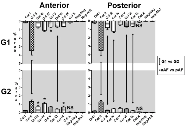
All tested collagen types stained an area larger than that stained in either negative controls in every disc sector in bothG1andG2with the exception of collagen X (Fig 5). Qualitative
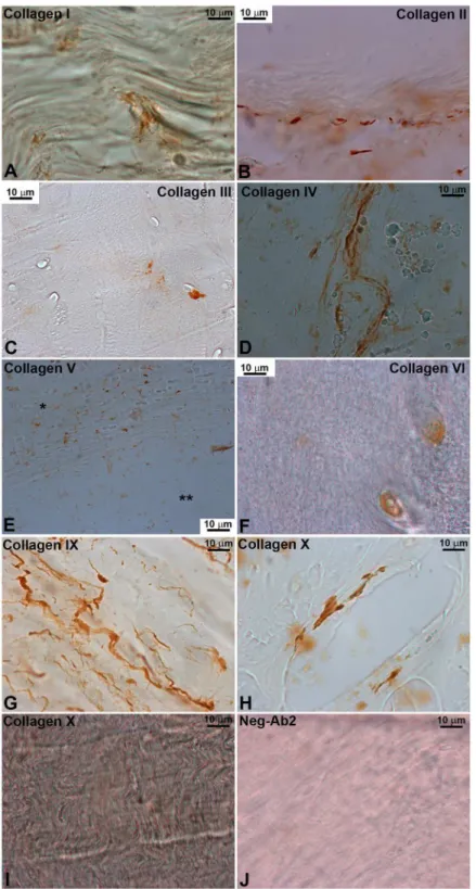
analy-sis demonstrated two main staining patterns. Collagens I, II, IV and IX had predominantly extracellular, filiform reactivity within the structural bundles composing the lamellae of the AF (Fig 6). Intra- and peri-cellular reactivity was seen with collagens III, V and VI, although Fig 3. Light microscopy slides.A well-delimited structure of fibrous lamellae in both anterior and posterior AF ofG1discs is substituted for a degenerated phenotype inG2with compact, fibrocartilaginous matrix populated with numerous chondrocyte clusters. Sharpey-type insertion of AF fibers (arrows) into the endplate (asterisks) is well visualized inG1discs and maintained inG2. In the anterior AF, fibers insert directly into the endplate while in the posterior sector this insertion is at an obtuse angle (arrows). Elastic fibers were rarely found inG2discs, regardless of Thompson (Th) score. SR, Sirius Red. HE, hematoxylin and eosin.
Fig 4. Scanning electron microscopy. G1(A-D) andG2(E-H) cervical specimens. AF ofG1composed of alternating lamellae while longitudinal bundles predominate in the posterior AF. Insertion of the AF into the endplate (*) is perpendicular in the anterior AF and obtuse in the posterior AF. A central area of loose connective tissue without endplate insertion is present in bothG1andG2specimens (**). Th, Thompson grade.
collagen V was mainly present next to the vertebral endplates. The area stained by collagen X antibodies was not significantly different than the negative controls except in the anterior AF ofG1discs; when present, its expression was similar to the filiform pattern.
Overall, collagen reactivity inG2was greatly decreased when compared toG1. Collagen II
was the most common collagen in both young and elderly discs but its expression was reduced by approximately 60% inG2. Collagen VI was also reduced in both anterior and posterior
sec-tors fromG1toG2, while types III, IV and IX were reduced in the posterior sector only.
Regional differences (anteriorvs.posterior) were rare and usually small; inG1, collagen V was
seen in a larger area in the anterior disc while inG2the expression of collagens III, IV and IX
was higher in the anterior disc as well.
Discussion
The morphological and collagen profile modifications described above are the phenotypical and ultrastructural manifestations of a multifactorial process generically described as interver-tebral disc degeneration. Based on the observation that endplate vascular channels close in the first two decades of life, a very early theory was conceived in that IVD degeneration was sec-ondary to decreased availability of oxygen and nutrients to the cells of the adult disc, further substantiated by description of biochemical pathways that mimic changes in the proteoglycan Fig 5. Stained area per collagen type (%).Predominance of collagen type II inG1and an approximate 60% reduction inG2. Vertical arrows indicatep<.05 inG1vs.G2comparison; black asterisks markp<.05 inanteriorvs.posteriorcomparison and are located in the frame with the higher expression. NS, staining not significant. Error bars represent standard error of the mean.
Fig 6. Immunohistochemistry of the different collagen subtypes.Two main patterns were seen: extracellular, filiform reactivity with collagens I, II, IV, VI and IX, and intra-/peri-cellular reactivity with collagens III, V and VI. Staining of collagen X was not significant in most areas but a rare positive example is included with filiform aspect (H). Endplate (*) and disc (**) are shown inE; detection of collagen V was higher in the endplate. Neg-Ab2, negative control with secondary antibody only.
structure of the disc [6,14–18]. How the collagen content is modified is less clear and a rela-tively more recent research interest. Axial and torsional stresses have been shown to play a role as well as the modulating actions of fibroblasts, chondrocytes and even structural molecules such as collagen IX. Also less clear is whether this process occurs in the same manner in cervi-cal discs—since axial and torsional loads are different in relation to the lumbar spine, it would be natural to suppose that degeneration is different too [19,20]. Only a small fraction of all research generated on disc degeneration is based on cervical material, despite the clinical importance of neck pain [21]. A possible explanation for this fact is that lumbar discs naturally yield more material for analyses due to their larger size, which may be important when dealing with small animal models. In any case, there is no reason to assume disc degeneration occurs in the same manner in the cervical spine—despite a common embryological origin, anatomical and functional differences are well-known [5]. The same principles apply to the aAF and pAF in cervical discs—not only the anatomy is different, but the loads and intradiscal pressures borne by the aAF and pAF are markedly different [5,22].
Our morphological, MR and histology data largely confirms literature data that degenera-tive findings can be found in young individuals and are significantly increased in older, asymp-tomatic individuals. Bodenet al. demonstrated in the early 1990s that disc degeneration may be seen in MRs of approximately 60% of asymptomatic subjects in their 60s; these numbers have increased into the 85–90% range as MR technology improved. Asymptomatic progression of these changes over a long period has also been demonstrated in upwards of 80% of individu-als from a Japanese cohort [8,23]. These numbers probably reflect a limitation of the examining method rather than the existence of naïve discs in late adulthood. Christeet al. have already shown that MR vastly underestimates the extent of degenerative alterations in cervical discs. This is corroborated by our findings regarding the cervical NP, which through SEM was dem-onstrated to persist in older discs as a separate structure although a clear boundary was not seen through MR or light microscopy [24]. It is rare, however, that a disc would be consider-ably more degenerated than the rest of the segment in the same individual—Thompson scores never differed by more than one grade, which validates our decision to use C4-5 and C5-6 results jointly in statistical analyses and support diagnostic and treatment algorithms that con-sider discs with dissonant degeneration (when compared to other discs in the same individual) as targets for therapeutic intervention [25].
Light microscopy results demonstrate the loss of the loose lamellar arrangement in the AF and substitution by a more compact arrangement with numerous chondrocyte clusters. Although these are findings described a long time ago in the lumbar spine, only recently Sitte et al. have demonstrated it in the anterior part of cervical discs [26]. The Sharpey-type insertion of aAF fibers into the endplate as well as the obtuse manner in which it takes place in pAF had not been described before in the human cervical spine. These may represent a consequence of the lordotic configuration of the cervical spine—ideally this would be investigated utilizing discs of neonates, before a lordotic curvature is developed but this material is even more diffi-cult to obtain. To the extent of our knowledge, it is also the first time that the fiber arrangement of cervical AF is studied with SEM, also demonstrating the Sharpey-type insertions and substi-tution of the loose matrix of the AF by a much more compact one with predominantly cartilag-inous characteristics.
Collagen profiling revealed that type II collagen is the most common in the cervical disc, fol-lowed by collagen IV. Detection of all collagen types was decreased inG2but in the aAF only
types II and VI reached statistical significance, while in the pAF types I, II, IV, VI and IX were significantly decreased. A single regional difference was seen inG1(collagen V) but inG2
demonstrate a different degeneration pattern, these results probably reflect the same process occurring faster in the pAF than in the aAF. It further suggests that the load each disc sector is subjected to alters its collagen content, as suggested by Brickley-Parsons et al. in young scoliotic spines [19].
Regarding the contribution of each collagen type to disc structure, there is no comparable data for human cervical discs in the literature; lumbar data utilizing semi-quantitative or chro-matography methods pointed towards a predominance of collagen II in the interior of the lum-bar disc with less significant components of collagens III (in the AF), V, VI and IX (diffusely) [6,27,28]. Collagen II as the main type within the disc had been first suggested by Eyre and Muir in the lumbar spine of suids and further demonstrated in humans of different ages [29–31]. Eyre and Muir described collagen II as making up at least 85% of all collagen in the lumbar spine at different ages but only analyzed types I and II using CNBr digestion and electrophoresis [31]. Previous studies utilizing immunohistochemistry had significantly different results—for example, Nerlich et al. describe type III as more frequent in the young NP—and it is the first time Eyre and Muir’s results are replicated with an area-specific method. Collagen IV expression was thought previously to not occur in most age ranges—Nerlichet al. had suggested expression could happen in advanced degeneration [6]. Several methodological differences may account for these different results—different vertebral region, quantitative method, utilization of surro-gates for normal discs (such as samples from scoliotic patients in Beardet al.[27]–so our results are actually in agreement with the vast majority of published collagen literature in the human IVD, although widely different than the biggest study utilizing the same method [6,31,19].
There are a number of limitations to our study. Firstly, despite the screening interview, there is always a possibility the studied individuals were indeed symptomatic. The rarity of ade-quate human material is the biggest impediment to properly study morphological alterations in asymptomatic individuals and there is no truly equivalent bipedal animal model [32,33]. This single reason may be responsible for several of the deficiencies of prior studies. On the other hand, short of prospectively enrolling a massive number of individuals and waiting decades for their deaths, we see no better alternative to this model and it has been employed in numerous studies [7]. Secondly, when this study was designed we imagined that degeneration would affect not only disc regions but also vertebral segments differently (hypothesis #2). Therefore, we tried to analyze as many disc regions as possible and that may have resulted in a seemingly small number of studied samples but in fact this corresponds to the largest study of IVD material with SEM and IHC in human specimens [34–36]. Once results were analyzed, it was satisfactorily demonstrated that the extracellular matrix of cervical discs is significantly modified during normal aging (proving hypothesis #1) but we realized the degenerative process is very similar in both the aAF and pAF and thus we are unable to prove hypothesis #2. We would thus favor analyzing fewer disc regions in future studies while increasing the number of repetitions per experiment.
Conclusions
Acknowledgments
The authors would like to thank Hernan Joel Cervantes Rodriguez, PhD and Thiago Habacu-que da Silva, MS, for assistance with MR imaging and the morphological study, respectively.
Author Contributions
Conceived and designed the experiments: RBVF JSB SRR VCT EAL. Performed the experi-ments: RBVF JSB. Analyzed the data: RBVF JSB EAL. Contributed reagents/materials/analysis tools: RBVF. Wrote the paper: RBVF JSB SRR VCT EAL.
References
1. Luschka H. Die Halbegelenke des Menschlichen Körpers. 2nd ed. Berlin: Reimer; 1858.
2. Schmorl G. Über die an den Wirbelbandscheiben vorkommenden Ausdehnungs- und Zerreissungsvor-gänge und die dadurch an ihnen und der Wirbelspongiosa hervorgerufenen Veränderungen. Verhan-dlungen Dtsch Ges Für Pathol. 1927; 22:250–62.
3. Coventry MB, Ghormley RK, Kernohan JW. The intervertebral disc: its microscopic anatomy and pathology. Part II: Changes in the intervertebral disc concomitant with age. J Bone Jt Surg Am. 1945; 27:233–47.
4. Gruber HE, Hanley EN. Ultrastructure of the human intervertebral disc during aging and degeneration: comparison of surgical and control specimens. Spine. 2002 Apr 15; 27(8):798–805. PMID:11935100
5. Mercer S, Bogduk N. The ligaments and annulus fibrosus of human adult cervical intervertebral discs. Spine. 1999; 24(7):619–28. PMID:10209789
6. Nerlich AG, Schleicher ED, Boos N. 1997 Volvo Award Winner in Basic Science Studies: Immunohisto-logic markers for age-related changes of human lumbar intervertebral discs. Spine. 1997; 22
(24):2781–95. PMID:9431614
7. Le Maitre C, Hoyland J, Freemont AJ. Catabolic cytokine expression in degenerate and herniated human intervertebral discs: IL-1βand TNFαexpression profile. Arthritis Res Ther. 2007; 9(4):R77. PMID:17688691
8. Okada E, Matsumoto M, Ichihara D, Chiba K, Toyama Y, Fujiwara H, et al. Aging of the cervical spine in healthy volunteers: a 10-year longitudinal magnetic resonance imaging study. Spine Phila Pa 1976. 2009; 34(7):706–12.PMID:19333104
9. Okada E, Matsumoto M, Fujiwara H, Toyama Y. Disc degeneration of cervical spine on MRI in patients with lumbar disc herniation: comparison study with asymptomatic volunteers. Eur Spine J. 2010 Dec; 20(4):585–91. doi:10.1007/s00586-010-1644-yPMID:21127918
10. Thompson JP, Pearce RH, Schechter MT, Adams ME, Tsang IK, Bishop PB. Preliminary evaluation of a scheme for grading the gross morphology of the human intervertebral disc. Spine. 1990; 15(5):411–5. PMID:2363069
11. Fontes RB, Saad F, Soares MS, de Oliveira F, Pinto FC, Liberti EA. Ultrastructural study of the filum ter-minale and its elastic fibers. Neurosurgery. 2006; 58(5):978–84; discussion 978–84. PMID:16639335
12. Lehr HA, van der Loos CM, Teeling P, Gown AM. Complete chromogen separation and analysis in dou-ble immunohistochemical stains using Photoshop-based image analysis. J Histochem Cytochem Off J Histochem Soc. 1999 Jan; 47(1):119–26.
13. Rothman KJ. No adjustments are needed for multiple comparisons. Epidemiol Camb Mass. 1990 Jan; 1(1):43–6.
14. Adams P, Muir H. Qualitative changes with age of proteoglycans of human lumbar discs. Ann Rheum Dis. 1976 Aug; 35(4):289–96. PMID:135533
15. Pattappa G, Li Z, Peroglio M, Wismer N, Alini M, Grad S. Diversity of intervertebral disc cells: phenotype and function. J Anat. 2012 Dec; 221(6):480–96. doi:10.1111/j.1469-7580.2012.01521.xPMID: 22686699
16. Taylor JR, Scott JE, Cribb AM, Bosworth TR. Human intervertebral disc acid glycosaminoglycans. J Anat. 1992 Feb; 180 (Pt 1):137–41. PMID:1452468
17. Übermuth H. Altersverändungen der menäschlichen Bandscheiben in der Wirbelsäule. Arch Klin Chir. 1930; 156:567–77.
19. Brickley-Parsons D, Glimcher MJ. Is the chemistry of collagen in intervertebral discs an expression of Wolff’s Law? A study of the human lumbar spine. Spine. 1984 Mar; 9(2):148–63. PMID:6729579
20. Gantenbein-Ritter B, Chan SCW. The evolutionary importance of cell ratio between notochordal and nucleus pulposus cells: an experimental 3-D co-culture study. Eur Spine J Off Publ Eur Spine Soc Eur Spinal Deform Soc Eur Sect Cerv Spine Res Soc. 2012 Aug; 21 Suppl 6:S819–25.
21. Fejer R, Kyvik KO, Hartvigsen J. The prevalence of neck pain in the world population: a systematic criti-cal review of the literature. Eur Spine J Off Publ Eur Spine Soc Eur Spinal Deform Soc Eur Sect Cerv Spine Res Soc. 2006 Jun; 15(6):834–48.
22. Park J, Shin JJ, Lim J. Biomechanical analysis of disc pressure and facet contact force following simu-lated two-level cervical surgeries (fusion and arthroplasty) and hybrid surgery. World Neurosurg. 2014 Jun 14;
23. Boden SD, McCowin PR, Davis DO, Dina TS, Mark AS, Wiesel S. Abnormal magnetic-resonance scans of the cervical spine in asymptomatic subjects. A prospective investigation. J Bone Joint Surg Am. 1990 Sep; 72(8):1178–84. PMID:2398088
24. Christe A, Läubli R, Guzman R, Berlemann U, Moore RJ, Schroth G, et al. Degeneration of the cervical disc: histology compared with radiography and magnetic resonance imaging. Neuroradiology. 2005 Sep; 47(10):721–9. PMID:16136264
25. Zheng Y, Liew SM, Simmons ED. Value of magnetic resonance imaging and discography in determin-ing the level of cervical discectomy and fusion. Spine. 2004 Oct 1; 29(19):2140–5; discussion 2146. PMID:15454705
26. Sitte I, Kathrein A, Pedross F, Freund MC, Pfaller K, Archer CW. Morphological changes in disc hernia-tion in the lower cervical spine: an ultrastructural study. Eur Spine J. 2012 Jul; 21(7):1396–409. doi:10. 1007/s00586-012-2212-4PMID:22407261
27. Beard HK, Roberts S, O’Brien JP. Immunofluorescent staining for collagen and proteoglycan in normal and scoliotic intervertebral discs. J Bone Joint Surg Br. 1981; 63B(4):529–34. PMID:6170646
28. Roberts S, Eisenstein SM, Menage J, Evans EH, Ashton IK. Mechanoreceptors in intervertebral discs. Morphology, distribution, and neuropeptides. Spine. 1995 Dec 15; 20(24):2645–51. PMID:8747242
29. Eyre DR, Muir H. Collagen polymorphism: two molecular species in pig intervertebral disc. FEBS Lett. 1974 Jun 1; 42(2):192–6. PMID:4851074
30. Eyre DR, Muir H. Types I and II collagens in intervertebral disc. Interchanging radial distributions in annulus fibrosus. Biochem J. 1976 Jul 1; 157(1):267–70. PMID:962859
31. Eyre DR, Muir H. Quantitative analysis of types I and II collagens in human intervertebral discs at vari-ous ages. Biochim Biophys Acta. 1977 May 27; 492(1):29–42. PMID:577186
32. Alini M, Eisenstein SM, Ito K, Little C, Kettler AA, Masuda K, et al. Are animal models useful for studying human disc disorders/degeneration? Eur Spine J. 2007 Jul; 17(1):2–19. PMID:17632738
33. An HS, Masuda K. Relevance of in vitro and in vivo models for intervertebral disc degeneration. J Bone Joint Surg Am. 2006 Apr; 88 Suppl 2:88–94. PMID:16595451
34. Paietta RC, Burger EL, Ferguson VL. Mineralization and collagen orientation throughout aging at the vertebral endplate in the human lumbar spine. J Struct Biol. 2013 Nov; 184(2):310–20. doi:10.1016/j. jsb.2013.08.011PMID:23999190
35. Nosikova YS, Santerre JP, Grynpas M, Gibson G, Kandel RA. Characterization of the annulus fibrosus-vertebral body interface: identification of new structural features. J Anat. 2012 Dec; 221(6):577–89. doi: 10.1111/j.1469-7580.2012.01537.xPMID:22747710
36. Zar J. Biostatistical analysis. 4th ed. Upper Saddle River N.J.: Prentice Hall; 1999.
37. Bach FC, Willems N, Penning LC, Ito K, Meij BP, Tryfonidou MA. Potential regenerative treatment strat-egies for intervertebral disc degeneration in dogs. BMC Vet Res. 2014; 10(1):3.