Morphogenesis Switch and Biofilm Formation through
Upregulation of
DPP3
in
Candida albicans
Li Zhang1, Wenqiang Chang1, Bin Sun1, Matthias Groh2, Andreas Speicher2, Hongxiang Lou1*
1School of Pharmaceutical Sciences, Shandong University, Jinan, Shandong, China,2Chemistry – Organic Chemistry, Saarland University, Saarbru¨cken, Germany
Abstract
The yeast-to-hypha transition plays a crucial role in the pathogenesis ofC. albicans. Farnesol, a quorum sensing molecule
(QSM) secreted by the fungal itself, could prevent the formation of hyphae and subsequently lead to the defect of biofilm formation. TheDPP3, encoding phosphatase, is a key gene in regulating farnesol synthesis. In this study, we screened 24
bisbibenzyls and 2 bibenzyls that were isolated from bryophytes or chemically synthesized by using CLSI method for antifungal effect. Seven bisbibenzyls were found to have antifungal effects with IC80less than 32mg/ml, and among them,
plagiochin F, isoriccardin C and BS-34 were found to inhibit the hyphae and biofilm formation ofC. albicans in a dose-dependent manner. To uncover the underlying relationship between morphogenesis switch and QSM formation, we measured the farnesol production by HPLC-MS and quantified Dpp3 expression by detecting the fluorescent intensity of green fluorescent protein tagged strain using Confocal Laser Scanning microscopy and Multifunction Microplate Reader. TheDPP3transcripts were determined by real-time PCR. The data indicated that the bisbibenzyls exerted antifungal effects through stimulating the synthesis of farnesolviaupregulation of Dpp3, suggesting a potential antifungal application of bisbibenzyls. In addition, our assay provides a novel, visual and convenient method to measure active compounds against morphogenesis switch.
Citation:Zhang L, Chang W, Sun B, Groh M, Speicher A, et al. (2011) Bisbibenzyls, a New Type of Antifungal Agent, Inhibit Morphogenesis Switch and Biofilm Formation through Upregulation ofDPP3inCandida albicans. PLoS ONE 6(12): e28953. doi:10.1371/journal.pone.0028953
Editor:Robert Alan Arkowitz, Institute of Developmental Biology and Cancer Research, France
ReceivedAugust 4, 2011;AcceptedNovember 17, 2011;PublishedDecember 12, 2011
Copyright:ß2011 Zhang et al. This is an open-access article distributed under the terms of the Creative Commons Attribution License, which permits unrestricted use, distribution, and reproduction in any medium, provided the original author and source are credited.
Funding:Funding was provided by the National Natural Science Foundation of China (no. 30925038, 30730109; http://www.nsfc.gov.cn/Portal0/default106.htm) and the Shandong Provincial Foundation (JQ200806; http://www.sdnsf.gov.cn/portal/). The funders had no role in study design, data collection and analysis, decision to publish, or preparation of the manuscript.
Competing Interests:The authors have declared that no competing interests exist.
* E-mail: louhongxiang@sdu.edu.cn
Introduction
Candida albicans, which can change from symbiotic form to pathogenic form upon an alteration in the external environment, causes shallow surface infections as well as deep infections [1]. In recent years, the over use of wide-spectrum antibiotics, long and repeated treatment have caused pathogenic fungi resistant to anti-fungal agents, especially to fungistatic drugs [2], and thereby more effective therapeutic drugs or alternative treatments are needed to alleviate the situation. Studies have demonstrated that filamenta-tion plays a crucial role in the pathogenic process [3,4,5]. The transition from yeast to hyphae could improve the virulence ofC. albicans during the course of infection [6]. Filaments generate strong top pressure for permeation into host body and have the advantage to escape from immune cells [7]. In addition, hyphae serving as the skeleton of biofilms play a critical role for the formation of highly heterogeneous architecture, which protects microorganisms from antibiotic treatment and also creates a source of persistent infection [7,8,9]. Therefore, prevention of the hyphae formation could be an effective means to reduce the biofilm formation and virulence in the pathogenesis ofC. albicans. Farnesol, a quorum sensing molecule (QSM) secreted by C. albicans, could inhibit yeast-to-hypha conversion and biofilm formation [10,11,12,13]. Some antifungal agents could stimulate farnesol production through inhibiting sterol synthesis in fungi
[14].DPP3, encoding phosphatase, has been demonstrated to be involved in farnesol synthesis by converting farnesyl pyrophos-phate to farnesol [15].
Bisbibenzyls, a group of phenolic compounds exclusively occurred in liverwort, have potent bioactivities serving as antifungal, antioxidant and antitumor compounds [16]. We have previously shown that plagiochin E (A6) [17] extracted from
Marchantia polymorphacould reverse fungal resistance to fluconazole and induce apoptosis of C. albicans; Riccardin D (A4) [18] from
Methods
Media and strains
C. albicansstrains BWP17 [19], SC5314 [20], CA2 [21], CA10 [22] and YEM30 [23] were used in MIC80 detection, and the transformant of BWP17 (BWP17-DPP3-GFP) was used throughout the studies, stored at 280uC. The C. albicanstrain SC5314 and YEM30 are wild types, CA2 is a clinical azole-sensitive strain while CA10 is clinical azole-resistant [18]. BWP17-DPP3-GFPwas constructed by transforming GFP into the strain of BWP17. The strains were grown in yeast extract peptone dextrose (YPD, 2% tryptone, 1% yeast extract, 2% glucose) solid medium containing
2% agar, and subcultured overnight to stationary phase with shaking at 30uC in YPD medium at least twice. Cells were harvested and washed twice in sterile water and re-suspended in RPMI 1640 medium plus morpholinepropanesulfonic acid (MOPS) and adjusted to the desired density after counting with a hematocytometer.
Antifungal agents
The compounds were naturally isolated from liverwort in our laboratory except the synthesized compounds including A1, A6, plagiochin G (A8), polymorphatin A (A9), plagiochin H (A11) and isoriccardin D (A17) (Fig. 1). All the compounds were diluted with
dimethylsulfoxide (DMSO) to 20480mg/ml as stock solutions and stored at 220uC. Final desired concentration was diluted with RPMI 1640 medium.
Construction of green fluorescent protein tagged strain
The C. albicans DPP3-GFP strain (MG1000) was created by homologous recombination of green fluorescent protein (GFP) sequences into the 39end of theDPP3open reading frame (ORF). The DNA used for the transformation was created by PCR using primers containing,70 base pairs of sequence homologous to the 39 end of the DPP3 open reading frame to amplify a cassette containing GFP and a URA3 selectable marker using pGFP-URA3 as the template [24]. The DPP3-GFP fragment was amplified using the primers (59 -TAAAAGGAAAGAAGAAAT- TAAACAAGATGAAAACAATTATAGAAGAATATCAGACA- TTTCCACTAATGTGGGTGGTGGTTCTAAAGGTGAAGA-ATTATT-39 and 59 -GAAAAGTAAAGCCCAACCAGACAGA- CGACTAGACCAAGACTAGCCCTGGGGGGGAGCTAAGT-AGTATCTAGAAGGACCACCTTTGATTG-39). The PCR pro-duct was transformed into auxotrophic mutant strain BWP17. The transformed strain was then spotted on SD minus uracil (SD 2
Ura) solid medium and grown at 37uC for 3 d. The colonies resulting from the transformation were then screened for GFP-positive cells by fluorescent microscopy.
Antifungal susceptibility study
The minimal inhibitory concentration (MIC) of the tested agents were determined by the broth microdilution procedure recommended by the Clinical and Laboratory Standards Institute (formerly the National Committee for Clinical Laboratory Standards) [25]. Cells were cultured in 96-wells plate at a density of (3–5)6103cells/ml. The yeast suspension was treated with the tested agents on several concentrations with two-fold increase, while the negative control was conducted with the same amount of vehicle. Testing of these compounds was performed in duplicate and incubated for 24 h at 35uC.
Anti-hyphal and biolfilm inhibition test
The measurement of hyphal growth was carried out with a modified broth micro-dilution method [18], cells were diluted with RPMI 1640 to 105cells/ml, and then the tested agents were added into the yeast suspension to reach the final concentration ranging from 0–64mg/ml with 2-fold increase. They were incubated at 37uC and photographed at indicated time. And the fluorescence images were captured with the same exposure time. The morphology ofC. albicanscells was assessed directly by microscopy analysis. To detect hyphae formation on solid media [26], Spider (1% [wt/vol] nutrient broth, 1% [wt/vol] mannitol, 0.2% [wt/vol] K2HPO4, with pH 7.2) agar plates containing different concen-trations of A1, A3 and A5 (Fig. 1) (ranging from 8mg/ml to 64mg/ml with 2-fold increase) were used to test morphology alteration of BWP17-DPP3-GFP cells, the images were captured from 2 d to 5 d using the 46objectives of an inverted fluorescent microscope. The effect of identified agents on biofilm formation were performed as previously described [27]. The final volume of 150ml of yeast suspension with initial cell density of 16106cells/ ml was seeded into microtiter plates with various concentrations (0, 4, 8, 16, 32 and 64mg/ml) of the compounds. 48 hours later, the supernatant was discarded, the biofilm was washed with sterile water at least three times to remove the planktonic cells, and then subject to XTT reduction assay [28]. Testing of these compounds was performed in quadruplicate. Besides, a visualized way was utilized for biofilm detection. The supernatant of performed biofilms was discarded and 100ml PBS plus 10ml alamar blue was
added into the well, and then the color change was photographed after 4–6 hours’ incubation in dark.
Measurement of farnesol using HPLC-MS
In each group, thirteen milliliters of yeast suspension with concentration of 105cells/ml, was treated with the tested agents at the final concentration of 26MIC, 16MIC, 0.56MIC and 0mg/ ml, and incubated at 37uC with shaking. Cells were harvested after 12 hours. The cell debris was dried and weighed with an electronic balance. Farnesol was extracted as previously described [29] and detected using HPLC-MS (LCMS-2020, SHIMADZU). The detection was performed with absorbance at 210-nm and a 5-mm C18 reversed-phase column (4.6 by 250 mm) was utilized with a mobile phase of 4:1 methanol-H2O eluted at 1 ml/min flow rate. In order to obtain the standard curve for figuring out the concentration of farnesol secreted byC. albicanscells, six farnesol standard samples with different concentrations were applied to receive the integral areas eluting at its retention time. The farnesol extracted from the supernatant of each sample was calculated based on the HPLC integral area compared with standard curve.
Detection of Dpp3 production using Confocal Laser Scanning microscopy and Multifunctional Microplate Reader
The Dpp3 expression was measured by detecting the fluorescent intensity of BWP17-DPP3-GFP strain with Confocal Laser Scanning microscopy (CLSM). C. albicanscells were cultured at 37uC and treated with different concentrations of the tested agents or fluconazole (FLC) (positive control). 12 hours later, the images were captured by CLSM and the fluorescence of GFP was excited by the laser of 488 nm with emission of 500–560 nm. The fluorescent intensity of the sample was also measured using a Multifunctional Microplate Reader (Berthold Biotechnologies, Bad Wildbad, Germany). Here the GFP was used as a marker for Dpp3 expression.
DPP3expression detected by Real-Time Reverse
Transcription Polymerase Chain Reaction (RT-PCR)
BWP17-DPP3-GFPcells were adjusted to 56106cells/ml with RPMI 1640 medium, 5 ml of cells were incubated with A1 and A3 (32mg/ml, 16mg/ml and 8mg/ml) and A5 (64mg/ml, 32mg/ml and 16mg/ml) with vehicle as control at 37uC for 12, 24 and 48 hours. The total RNAs were isolated as previously described [30].GSP1was used as the internal control. Measurement of the relative quantitative expression of DPP3 levels was conducted through using an Eppendorf Mastercycler Real Time PCR System. And the gene relative expression was calculated using the formula 22DDCT.
Statistical analysis
Values are mean6standard deviation (SD). Statistical signifi-cances were determined by Student’s t-test and statistical significance was determined byp,0.05.
Results
In vitro antifungal activity
Antifungal activity of mycelium growth and biofilm formation
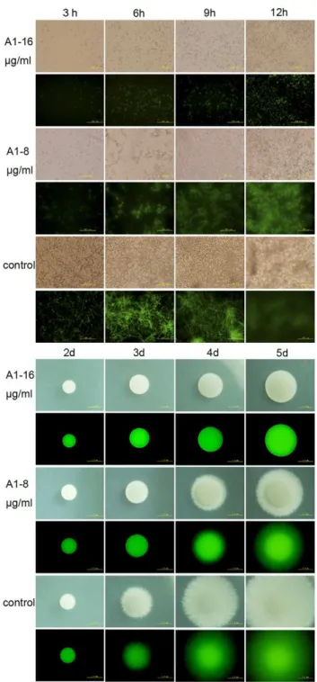
We next evaluated the activity of A1, A3 and A5 against morphological conversion and biofilm formation ofC. albicans. Cells of BWP17-DPP3-GFP were cultured in RPMI 1640 medium or Spider agar plates containing gradient concentrations of agents at 37uC and then observed at indicated time under fluorescent microscope (Olympus IX71, Olympus, Tokyo, Japan). A1, A3 and A5 at the concentration of 16mg/ml, completely inhibited hyphal formation in liquid RPMI 1640 medium within 12 hours and on Spider agar plates within 5 days (Fig. 2). The inhibitory effects were shown in a dose-dependent manner. A1 was chosen as an example to show the fungal morphological change as the time elapses. During 12 hours in liquid RPMI 1640 medium, cells were treated with 16mg/ml of A1 maintained the yeast form with only a slight increase in the number. A suboptimal concentration of A1 (8mg/ml) inhibited the hyphae growth but had no effect on germ tube formation. By contrast, cells in the negative control group formed the true hyphae after three hours and the filaments formed a criss-cross network architecture after 9 hours (Fig. 2 A). On Spider agar plates, the morphology of the colony was photographed from 2 to 5 days using 46objective of an inverted fluorescent microscope. A1 at its MIC could block the yeast-to-hypha transition revealed by the colony that showed a very smooth round plaque without mycelium around the edges throughout the five days. When the dose of A1 was reduced to half of its MIC, the hyphae growth could only be partially inhibited. However, the negative control formed hyphae within 3 days (Fig. 2 B). Collectively, the data suggested the compounds could completely block the morphogenesis switch at the concentration of one MIC, and reduce the hyphae growth at half of the MICs.
Biofilm formed by BWP17-DPP3-GFPwas monitored by CLSM and its metabolism activity was evaluated using XTT reduction assay. The XTT results showed the biofilm inhibitory effect displayed a dose-dependent manner in the presence of detected agents. A1 and A3 reduced biofilm formation by about eighty percent at 32mg/ml. And the biofilm mass was reduced to sixty percent by A5 at 32mg/ml, approximately thirty percent at 64mg/ml (Fig. 3A). CLSM analysis showed that cells within treated biofilm displayed yeast morphology when the concentra-tion of A1 was 32mg/ml or above. When the dose of A1 was reduced to 16mg/ml or in control group, the biofilm predomi-nantly composed of hyphae was formed (Fig. 3 B). We also utilized an alamar blue assay to evaluate the efficacy of compounds that inhibit the biofilm formation. The assay supported the conclusion that A1 (32mg/ml or above) inhibited the biofilm formation, demonstrated by the blue color (Fig. 3 C). When the organism increased, the color changed from blue to pink as shown in low doses of A1 treated or negative control groups (Fig. 3 C).
Enhancement of farnesol production by the tested compounds
To uncover the underlying mechanism of the tested agents in regulating the morphogenesis, we examined farnesol secreted by
C. albicansby using HPLC-MS. Cells treated with the tested agents
Table 1.MIC80of Biol Pharm Bull bisbibenzyls in different Candida albicansstrains.
Strains MIC80(mg/ml)
A1 A3 A5
BWP17-DPP3-GFP 16 16 16
SC5314 16 16 16
CA2 16 32 32
CA10 16 16 16
YEM30 16 16 16
doi:10.1371/journal.pone.0028953.t001
Figure 2. Phase-contrast micrographs of BWP-DPP3-GFPcells challenged by bisbibenzyls compounds within 12 hours. A) Candida albicanscells grown in RPMI 1640 broth medium was captured using an Olympus fluorescent microscope. The results showed that cells co-incubated with A1 (16mg/ml) displayed yeast cells, cells with no drugs formed mycelium after three hours and maintained substantial growth of hyphae. B) Cells grown on Spider solid media were photographed from 2 d to 5 d using 46objectives of an Olympus
fluorescent microscope.
showed a higher amount of extracellular farnesol. The retention time of farnesol was at 20.29 min (Fig. 4 A). The peak eluting at 20.29 min was subjected to MS and showed a molecular ion of m/ z 221.2 and 222.2, which further confirmed the evidence of farnesol (Fig. 4 B). The farnesol production increased as the dose of agents rose (Fig. 4 C). At 26MIC, A1, A3, A5 stimulated farnesol production by 27.9, 12.7 and 6.9-fold increases, respectively, compared with that in negative control. The farnesol production showed 5, 3.3, and 3.1-fold increase at 16MIC, and 3.1, 3, and 2.6 fold-increase at 0.56MIC, respectively. The positive control (FLC) could only promote the production of farnesol by 4.6, 4.2 and 1.9-fold respectively, at 26MIC, 16MIC and 0.56MIC (Fig. 4 C).
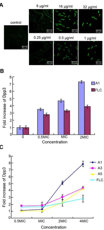
Dpp3 andDPP3expression induced by the tested agents Given the results of farnesol production, we next utilized BWP17-DPP3-GFP strain to detect the expression of Dpp3, an enzyme converting farnesyl pyrophosphate to farnesol. The fluorescent intensity of GFP, as the indicator of Dpp3 expression, was measured by CLSM as well as Multifunctional Microplate Reader. CLSM analysis suggested Dpp3 expression displayed a dose-dependent manner under the treatment of tested agents after the cells were incubated for 12 hours at 37uC. The Dpp3 expression was augumented by A1 with almost 3.7-fold at 8mg/ ml, 4.7-fold at 16mg/ml and 7.3 –fold at 32mg/ml compared with control (Fig. 5 A, B). The results obtained from Multifunctional Microplate Reader also showed that the tested agents promoted the expression of Dpp3 in biofilm. Fluorescent intensity of cells exposed to A1, A3 and A5 from 64mg/ml to 8mg/ml showed a downward trend, while the biofilm formation changed from scattered form to mature one, suggesting that Dpp3 expression was negatively correlated with biofilm formation (Fig. 5 C). To determine whether the agents regulate Dpp3 in transcriptional level, we measured DPP3 expression with real-time PCR and showedDPP3mRNA could be induced by the tested agents when the cells were cultured for 12 to 48 h, suggesting the antifungal
activity of the tested compounds by upregulation of DPP3
expression (Fig. 6).
Discussion
As the earliest land plants, bryophytes grow in an unfavorable environment and inevitably produce secondary metabolites against different surviving stresses [31]. Among those diverse suite of fungi interacts with bryophytes as pathogens, which exhibit different methods of host cell disruption such as invading the host cell by hyphae formation or causing host protoplast degeneration [32]. Previous studies suggest these plants could biosynthesize some chemicals to control the plant diseases caused by fungi or bacteria [1].
C. albicans, as a pathogenic fungal, switches from yeast morphotype to filaments and develop the formation of biofilm to colonize in the host. Based on the aforementioned property of bryophytes, we evaluated the antifungal activity of their extracts. Our lab previously reported that bisbibenzyls including plagiochin E and riccardin D derived from bryophytes displayed a moderate antifungal action [33,34]. We presume bisbibenzyls are the active agents in bryophytes to combat fungal invasion. In our present study, 26 bisbibenzyls isolated from liverworts and chemical synthesizes were screened for antifungal activities. Among them three compounds (A1, A3 and A5) showed a good effect in inhibiting the transition of yeast-to-hypha and biofilm formation (Fig. 2, 3). The effects of these compounds against biofilm formation were observed at or above their MICs. And they were the combination result of inhibitory growth and retarded the yeast to hyphal transition. Farnesol, a QSM, was found to regulate the morphogenesis switch and the inhibitory effect was positively correlated with farnesol formation as detected by HPLC-MS (Fig. 4). When the farnesol production was divided by the volume of culture, the concentration of farnesol could reach to 2.44– 14.38mM. At these concentrations, exogenous added farnesol could partially inhibit the hyphae formation or growth of C. albicans[13], but the inhibitory effect is less than that displayed by
Figure 3. Biofilm formed of BWP17-DPP3-GFPcells pretreated with bisbibenzyls compounds.A) Quantitative measurement of inhibition of biofilm formation with Microplate Reader. B) Confocal micrographs of BWP17-DPP3-GFPcells cultured in RPMI 1640 for 48 hours. C) Cells were stained with alamar blue and cultured for 4–6 hours in dark, the supernatant would become pink when cells grow into biofilm, otherwise turn into blue. The test was performed in quadruplicate. A11 represents the compounds that biofilm inhibitory activity.64mg/ml, bk represents blank control and nk represents negative control.
the tested agents. We speculated that it might be attributed to the better action of endogenous farnesol.DPP3 encodes phosphatase which converts farnesyl pyrophosphate to farnesol [15]. To determine whether the agents stimulated farnesol production by upregulation of Dpp3, C. albicans strain BWP-DPP3-GFP was constructed. The results obtained from CLSM and a Multifunc-tional Microplate Reader showed Dpp3 was stimulated by the above agents, which was in accordance with the enhanced farnesol production (Fig. 5). To further investigate whether the upregula-tion of Dpp3 was the result ofDPP3variation in transcript level, real-time PCR was performed. The results suggested thatDPP3
expression ofC. albicans cells was promoted by the tested agents throughout the 48 h culture (Fig. 6), Based on above analysis, we concluded that the above agents induced the expression ofDPP3
encoding Dpp3, which synthesized more farnesol, to regulate hyphae and biofilm formation.
The clinical used antifungal agents were discovered based on killing the cells, at least inhibiting the growth. Azoles displayed fungistatic action by reducing the sterol synthesis; Echinocandins inhibit b-glucan synthase to synthesize the cell wall component and then kill the fungal cells; Rapamycin exerts its antifungal action by targeting the kinase Tor; Icofungipen functions as tRNA
Figure 4. Farnesol production of BWP17-DPP3-GFPcells stim-ulated by bisbibenzyls.The cells were pretreated with A1, A3, A5 and FLC at 37uC for 12 hours and the farnesol amounts were determined by HPLC-MS. A) The retention time of farnesol eluting at 20.29 min. B) MS profile showed a molecular ion of m/z 221.2 and 222.2. C) Values of ordinate represents the increased folds of farnesol treated by tested agents compared with control.
doi:10.1371/journal.pone.0028953.g004
Figure 5. Dpp3 expression levels of BWP17-DPP3-GFP cells induced by bisbibenzyls.Values represent the increased folds of Dpp3 treated by tested agents compared with control. A) and B) Cells were treated with A1 and FLC at 37uC for 12 hours and then observed by CLSM and the Dpp3 expresson was quantitatively measured based on the fluorescence intensity. C) Fluorescence intensity ofC.albicans cells pretreated with the tested agents was measured using a Multifunctional Microplater Reader after biofilm formation.
synthetase inhibitor to inhibit the cell growth [35]. However, agents targeting the virulence were less developed, although related genes of virulence were well investigated [36]. In this study, we investigated the application of bisbibenzyls against fungal invasion through regulating the known molecular mechanism of morphogenesis switch. Moreover, the structure of each active chemical agent gives a clue for further modification.
Taken together, we provided an alternative way to combat pathogenic fungi infection by targeting the step of morphogenesis switch. In addition, an effective assay for screening potential antifungal agents targeting farnesol synthesis is established through
evaluating hyphal and biofilm formation by measuring Dpp3 expression and farnesol production.
Acknowledgments
We would like to thank Dr. Chen Wanjun at the National Institutes of Health.
Author Contributions
Conceived and designed the experiments: HXL WQC. Performed the experiments: LZ. Analyzed the data: LZ WQC. Contributed reagents/ materials/analysis tools: BS AS MG. Wrote the paper: LZ HXL.
References
1. Sullivan DJ, Moran GP, Pinjon E, Al-Mosaid A, Stokes C (2004) Comparison of the epidemiology, drug resistance mechanisms, and virulence of Candida dubliniensisandCandida albicans. FEMS Yeast Res 4: 369–376.
2. Balkis MM, Leidich SD, Mukherjee PK, Ghannoum MA (2002) Mechanisms of fungal resistance: an overview. Drugs 62: 1025–1040.
3. Southern P, Horbul J, Maher D, Davis DA (2008)C. albicanscolonization of human mucosal surfaces. PLoS One 3: e2067.
4. Kobayashi SD, Cutler JE (1998)Candida albicanshyphal formation and virulence: is there a clearly defined role? Trends Microbiol 6: 92–94.
5. Phan QT, Belanger PH, Filler SG (2000) Role of hyphal formation in interactions of Candida albicans with endothelial cells. Infect Immun 68: 3485–3490.
6. Carlisle PL, Banerjee M, Lazzell A, Monteagudo C, Lo´pez-Ribot JL, et al. (2009) Expression levels of a filament-specific transcriptional regulator are sufficient to determineCandida albicansmorphology and virulence. Proc Natl Acad Sci U S A 106: 599–604.
7. Kumamoto CA, Vinces MD (2005) Contributions of hyphae and hypha-co-regulated genes toCandida albicansvirulence. Cell Microbiol 7: 1546–1554. 8. Lo´pez-Ribot JL (2005)Candida albicansbiofilms: more than filamentation. Curr
Biol 15: R453–R455.
9. Chandra J, Kuhn DM, Mukherjee PK, Hoyer LL, McCormick T, et al. (2001) Biofilm formation by the fungal pathogen Candida albicans: development, architecture, and drug resistance. J Bacteriol 183: 5385–5394.
10. Enjalbert B, Whiteway M (2005) Release from quorum-sensing molecules triggers hyphal formation during Candida albicans resumption of growth. Eukaryot Cell 4: 1203–1210.
11. Ramage G, Saville SP, Wickes BL, Lo´pez-Ribot JL (2002) Inhibition ofCandida albicansbiofilm formation by farnesol, a quorum-sensing molecule. Appl Environ Microbiol 68: 5459–5463.
12. Koo H, Hayacibara M, Schobel B, Cury J, Rosalen P, et al. (2003) Inhibition of
Streptococcusmutans biofilm accumulation and polysaccharide production by apigenin and tt-farnesol. J Antimicrob Chemother 52: 782–789.
13. Sato T, Watanabe T, Mikami T, Matsumoto T (2004) Farnesol, a morphogenetic autoregulatory substance in the dimorphic fungus Candida albicans, inhibits hyphae growth through suppression of a mitogen-activated protein kinase cascade. Biol Pharm Bull 27: 751–752.
14. Nickerson KW, Atkin AL, Hornby JM (2006) Quorum sensing in dimorphic fungi: farnesol and beyond. Appl Environ Microbiol 72: 3805–3813.
15. Navarathna DH, Hornby JM, Krishnan N, Parkhurst A, Duhamel GE, et al. (2007) Effect of farnesol on a mouse model of systemic candidiasis, determined by use of a DPP3knockout mutant of Candida albicans. Infect Immun 75: 1609–1618.
16. Dai Y, Sun LR (2008) Advances in studies on bibenzyls naturally occurred in plant. Chin Tradit Herb Drugs 39: 1753–1756.
17. Guo XL, Leng P, Yang Y, Yu LG, Lou HX (2008) Plagiochin E, a botanic-derived phenolic compound, reverses fungal resistance to fluconazole relating to the efflux pump. J Appl Microbiol 104: 831–838.
18. Sun L, Sun S, Cheng A, Wu X, Zhang Y, et al. (2009) In vitro activities of retigeric acid B alone and in combination with azole antifungal agents against
Candida albicans. Antimicrobl Agents Chemother 53: 1586–1591.
19. Wilson RB, Davis D, Mitchell AP (1999) Rapid hypothesis testing withCandida albicansthrough gene disruption with short homology regions. J Bacteriol 181: 1868–1874.
20. Gillum A, Tsay E, Kirsch D (1984) Isolation of theCandida albicansgene for orotidine-59-phosphate decarboxylase by complementation ofS. cerevisiae ura3
andE. coli pyrF mutations. Mol Gen Genet 198: 179–182.
21. Mason RE, Likar I (1966) A new system of multiple-lead exercise electrocar-diography. Am Heart J 71: 196–205.
22. Frade JP, Arthington-Skaggs BA (2011) Effect of serum and surface characteristics onCandida albicansbiofilm formation. Mycoses 54: e154–e162. 23. Chang WQ, Wu XZ, Cheng AX, Zhang L, Ji M, et al. (2011) Retigeric acid B
exerts antifungal effect through enhanced reactive oxygen species and decreased cAMP. Biochim Biophys Acta 1810: 569–576.
24. Gerami-Nejad M, Berman J, Gale CA (2001) Cassettes for PCR-mediated construction of green, yellow, and cyan fluorescent protein fusions inCandida albicans. Yeast 18: 859–864.
25. NCCLS (2002) Reference method for broth dilution antifungal susceptibility testing of yeasts: Approved standard M27-A2. National Committee for Clinical Laboratory Standards, Wayne, PA.
26. Rashki Ghalehnoo Z, Rashki A, Najimi M, Dominguez A (2010) The role of diclofenac sodium in the dimorphic transition inCandida albicans. Microb Pathog 48: 110–115.
27. Pierce CG, Uppuluri P, Tristan AR, Wormley Jr. FL, Mowat E, et al. (2008) A simple and reproducible 96 well plate-based method for the formation of fungal biofilms and its application to antifungal susceptibility testing. Nat Protoc 3: 1494–1500.
Figure 6.DPP3expression levels of BWP17-DPP3-GFPcells challenged with bisbibenzyls.
28. Ramage G, Vande Walle K, Wickes BL, Lopez-Ribot JL (2001) Standardized method for in vitro antifungal susceptibility testing ofCandida albicansbiofilms. Antimicrob Agents Chemother 45: 2475–2479.
29. Hornby JM, Jensen EC, Lisec AD, Tasto JJ, Jahnke B, et al. (2001) Quorum sensing in the dimorphic fungusCandida albicansis mediated by farnesol. Appl Environ Microbiol 67: 2982–2992.
30. Cao YY, Cao YB, Xu Z, Ying K, Li Y, et al. (2005) cDNA microarray analysis of differential gene expression in Candida albicans biofilm exposed to farnesol. Antimicrobl Agents Chemother 49: 584–589.
31. Xie CF, Lou HX (2009) Secondary metabolites in bryophytes: an ecological aspect. Chem Biodivers 6: 303–312.
32. Davey ML, Currah RS (2006) Interactions between mosses (Bryophyta) and fungi. Can J Bot 84: 1509–1519.
33. Wu XZ, Chang WQ, Cheng AX, Sun LM, Lou HX (2010) Plagiochin E, an antifungal active macrocyclic bis (bibenzyl), induced apoptosis inCandida albicans
through a metacaspase-dependent apoptotic pathway. Biochim Biophys Acta 1800: 439–447.
34. Cheng A, Sun L, Wu X, Lou H (2009) The Inhibitory Effect of a Macrocyclic Bisbibenzyl Riccardin D on the Biofilms ofCandida albicans. Biol Pharm Bull 32: 1417–1421.
35. Paga´n-Mercado G, Rivera-Ruiz ME, Segarra-Roma´n F, Rodrı´guez-Medina JR (2009) antifungal research Strategies aiming for New targets. Puerto Rico Health Sciences Jo 28: 220–226.