ABSTRACT
The principal function of the parathyroid hormone (PTH) is maintenance of calcium plasmatic levels, withdrawing the calcium from bone tissue, reabsorbing it from the glomerular filtrate, and indirectly increasing its intestinal absorption by stimulating active vitamin D (calcitriol) produc-tion. Additionally, the PTH prompts an increase in urinary excretion of phosphorus and bicarbonate, seeking a larger quantity of free calcium available in circulation. Two mechanisms may alter its function, limiting its control on calcium: insufficient PTH production by the parathyroids (hypoparathyroidism), or a resistance against its action in target tissues (pseudohypoparathyroidism). In both cases, there are significantly reduced levels of plasmatic calcium associated with hyperphos-phatemia. Clinical cases are characterized by nervous hyperexcitability, with paresthesia, cramps, tetany, hyperreflexia, convulsions, and tetanic crisis. Abnormalities such as cataracts and basal ganglia calcification are also typical of these diseases. Treatment consists of oral calcium sup-plementation associated with increased doses of vitamin D derivatives. (Arq Bras Endocrinol Metab 2006;50/4:664-673)
Keywords: Hypoparathyroidism; PTH; Hypocalcemia; Pseudohy-poparathyroidism; PTH resistance; Albright hereditary osteodistrophy
RESUMO
Hipoparatiroidismo e Pseudohipoparatiroidismo.
A principal função do paratormônio (PTH) é a manutenção dos níveis plasmáticos de cálcio, retirando-o do tecido ósseo, reabsorvendo-o do filtrado glomerular e, indiretamente, aumentando sua absorção intesti-nal através do estímulo para a produção de vitamina D ativa (calcitriol). Além disso, o PTH promove um aumento na excreção urinária de fósforo e bicarbonato, objetivando uma maior quantidade de cálcio livre disponível na circulação. Dois mecanismos podem alterar sua função, limitando seu controle sobre o cálcio: produção insuficiente de PTH pelas paratiróides (hipoparatiroidismo), ou uma resistência à sua ação nos órgãos-alvo (pseudohipoparatiroidismo). Em ambos os casos, ocorre uma redução significativa dos níveis plasmáticos de cálcio em associ-ação com hiperfosfatemia. Manifestações clínicas características são: hiperexcitabilidade nervosa, com parestesia, cãimbras, tetania, hiper-reflexia, convulsões e crise tetânica. Catarata e calcificação dos gânglios basais são anormalidades típicas dessas doenças. O tratamen-to consiste da suplementação oral de cálcio, associada com doses ele-vadas de derivados da vitamina D. (Arq Bras Endocrinol Metab 2006;50/4:664-673)
Descritores: Hipoparatiroidismo; PTH; Hipocalcemia; Pseudo-hipoparatiroidismo; Resistência a PTH; Osteodistrofia hereditária de Albright
Sergio S. Maeda
Erika M. Fortes
Ulisses M. Oliveira
Victoria C.Z. Borba
Marise Lazaretti-Castro
Division of Endocrinology (SSM, EMF, UO & ML-C), UNIFESP/ EPM, São Paulo, SP; e SEMPR (VCZB), UFPr, Curitiba, PR.
HYPOCALCEMIA: CLINICAL MANIFESTATIONS AND DIFFERENTIAL DIAGNOSIS
S
ERUM CALCIUM CONCENTRATIONis kept within anar-row physiological range due to complex control mechanisms involving the parathyroid hormone (PTH), active vitamin D (1.25(OH)2D), and calcium sensor
receptors, in addition to the concentrations of calcium and phosphate, which act in the renal, intestinal, and bone tissues in order to maintain calcium homeostasis. Hypocalcemia occurs when homeostatic mechanisms fail or when they are not fully compensated.
The normal range of total serum calcium is between 8.5 and 10.2 mg/dL (2.12 to 2.55 mmol/L). Symptoms of hypocalcemia occur when the level of ionized calcium is below 2.8 mg/dL (0.7 mmol/L), equivalent to 7.0 to 7.5 mg/dl (1.75 to 1.87 mmol/l) of total calcium (1,2). The severity of symptoms and clinical signs of hypocalcemia correlates with the magnitude and speed at which calcium declines, influenced by acid-base status and presence of hypomagnesemia and/or sympathetic hyperactivity.
Acute hypocalcemia
In acute and/or severe symptomatic hypocalcemia there is a predominance of neuromuscular, neuropsychiatric, and cardiovascular abnormalities. There is an increase in neuromuscular excitability, latent or evident, with senso-ry and motor disruption. Perioral or extremity paresthe-sia, cramps, myalgia, and muscular weakness are mild to moderate symptoms. Smooth muscle spasms may cause biliary and intestinal cramps, dysphagia, bronchospasms, laryngeal stridor, premature birth, and detrusor muscle dysfunction. Severe hypocalcemia manifests as sponta-neous tetany, which may appear in the form of carpope-dal spasms and, more rarely, laryngospasms. Neurochiatric manifestations include irritability, anxiety, psy-chosis, hallucinations, dementia, depression, mental con-fusion, and extrapyramidal abnormalities. Increased intracranial pressure, papilledema, and convulsions can also be present, and must be differentiated from severe tetany muscular spasms (3-5).
Typical clinical signs of neuromuscular irritabil-ity associated with latent tetany include hyperreflexia and Chvostek’s and Trousseau’s signs, respectively. Chvostek’s sign is obtained by the percussion of the facial nerve approximately 2 cm anterior to the ear lobe, causing contraction of the ipsilateral facial mus-cles. It has low specificity and sensibility; 25% of healthy individuals present a positive sign, while 29% of hypocalcemic individuals present a negative sign. Trousseau’s sign is more reliable; 94% of hypocalcemic
individuals and only 1 to 4% of healthy individuals pre-sent a positive sign, which is obtained by pressurizing a sphygmomanometer approximately 20–30 mmHg above systolic pressure for 3 minutes. It is character-ized by carpal spasms, with adduction of the thumb, followed by flexion of the metacarpophalangeal joint, extension of the interphalangeal joints, and flexion of the wrist, creating the classic “obstetrician’s hand” pose, in addition to causing paresthesia, muscular ten-sion, and local cramps (6).
Severe hypocalcemia may result in bradycardia or ventricular arrhythmias, cardiovascular collapse, and hypotension that is non-responsive to fluids and vaso-pressors. A decrease in myocardial contractility occurs, as well as a typical electrocardiographic abnormality, which is the rate-corrected QT interval (QTc) prolongation.
Chronic hypocalcemia
Patients with chronic hypocalcemia may or may not have symptoms of discreet neuromuscular irritation, even with markedly low calcium levels. Asymptomatic cases may be detected by chance, by the dosage of cal-cium in routine exams, during periods of greater calci-um demand (i.e.: gestation, lactation, menstrual cycle and states of alkalosis), or during the use of hypocal-cemic drugs (i.e.: bisphosphonates). Ectodermic abnormalities from dry skin to dermatitis and even alopecia are common. Dental abnormalities suggest the time of hypocalcemic onset. In infancy, it may lead to dental or enamel hypoplasia, the delay or absence of permanent tooth eruption, an increase in cavity occur-rence, the shortening of molar roots, and, in some cases, the loss of all teeth (7).
Significant cognitive deficits, neuropsychiatric abnormalities, and extrapyramidal symptoms that resemble Parkinson’s disease or chorea are associated with the calcification of basal ganglia, which occurs in all forms of chronic hypocalcemia and may be detect-ed with greater sensibility using computerizdetect-ed tomog-raphy (8).
Other findings of chronic hypocalcemia include sub-capsular cataracts, an increase in bone mineral density (BMD), and greater susceptibility to dystonic reactions induced by phenothiazines (4).
Differential diagnosis of hypocalcemia
Corrected Serum Ca= Total Serum Ca mg/dL (0.8 x [(4 – albumin mg/dL)].
Differential diagnosis of hypocalcemia will depend largely upon PTH and phosphorus levels, eval-uated along with other clinical and laboratory data (table 1). Cases presenting hypophosphatemia should include differential diagnosis of vitamin D, while cases associated with hyperphosphatemia are determined according to PTH levels.
HYPOPARATHYROIDISM
Hypoparathyroidism is an abnormality caused by a parathyroid hormone (PTH) secretion deficiency, and encompasses heterogeneous conditions (table 1), which makes etiological differentiation crucial to the detection of abnormalities associated with some of these diseases beforehand, thereby preventing compli-cations (4). Signs and symptoms are caused by cemia. Laboratory measurements present hypocal-cemia, hyperphosphatemia, and inappropriately low or undetectable PTH. Generally, levels of 1.25(OH)2D
are low and the alkaline phosphatase is normal. In the majority of cases, hypoparathyroidism is sporadic, but there are familial cases in which transmission may be autosomic recessive, dominant, or X-linked.
The most common cause of hypoparathy-roidism is after thyroid surgery. Incidence of
perma-nent hypoparathyroidism varies and depends on the root disease, extent of surgery, and surgeon’s experi-ence. Symptoms generally begin 1 or 2 days after the procedure, and in approximately 50% of the cases this abnormality is transient and caused by edema or glan-dular hemorrhage. After the removal of 1 or more parathyroids for the treatment of primary hyper-parathyroidism, hypocalcemia indicate 2 etiologies: Hungry Bone Syndrome or hypoparathyroidism. Dif-ferentiation is made via measurements of phosphorus, which will be low in Hungry Bone Syndrome and high in hypoparathyroidism. Hypoparathyroidism may be a rare complication after radioactive iodine treatment or external radiotherapy on the cervical region (9,10).
Autoimmunity may also be another cause of hypoparathyroidism, which may be isolated or associ-ated with other autoimmune diseases (11). Antibodies against parathyroids were detected in more than 30% of patients with isolated hypoparathyroidism and more than 40% of patients with hypoparathyroidism associ-ated with other endocrine deficiencies. Antibodies against the calcium sensor-receptor (CASR) have been found in more than 50% of patients with polyglandu-lar autoimmune syndrome type I or hypoparathy-roidism associated with autoimmune hypothyhypoparathy-roidism. The polyglandular autoimmune syndrome type I is characterized by the triad: hypoparathyroidism (which is the most frequent endocrinopathy), mucocutaneous candidiasis, and adrenal insufficiency. It is caused by a mutation in the autoimmune regulator gene (AIRE),
Low PTH levels (hypoparathyroidism)
Parathyroid Destruction
Surgery
Auto-immune (isolated or polyglandular) Cervical irradiation
Infiltration by metastasis or systemic diseases (Sarcoidosis, amyloidosis, hemochromatosis, Wilson’s disease, thalassemia)
Reduced parathyroid function
Hypomagnesemia PTH gene defects
Calcium sensing receptor mutations
Parathyroid agenesis
DiGeorge Syndrome
Isolated x-linked hypoparathyroidism Kenny-Caffey syndrome
Mitochondrial neuropathies
High PTH levels (secondary hyperparathyroidism)
PTH resistance
Pseudohypoparathyroidism Hypomagnesemia
Vitamin D deficiency
Nutritional Lack of sunlight Malabsorption
Vitamin D dependent rickets
type I (lack of activity of 1a-hydroxylase) type II (Vitamin D receptor resistance) Chronic renal disease
Drugs
Bisphosphonates, cisplatin, ketoconazole, gallium nitrate, anticonvulsants
Hyperphosphatemia
Renal insufficiency Massive tumor lysis Acute rhabdomyolysis
Acute pancreatitis Hungry bone
Toxic shock syndrome Acute severe illness
located in locus 21q22.3, which produces a protein that functions as a transcription regulator. This syn-drome may be sporadic or familial, with autosomic recessive transmission. Clinical signs begin to appear in patients approximately 5 years after birth with the emergence of candidiasis, followed by hypoparathy-roidism before the age of 10, and later adrenal insuffi-ciency, found in 100%, 79%, and 72% of the cases, respectively (11,12). Hypogonadism, hypothyroidism, insulin-dependent mellitus diabetes, alopecia, vitiligo, keratoconjunctivitis, intestinal malabsorption syn-drome, chronic hepatitis, and pernicious anemia may also be present.
Maternal hyperparathyroidism, which inhibits PTH secretion by newborns’ parathyroids, is another cause of hypoparathyroidism. Hypocalcemia general-ly develops in the third week of life and is self-con-tained (10). Variations in magnesium levels may also cause hypoparathyroidism. Hypermagnesemia stimu-lates the CASR and inhibits PTH secretion. In such circumstances, the clinical case of neuromuscular hyperexcitability is usually more discreet. Hypomag-nesemia, commonly seen in chronic alcoholism and burn victims, may also cause hypocalcemia because it promotes the diminution of PTH secretion levels in addition to causing renal and bone resistance to PTH action (13).
DiGeorge Syndrome is characterized by thymus and parathyroid dysgenesis, cardiac malformation, and facial dysmorphogenesis. There is a defect in the for-mation of the third and fourth bronchial arches, owed to the deletions of locus 22q11.2, but the gene responsible for the syndrome has yet to be identified. Some patients with phenotype similar to DiGeorge Syndrome present deletions in other chromosomes (10p13 and 10p14) (14). The agenesis or dysgenesis of isolated parathyroids have X-linked or autosomic recessive transmission. Cases with X-linked transmis-sion are related to a mutation in locus Xq26-27, which likely plays a role in the embryological development of parathyroids (10).
Another genetic cause is HDR Syndrome, char-acterized by hypoparathyroidism, neuro-sensorial deaf-ness, and renal dysplasia. HDR is related to haploinsuf-ficiency or function loss of gene GATA3 (GATA bind-ing protein 3), located in chromosome 10p14-10, which belongs to the family of transcription factors involved in embryological development (15).
Other complex syndromes associated with hypoparathyroidism are: Sanjat-Sakati syndrome (hypoparathyroidism, mental retardation, stunted growth, dysmorphism, and seizures, related to a
muta-tion in the chromosome region 1q43-44); Kenny-Caf-fey Syndrome (stunted growth, facial-cranial abnor-malities, long bone cortical thickening, with medullary stenosis and basal ganglia calcification); mitochondrial myopathies, such as Kearns-Sayre Syndrome (encephalomyopathy, oftalmoplegia, retinal pigment dystrophy and cardiomyopathy) and Pearson’s mar-row-pancreas syndrome (neutropenia, sideroblastic anemia, vacuolization of the bone marrow cells, and exocrine pancreas dysfunction) (10).
isolated hypoparathyroidism, placing added impor-tance on the screening of this gene, especially in hypocalcemic patients showing normal urinary calcium excretion (relative hypercalciuria). Since there is an excellent correlation between the presence of muta-tion in the CASR and hypocalcemia, the measurement of the serum calcium may be used as a sufficiently sen-sitive screening in the affected families with this abnor-mality (18). Patients are generally asymptomatic, the serum PTH is normal or low, and they receive the hypoparathyroidism diagnosis (15). It is important to distinguish these patients from those with hypoparathyroidism, because their treatment with cal-cium and vitamin D may lead to hypercalciuria, nephrocalcinosis, and loss of renal function (10).
Mutations of the PTH gene, located in locus 11p15, are uncommon causes of hypoparathyroidism, and they can present autosomic recessive or dominant modes of inheritance. They result in defective PTH molecule synthesis and undetectable PTH levels. Another gene related to hypoparathyroidism is GCMB (a human gene homologous to the Drosophila glial cell missing gene b), located in chromosome 6p24.2, expressed exclusively in the parathyroids, and which codifies an important transcription factor for the devel-opment of these glands (10). The Gcm2 (glial cells missing 2) is the murine gene homologous for GCMB. Homozygote knockout rats (-/-) for the Gcm2 do not present parathyroids and develop hypocalcemia and hyperphosphatemia. PTH levels are normal or low, but insufficient to control hypocalcemia. In these rats there may also be PTH production by parathyroid cells locat-ed in the thymus capsule, explaining the diminishlocat-ed, but detectable, PTH levels. In humans it is unknown if there is intrathymic PTH production or if this small PTH production is due to the gene’s residual activity (15,19).
PSEUDOHYPOPARATHYROIDISM
The term pseudo-hypoparathyroidism describes a group of abnormalities characterized by clinical and laborator-ial hypoparathyroidism findings (hypocalcemia, hyper-phosphatemia), but with high plasmatic levels of PTH due to a target tissue resistance, if chronic renal failure or magnesium deficiency were excluded (20).
Historical
In 1942, Fuller Albright and cols. described 3 patients with clinical and laboratorial hypocalcemia findings in which the administration of parathyroid tissue extracts
did not promote an increase in serum calcium or phos-phaturia (21). These individuals presented develop-ment abnormalities: rounded face, short stature, obe-sity, brachydactyly, short and low-set nasal bridge, strabismus, and ectopic calcifications; which were des-ignated Albright’s Hereditary Osteodystrophy (AHO). Due to biochemical similarities with hypo-parathyroidism, but associated with high levels of PTH was called pseudo-hypoparathyroidism (PHP). In 1966, Tashjian et al. (22) demonstrated an increase in serum PTH concentrations in these patients and, in 1969, Chase et al. (23) presented that there was no urinary AMPc generation in patients with PHP and the pathogenesis of this abnormality should be related to a renal defect in the generation of urinary AMPc by the PTH receptor.
PTH receptor type I
PTH receptor type I is expressed in the kidney and bone as a glycoprotein of approximately 75 kDa that connects to both PTH and PTH-related protein (PTHrP). This receptor, attached to regulatory heterotrimeric protein G (α, β, γ), presents a single polypeptide chain with seven transmembrane domains, 3 extracellular loops, and 3 or 4 intracellular loops (20).
Hormone binding leads to the activation of protein Gs, with disassociation of subunit α. This, when activated, disconnects itself from the receptor and functions as a primary modulator of the adenyl cyclase, promoting the intracellular formation of cyclic AMP (AMPc), which rapidly activates the protein kinase A (PKA) (21-25). The intrinsic GTPase activity of subunit αpermits the linking of βγdimer for a new activation cycle.
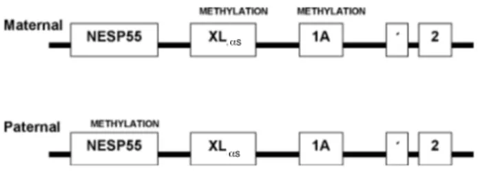
Patients with AHO present inactivating muta-tions in the GNAS1 gene, which codifies the subunit α of protein Gs, mapped in chromosome 20q13.2-20q13.3. The GNAS1 gene is codified by 13 exons, in addition to 3 initial exons that correspond to different promoters, allowing, by alternative splicing, the for-mation of 4 different isoforms. Exons 2-13 are com-mon to all transcripts. Mutations were described in all exons, except in 3, which despite suffering splicing out still produced a functional protein (26).
55), which is expressed exclusively by the maternal allele, while exons XLαs (extra large as like protein) and 1A (1 alternative) are expressed by the paternal allele (figure 1). Despite simultaneous imprinting (paternal and maternal), expression of Gsαappears to be biallelic in the majority of examined human tissues, but expressed maternally in only some tissues such as the proximal renal tubules (25).
Pathogenesis and classification
The characterization of the molecular bases of pseudo-hypoparathyroidism began with the observation that the AMPc is the mediator of various PTH actions in the kidney and bone fiber.
PTH infusion remains the most consistent test for the distinction of the diverse variants of this syn-drome. Based in the abnormalities of the various com-ponents of the transduction of the cell membrane sig-nal, the pseudo-hypoparathyroidism may be divided in distinct forms.
Pseudo-hypoparathyroidism type 1a (PHP1a)
Presents AHO phenotype. There is a reduction in activity of the stimulatory protein G (Gs), limiting AMPc synthesis. In this situation, patients are not only resistant to PTH, but to other peptide hormones like TSH, gonadotrophins, and glucagon. Transmission is autosomic dominant (29). Hypocalcaemia and hyper-phosphatemia are not typically present until five years of age, but PTH elevations may be documented much earlier and sometimes are associated with light hyper-calcemia. Primary hypoparathyroidism frequently pre-cedes the development of functional hypoparathy-roidism and may be detected during neonatal screen-ing. Gonadal dysfunction (hypogonadism or delayed puberty) is very frequent, especially in women (30). There are accounts of resistance to GHRH and pro-lactin deficiency in PHP1a cases (25,31,32). There is considerable variability in relation to tissue
responsive-ness to Gs protein deficiency, which may also be
explained by the differences in the quantity of AMPc necessary in order to activate the kinase proteins and generate physiological responses in each tissue. The absence of imprinting in other target tissues would be another explication for patients with PHP1a who do not develop resistance to other hormones that stimu-late the Gsαprotein and paths linked to the AMPc (for example: ACTH and vasopressin) (25,26).
Pseudo-hypoparathyroidism type 1b (PHP1b)
Presents normal appearance, normal Gs activity, and isolated resistance to PTH. Molecular studies do not show an intrinsic defect in the PTH receptor, and pre-sent normal Gsfunction in the erythrocytes (25). The
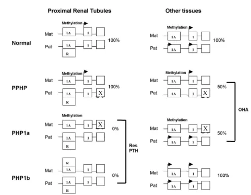
majority of cases appear to be sporadic, but some familial cases were described as having autosomic dominant transmission (29,33). As in PHP1a, a defec-tive nephrogenic answer to AMPc occurs in relation to PTH. However, these individuals present elevated PTH concentrations and frequently manifest skeletal abnormalities similar to those that occur in patients with hyperparathyroidism (30). Fibroblast studies show small accumulation of AMPc in response to PTH in some cases associated with lower quantities of the PTH type I Receptor RNAm. A molecular defect was mapped in a small region of chromosome 20q13.3 (exon 1A) next to the GNAS1 gene. Exon 1A is important to maintain the tissue-specific imprinting of Gsα and is normally methylated in the maternal allele and not methylated in the paternal allele (25). One of the proposed mechanisms to explain PHP1b cases was suggested by Weinstein and cols. (figure 2). Exon 1A contains a cis-acting, negative regulatory element (silencer) that is both methylation-sensitive and tissue-specific (25,26). A tissue-tissue-specific trans-acting repres-sor (R) binds to the silencer and suppresses Gs expres-sion on the paternal allele, but is unable to bind the maternal allele due to methylation, allowing Gsαto be
expressed from the maternal allele. In the majority of tissues, exon 1A is methylated but the repressor is not expressed, and as a result the Gsαis expressed
bialleli-cally. In PHP1b, the methylation of this exon is absent in the maternal allele, allowing the repressor to link to both alleles and suppress the Gsα expression in the
renal proximal tubules, raising the Gsα deficiency and
PTH resistance (26,29,34), which normally only express the maternal allele. These individuals do not present AHO, because the defective imprinting does not have an effect in the Gsαexpression of the major-ity of the tissues where it is expressed biallelically.
Figure 1. General organization and imprinting patterns of the GNAS1 gene. Promoter NESP55 is only expressed by the maternal allele, while promoters XLαsand 1A are expressed
by the paternal allele.
Pseudo-hypoparathyroidism type 1c (PHP1c)
Presents Albright’s phenotype and resistance to multi-ple hormones (PTH, TSH, gonadotrophins, and glucagon). Gsactivity is normal, and so is Gi. Studies
show reduced activity of the membrane’s adenyl cyclase catalytic subunit. It has yet to be clarified which molecular defect is responsible (29).
Pseudo-hypoparathyroidism type 2 (PHP2)
Some patients present normal phenotype, normal AMPc urinary excretion but absent phosphaturic response to PTH. It is probably associated with defects in stages posterior to AMPc formation, because Gs
activity is normal. Its defect has yet to be identified and suggests that there is not any evidence of familial transmission and that it may be an acquired defect.
Pseudo-pseudohypoparathyroidism (PPHP)
The term was used by Albright to describe patients with the Albright phenotype, but without the
labora-torial findings (35). Some clinical syndromes, howev-er, may present the same skeletal abnormalities as AHO without evidence of PTH resistance. PPHP car-riers are often times relatives of PHP patients, and may present periods of hypocalcemia and normocalcemia. AHO patients who inherit Gsα mutations from their
mothers also develop resistance to TSH, PTH, and gonadotrophins (PHP1a), while those who inherit it from their fathers develop only OHA (PPHP). This is owed to the fact that the Gsα gene is expressed
pri-marily from the maternal allele in target-tissues for the respective hormones (renal proximal tubules, thyroid, and ovaries). Mutations in the active maternal allele lead to Gsαexpression deficiency and hormonal resis-tance, while mutations of the paternal allele have little or no effect on genetic expression or hormonal signal-ing (26). The presence of a Gsαallele, however, is not sufficient in all tissues. Different phenotypes probably result from tissue-specific combinations of haploinsuf-ficiency to paternal imprinting (maternal allele
expres-Figure 2. Mechanisms that explain the molecular pathogenesis of PTH resistance (res PTH) and Albright’s Hereditary Osteodystrophy (AHO) by inactivation and imprinting of the GNAS1 gene. Exon 1A is only methylated in the maternal allele and is transcriptionally active in the paternal allele in all tissues. In the majority of tissues, exon 1A does not affect Gsα expression, which is biallelically expressed. In the proximal renal tubules, the non-methylated exon possibly suppresses Gsαexpression in the paternal allele via a repressor (R), leading to a maternal-specific expression of Gsα. In PPHP and PHP1a, the inactive mutations in the paternal and maternal alleles, respectively, reduce the Gsα
expression by 50%, and this haploinsufficiency produces the Albright phenotype. In the proximal tubules, mutations in the inactive paternal allele do not affect Gsαexpression, while mutations in the active maternal allele produce PTH resistance. In PHP1b, the methylation defect in the maternal allele leads to a paternal-specific imprinting model of exon 1A in both alleles. When this happens, Gsαloses expression in the proximal tubules and there is a PTH resistance, but not affecting the expression in other tissues, which explains why these individuals do not possess Albright’s phenotype (25).
X
X X
sion). The AHO phenotype results from deficient sig-naling in cells in which the Gsα gene is haploinsuffi-cient but does not suffer imprinting (both alleles are expressed) (24). The diminution of signaling Gsα
appears to promote osteoblastic differentiation, which may explain ectopic calcifications and premature clos-ing of growth plates (25).
Defects outside the endocrine system
Although it is characterized by endocrine dysfunction, it may have abnormalities in organs whose functions are mediated by the AMPc path. Weinstock et al. (36) showed that patients with PHPIA have altered olfac-tory function if compared with PHPIB patients. The mental deficiency that occurs 50–75% of PHP-carrying individuals is also associated with Gsdeficiency.
Stud-ies also show evidence of paternal imprinting in the brown and white adipose tissue, suggesting that obe-sity may be resultant of decreased Gsα expression in
the adipose tissues, with diminution of the AMPc and lipolysis formation in response to β-adrenergic stimu-lus in the adipocyte (25,29).
TREATMENT OF HYPOCALCEMIA
Management of acute or severe symptomatic hypocal-cemia must be made with intravenous calcium, with the goal of interrupting symptoms, preventing laryn-geal spasm, and maintain total calcium levels above 7.0–7.5 mg/dL (ionized calcium greater than 0.7 mmol/L). Hyperphosphatemia, alkalosis, and hypo-magnesemia, when present, must be corrected. At that moment, the 10 to 30 mL of 10% calcium gluconate should be made slowly (10 minutes – maximum of 30 mg/min) in bolus IV (93 to 279 mg of elementary calcium), and repeated as many times as necessary. After the grave acute symptoms have been suspended, a maintenance of calcium levels via continuous endovenous infusion of 0.5 to 1.5 mg/Kg/h (maxi-mum of 100 mg/h) of elementary calcium for 4 to 6 hours should be made, using a solution with SG5% 900 ml + 100 ml of 10% gluconate (930 mg elemen-tary calcium/liter). Serum calcium levels must be mea-sured frequently in this period, and electrocardio-graphic monitoring must be done, especially in those patients using digitalics, due to growing levels or cal-cium predisposed to digitalis intoxication. Endove-nous transition to the oral must be made as soon as possible (2,3,38).
Long-term treatment of patients with chronic hypocalcemia is done with 1 to 3 g of elementary
cal-cium per day in the various forms of salts available (table 2). Although calcium carbonate is the most used salt, it needs gastric acid so that it may be solu-bilized and absorbed, and causes more collateral gas-trointestinal effects than other salts. (39). All patients with hypoparathyroidism or pseudohypoparathy-roidism who become hypocalcemic must use vitamin D or analogous in addition to calcium (table 3). The vast majority of patients obtain control with calcitriol in dosages of 0.25 µg, taken twice daily, up to 0.5 µg four times daily. High doses of oral Cholecalciferol can also be used (50.000 to 150.000 UI/day), but the risk of intoxication after years of treatment is ele-vated due to their long half-life. Treatment objectives are to maintain free ionized calcium levels within the normal interval, to avoid hypercalciuria, and to sup-press PTH levels in patients with pseudohypoparathy-roidism. Hypoparathyroidism causes increased excre-tion of urinary calcium in relaexcre-tion to serum calcium and predisposes hypercalciuria, nephrolithiasis, and nephrocalcinosis. On the other hand, patients with pseudohypoparathyroidism that have low urinary cal-cium in relation to serum calcal-cium may tolerate serum calcium levels within the normal interval without developing hypercalcemia. Vitamin D intoxication must be remembered in patients with hypercalcemic symptoms. Thiazide diuretics are used in cases of hypercalciuria and nonabsorbable antacid can be added to reduce hyperphosphatemia and prevent metastatic calcification. The product of calcium x phosphatemust be kept below 55. Those patients must have their kidneys radiologically evaluated regularly in order to rule out nephrocalcinosis (4).
PTH1-34 treatment has the advantage of
nor-malizing calcemia without increasing calciuria, reduc-ing the risk of nephrocalcinosis and renal insufficiency, and, theoretically, the antagonists of the calcium sen-sor receptor may be utilized in the treatment to pro-mote the inactivation of the receptor and, conse-quently, increase PTH secretion, but there still have not been sufficient studies (40).
REFERENCES
1. Broadus AE. Mineral balance and homeostasis. In: Favus MJ, editor. Primer on the metabolic bone diseases and disorders of mineral metabolism, 5thed. American
Soci-ety for Bone and Mineral Research; 2003. p. 105-11. 2. Bushinsky DA, Monk RD. Electrolyte quintet: calcium.
Lancet 1998;352:306-11.
3. Tohme JF, Bilezikian JP. Hypocalcemic emergencies.
4. Levine M. Hypoparathyroidism and pseudohypopa-rathyroidism. In: DeGroot LJ, Jameson JL. Endocrinology, 4thed. Philadelphia: WB Saunders, 2001. pp. 1133-53.
5. Weiss-Guillet EM, Takala J, Jakob SM. Diagnosis and management of electrolyte emergencies. Best Pract Res Clin Endocrinol Metab 2003;17:623-51.
6. Urbano FL. Signs of hypocalcemia: Chvosteck’s and Trousseau’s signs. Hosp Physician 2000:43-5.
7. Abugassa S, Nordenstrom J, Eriksson S, Sjoden G. Bone mineral density in patients with chronic hypoparathy-roidism. J Clin Endocrinol Metab 1993;76:1617.
8. Kowdley KV, Coull BM, Orwoll ES. Cognitive impairment and intracranial calcification in chronic hypoparathy-roidism. Am J Med Sci 1999;317:273-7.
9. Chandran M, Deftos LJ, Stuenkel CA, Haghighi P, Orloff LA. Thymic parathyroid carcinoma and postoperative hungry bone syndrome. Endocr Pract 2003;9:152-6. 10. Garfield N, Karaplis AC. Genetics and animals models of
hypoparathyroidism. Trends Endocrinol Metab 2001; 12:288-94.
11. Goswami R, Brown EM, Kochupillai N, Gupta N, Rani R, Kifor O, et al. Prevalence of calcium sensing receptor autoantibodies in patients with sporadic idiopathic hypoparathyroidism. Eur J Endocrinol 2004;150:9-18. 12. Mayer A, Ploix C, Orgiazzi J, Desbos A, Moreira A, Vidal
H, et al. Calcium-sensing receptor autoantibodies are relevant markers of acquired hypoparathyroidism. J Clin Endocrinol Metab 2004;89:4484-8.
13. Graber ML. Magnesium deficiency: pathophysiologic and clinical overview. Am J Kidney Dis 1995;25:973. 14. Goldmuntz E. DiGeorge syndrome: new insights. Clin
Perinatol 2005;32:963-78.
15. Thakker RV. Genetic developments in hypoparathy-roidism. Lancet 2001;357:974-6.
16. Hauache OM. Extracellular calcium-sensing receptor: structural and functional features and association with diseases. Braz J Med Biol Res 2001;34:577-84.
17. Thakker RV. Diseases associated with the extracellular calcium-sensing receptor. Cell Calcium 2004 ;35:275-82.
Table 2.Most used calcium salt forms.
Salt Element Milligrams of salt needed Parenteral content in order to obtain 1 g preparation
elementary calcium
Calcium carbonate 40% 2500
Calcium phosphate 38% 2631
Calcium chloride 27% 3700 10% Solution= 273 mg/10 ml
Calcium citrate 21% 4762
Calcium lactate 13% 7700
Calcium gluconate 9% 11100 10% Solution= 93 mg/10 ml
Table 3.Vitamin D analogues.
Calciferol D3/ D2 Calcidiol Calcitriol Alfacalcidol
25(OH)D 1,25(OH)2D 1αOHD
25-hydroxylation necessary + – – –
1α-hydroxylation necessary + + – –
Physiological dose/dosage 2.5–10 µg 1–5 µg 0.25–0.5 µg 1–3 µg
(1 µg= 40 UI) (1 µg= 40 UI)
Pharmacological dose/dosage 0.625–5 mg 20–250 µg 0.5–3 µg 1–3 µg Time needed to normalize calcium 4–8 weeks 2–4 weeks 3–7 days 7–14 days
Duration of effect 6–12 weeks 2–6 weeks 3–7 days 7–14 days
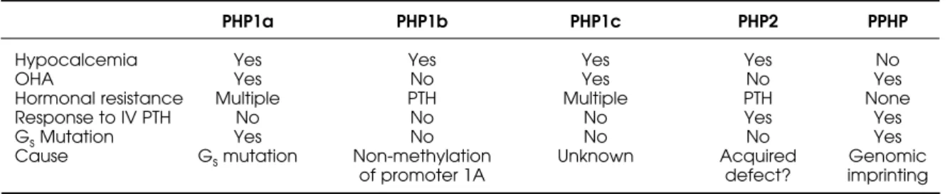
Table 4.Classification and clinical features of Pseudohypoparathyroidism subtypes.
PHP1a PHP1b PHP1c PHP2 PPHP
Hypocalcemia Yes Yes Yes Yes No
OHA Yes No Yes No Yes
Hormonal resistance Multiple PTH Multiple PTH None
Response to IV PTH No No No Yes Yes
GsMutation Yes No No No Yes
Cause Gsmutation Non-methylation Unknown Acquired Genomic
18. Lienhardt A, Bai M, Lagarde JP, Rigaud M, Zhang Z, Jiang Y, et al. Activating mutations of the calcium-sens-ing receptor: management of hypocalcemia. J Clin Endocrinol Metab 2001;86:5313-23.
19. Thomee C, Schubert SW, Parma J, Le PQ, Hashemolhos-seini S, Wegner M, et al. GCMB mutation in familial iso-lated hypoparathyroidism with residual secretion of parathyroid hormone. J Clin Endocrinol Metab 2005; 90:2487-92.
20. Breslau NA. Pseudohypoparathyroidism: Current con-cepts. Am J Med Sci 1989;298:130-40.
21. Albright F, Burnett CH, Smith PH. Pseudohypoparathy-roidism: An example of ‘Seabright-Bantam syndrome’.
Endocrinology 1942;30:922-32.
22. Tashjian Jr AH, Frantz AG, Lee JB. Pseudohypoparathy-roidism: assays of parathyroid hormone and thyrocalci-tonin. Proc Natl Acad Sci 1966;56:1138-42.
23. Chase LR, Melson GL, Aurbach GD. Pseudohy-poparathyroidism: defective excretion of 3’,5’-AMP in response to parathyroid hormone. J Clin Invest 1969;48:1832-44.
24. Farfel Z, Bourne HR, Iiri T. The expanding spectrum of G protein diseases. N Engl J Med 1999;340:1012-20. 25. Weinstein LS, Shuhua Y, Warner DR, Liu J. Endocrine
manifestations of stimulatory G protein a-subunit muta-tions and the role of genomic imprinting. Endoc Rev 2001;22:675-705.
26. Weinstein LS, Liu J, Sakamoto A, Xie T, Chen M. Minire-view: GNAS: Normal and abnormal functions.
Endocrinology 2004;145:5459-64.
27. Davies SJ, Hughes HE. Imprinting in Albright’s Hereditary Osteodistrophy. J Med Genet 1993;30:101-3.
28. Lalande M. Imprints of disease at GNAS1. J Clin Invest 2001;107:793-4.
29. Lania A, Mantovani G, Spada A. G protein mutations in endocrine diseases. Eur J Endocrinol 2001;145:543-59. 30. Levine MA. Clinical Review: Pseudohypoparathyroidism:
From bedside to bench and back. J Bone Miner Res 1999;14:1255-60.
31. Mantovani G, Maghnie M, Weber G, DeMenis E, Brunel-li V, Cappa M, et al. Growth hormone-releasing hor-mone resistance in pseudohypoparathyroidism type 1a: new evidence for imprinting of the Gsα gene. J Clin Endocrinol Metab 2003;88:4070-4.
32. Carlson HE, Brickman AS, Botazzo GF. Prolactin deficien-cy in pseudohypoparathyroidism. N Engl J Med 1977; 296:140-4.
33. Allen DB, Friedman AL, Greer FR, Chesney RW. Hypo-magnesemia masking the appearance of elevated parathyroidism hormone concentrations in familial pseudohypoparathyroidism. Am J Med Genet 1988; 31:153-8.
34. Liu J, Yu S, Litman D, Chen W, Weinstein LS. Identification of a methylation imprint mark within the mouse Gnas locus. Mol Cell Biol 2000;20:5808-17.
35. Albright F, Forbes AP, Henneman PH. Pseudo-pseudohy-poparathyroidism. Trans Assoc Am Physicians 1952;65:337-50.
36. Weinstock RS, Wright HN, Spiegel AM, Levine MA, Moses AM. Olfactory dysfunction in humans with deficient gua-nine nucleotide-binding protein. Nature 1986;322:635-6. 37. Tartaglia F, Giuliani A, Sgueglia M, Biancari F, Juvonen T,
Campana F. Randomized study on oral administration of calcitriol to prevent symptomatic hypocalcemia after total thyroidectomy. Am J Surg 2005;190:424-9.
38. Edwards SL. Maintaining calcium balance: physiology and implications. Nurs Times 2005;101:58-61.
39. Harvey JA, Zobitz MM, Pak CYC. Dose dependency of calcium absorption: A comparison of calcium carbon-ate and calcium citrcarbon-ate. J Bone Miner Res 1988;3:253. 40. Winer KK, Ko CW, Reynolds JC, Dowdy K, Keil M,
Peter-son D, et al. Long-term treatment of hypoparathy-roidism: A randomized controlled study comparing parathyroid hormone-(1-34) versus calcitriol and calci-um. J Clin Endocrinol Metab 2003;88:4214-20.
41. Sarko J. Bone and mineral metabolism. Emerg Med Clin North Am 2005;23:703-21.
Address for correspondence:
Marise Lazaretti-Castro Division of Endocrinology