of Particle Size, Crystal Phase and Water Chemistry
Xiuchun Lin1,2, Jingyi Li2, Si Ma2, Gesheng Liu2, Kun Yang2,3, Meiping Tong4, Daohui Lin2,3*
1College of Environmental and Biological Engineering, Putian University, Fujian, China,2Department of Environmental Science, Zhejiang University, Hangzhou, China,
3Zhejiang Provincial Key Laboratory of Organic Pollution Process and Control, Zhejiang University, Hangzhou, China, 4College of Environmental Sciences and Engineering, Peking University, Beijing, P. R. China
Abstract
Controversial and inconsistent results on the eco-toxicity of TiO2 nanoparticles (NPs) are commonly found in recorded
studies and more experimental works are therefore warranted to elucidate the nanotoxicity and its underlying precise mechanisms. Toxicities of five types of TiO2 NPs with different particle sizes (10,50 nm) and crystal phases were investigated usingEscherichia colias a test organism. The effect of water chemistry on the nanotoxicity was also examined. The antibacterial effects of TiO2 NPs as revealed by dose-effect experiments decreased with increasing particle size and
rutile content of the TiO2NPs. More bacteria could survive at higher solution pH (5.0–10.0) and ionic strength (50–200 mg
L21NaCl) as affected by the anatase TiO2NPs. The TiO2NPs with anatase crystal structure and smaller particle size produced
higher content of intracellular reactive oxygen species and malondialdehyde, in line with their greater antibacterial effect. Transmission electron microscopic observations showed the concentration buildup of the anatase TiO2NPs especially those
with smaller particle sizes on the cell surfaces, leading to membrane damage and internalization. These research results will shed new light on the understanding of ecological effects of TiO2NPs.
Citation:Lin X, Li J, Ma S, Liu G, Yang K, et al. (2014) Toxicity of TiO2Nanoparticles toEscherichia coli: Effects of Particle Size, Crystal Phase and Water Chemistry. PLoS ONE 9(10): e110247. doi:10.1371/journal.pone.0110247
Editor:Elena A. Rozhkova, Argonne National Laboratory, United States of America
ReceivedJune 28, 2014;AcceptedSeptember 13, 2014;PublishedOctober 13, 2014
Copyright:ß2014 Lin et al. This is an open-access article distributed under the terms of the Creative Commons Attribution License, which permits unrestricted use, distribution, and reproduction in any medium, provided the original author and source are credited.
Data Availability:The authors confirm that all data underlying the findings are fully available without restriction. All relevant data are within the paper.
Funding:This work was supported by the 973 Program of China (2014CB441104), National Natural Science Foundation of China (21337004, 21477107), Natural Science Foundations of Zhejiang Province (LR12B07001) and Fujian Province (2014J01053) of China. The funders had no role in study design, data collection and analysis, decision to publish, or preparation of the manuscript.
Competing Interests:The authors have declared that no competing interests exist.
* Email: [email protected]
Introduction
Due to their unique chemical and physical properties, titanium dioxide (TiO2) nanoparticles (NPs) are produced at a large scale
for industrial applications to meet with ever-increasing market demands [1]. The annual production of TiO2NPs is predicted to
reach 2.5 million tons by 2025 [2]. The widely used TiO2 NPs
would find their way into aquatic environments [3–6] and interact with aquatic organisms [7]. Eco-toxicity of TiO2NPs is therefore
received worldwide research attentions [8–16].
Bacteria, e.g.,Escherichia coli(E.coli), as single cell organisms and ubiquitous in aquatic environments, are good model organisms for studying the eco-toxicity of NPs and the cell/ organism-NP interaction. Many research works [8] have investi-gated the toxicity of various TiO2NPs towardE.coli, with a focus
on the influencing factors such as: (1) Size. Many studies attributed the toxicity of TiO2NPs to their small particle size [17–22]. (2)
Crystal structure. It is generally concluded that anatase TiO2NPs
are more toxic than rutile NPs by inducing greater oxidative stress [15,23,24]. (3) Experimental matrix. Changes in water chemistry (e.g., pH and ionic strength) may influence the agglomeration and sedimentation characteristics of NPs and then their toxicity [12,21,25–28]. (4) Solar radiation, especially those in the UVA region, is also considered as a critical factor of aquatic nanotoxicity [13,29–35]. These researches substantially increased our knowl-edge on the eco-toxicity of TiO2NPs.
However, controversial and inconsistent results on the toxicity of TiO2NPs are commonly found in recorded studies and precise
mechanisms of the nanotoxicity warrant more specific researches. For example, Adams et al. (2006) reported 44% reduction in the growth ofE.coliby 1 g L21and 72% reduction by 5 g L21TiO2
NPs (66 nm, crystal structure not determined) [36]; Tong et al. (2013) [21] reported 70% reduction in the growth ofE.coli by 10 mg L21 TiO2 NPs while 30% reduction was observed by
Planchon et al. (2013) [15] with the same TiO2NPs at 10 mg L21
(P25, consisting of an 80:20 ratio of anatase:rutile). So an acute lack of emphasis on the environmental and nanoparticle param-eters prevents a meaningful comparative assessment from the hitherto available nanotoxicity data, and it highlights the necessity to provide additional eco-toxicological studies and physicochem-ical characterization of TiO2NPs to ensure consistency of research
results.
This study is aimed to elucidate the roles of particle size and crystal structure in the toxicity of TiO2NPs usingE.colias a model
organism. Five types of well characterized TiO2NPs with different
particle sizes and crystal phases were examined. The toxicity assays were conducted at different concentrations of the NPs and various solution pHs and ionic strengths. In addition, the interactions between TiO2NPs and bacteria and the cell reactive
(ROS) and malondialdehyde (MDA) to address the toxicity mechanism. The results are believed to increase our understanding of the nanotoxicology.
Materials and Methods
1. Nanoparticles and characterizations
Five types of TiO2NPs were purchased and used in this study.
They were anatase TiO2with particle sizes measured to be around
10 nm (TiO2-NP 10A), 25 nm (TiO2-NP 25A), and 50 nm (TiO2
-NP 50A) and rutile TiO2 of 50 nm (TiO2-NP 50R) and mixed
anatase and rutile TiO2of 25 nm (TiO2-NP25 AR). TiO2-NP 10A
and TiO2-NP 50R were from Hongsheng Material Sci & Tech
Co., Zhejiang, China and the other three TiO2 NPs from
Wangjing New Material Sci & Tech Co., Zhejiang, China. Morphologies of the NPs were examined using TEM (JEM-1230, JEOL Ltd., Tokyo, Japan). Powered X-ray diffraction analysis (XRD, X’Pert Pro, Holland) was carried out to characterize the crystal structure of the NPs. Elemental compo-sitions of the NPs were determined by using an X-ray energy dispersion spectroscope (EDS, GEN-ESIS 4000, EDAX Inc. America). Hydrodynamic diameters and zeta potentials of the NPs (50 mg L21) were measured with a Zetasizer (Nano ZS90, Malvern, UK) after being sonicated (100 W, 40 kHz, 30 min) into 100 mg L21NaCl solution at 25uC and various pH values. Points of zero charge (pHpzc) of the NPs were obtained from the
zeta potential versus pH curves. Specific surface areas of the NPs were determined using the multi-point Brunauer-Emmett-Teller (BET) method (Quantachrome NOVA 2000e, America).
2. Dose-effect experiments
E.coliO111 (Genbank access no. GU237022.1) isolated from a sewage water was used as the test organism, as reported in our previous studies [37,38]. The bacteria were maintained in Luria Bertani (LB) solid plates at 4uC and inoculated in LB broth (pH 7.2,7.4) at 37uC overnight (12,16 h) at 150 rpm. The bacteria were separated from the broth by centrifugation at 3000g
for 15 min and washed twice with 0.85% physiology salt-water. The bacterium stock suspension was prepared by resuspending the bacterial pellets in 0.85% NaCl physiology salt-water with the cell concentration determined by the absorbance at 600 nm (OD600)
being adjusted to 1.0.
The stock NP suspensions (500 mg L21) were obtained by sonicating (100 W, 40 kHz, 30 min) 50 mg of the TiO2NPs into
100 mL of ultra-pure water. The stock NP suspensions after sonication were diluted using ultra-pure water to the target test concentrations (10–500 mg L21).
One mL of theE.colistock suspension was added into 100 mL of the test NP suspensions. The mixtures were placed on a shaker at 37uC and 150 rpm for 3 h with natural light. The bacteria in the resultant mixtures were spread on LB agar plates and incubated at 37uC for 24 h, and the colonies were counted. The percentage viabilities of the bacteria in the NP suspensions were calculated by dividing their colony forming units (CFU) mL21by that in the NP-free control. All treatments including the control were repeated in triplicate.
3. TEM Observations
TEM was used to observe the direct contact between the NPs and the bacterial cells. A drop of the bacteria exposed to the NPs (50 mg L21, 3 h) and the NP-free control was air-dried onto a
copper grid and was then imaged by the TEM. To observe the internalization and localization of the NPs in the cells and the changes in cellular structure as affected by the NPs, the NPs-treated and unNPs-treated bacteria were fixed in 2.5% glutaraldehyde, dehydrated in graded concentrations of ethanol, embedded in Epon resin, and stained with OsO4 [12,39]. Ultrathin sections
were then cut and counterstained with Reynold’s and uranyl acetate for the TEM observation.
4. Reactive oxygen species (ROS) and lipid peroxidation measurements
The fluorescence probe 2979-dichlorodihydrofluorescein diace-tate (H2DCFDA) was used to quantify the formation of
intracellular ROS as described in our previous papers [12,39] with minor modifications. The bacterial cells after exposure to the test media were collected by centrifugation (8000g, 5 min). The pellet was resuspended in 0.85% physiology salt-water containing 10mM H2DCFDA and incubated on the shaker (150 rpm) for
30 min at 37uC. The bacteria were further pelleted and resuspended in 300mL of 0.85% physiology salt-water. The fluorescence values were measured in a 96-well plate using a
multifunctional microplate reader (M200 PRO, Ltd., Austria) with the excitation and emission wavelengths of 485 nm and 528 nm, respectively. Relative ROS levels were calculated by the fluores-cence ratio of the treatments to the control.
The concentration of malondialdehyde (MDA) was determined as an indicator of lipid peroxidation as described in previous works [12,39]. Briefly, the exposed bacteria were mixed into 1 mL of 10% (wt/vol) trichloroacetic acid and left at room temperature for 10 min; the supernatant of the mixture was collected after centrifugation at 11,000g for 40 min and then mixed with 1.5 mL of a freshly prepared 0.67% (wt/vol) thiobarbituric acid solution; the resultant mixture was incubated for 20 min in a boiling water bath and after cooling the absorbance was measured at 532 nm; and the MDA concentration was calculated using the Hodges’ equations.
5. Effects of pH and ionic strength
TiO2-NP 10A with a concentration fixed at 10 mg L21 was
selected as a type of representative NPs in examining the effect of water chemistry on the nanotoxicity. In the pH effect experiment, the suspension pHs were adjusted to 5.0, 7.0, 8.0 and 10.0 using 0.1 M HCl and NaOH; NaCl was added to maintain a constant background ionic strength (100 mg L21). For the ionic strength effect experiment, difference concentrations of NaCl (0, 50, 100, 150 and 200 mg L21) were added into the NP suspensions; the final suspension pH remained at about neutral without further adjustment. The bacterial exposure method in the pH and ionic strength effect experiments was the same as the above dose-effect experiments. Zeta potential of 1 mL of the stock bacterial suspension after being mixed into 100 mL of ultra-pure water at pH 5.0, 7.0, 8.0 and 10.0 was also measured by the Zetasizer.
6. Statistical analysis
Each treatment including the blank control was conducted in triplicate and the results were presented as mean6SD (standard deviation). For each datum point, data were normalized by the reference without NPs. This representation thus strictly reports the incremental impacts of TiO2NPs, excluding the medium stress.
The Student’sttest was performed to analyze the significance of difference between two groups of data. Origin 8.0 was used to make graphs. The concentration resulting in 50% mortality (LC50)
was calculated with SPSS 20.0.
Table 1.Characteristics of the nanoparticles.
Sample
Crystal
phase, % Zeta potential, mV pHpzc hydrodynamic size, nm SBET, m2g21 TEM size, nm
TiO2-NP10A Anatase,
100
221.6 6.2 31468 324 11.063.4
TiO2-NP25A Anatase,
99.2
25.48 5.6 251637 77 26.266.1
TiO2-NP25AR Anatase,
93.0
213.6 5.2 202657 66 26.765.0
TiO2-NP50A Anatase,
98.8
29.34 6.0 486612 105 57.1614.0
TiO2-NP50R Rutile, 100 233.8 3.6 260610 30 57.2617.8
Note: the crystal phase was determined by the XRD; zeta potential and hydrodynamic size were measured in the toxicity test medium at pH 6.5 by the Zetasizer; pHpzc was calculated from the zeta potential versus pH curves shown in Figure 2; SBET(specific surface area) measured using the BET (Brunauer-Emmett-Teller) method; TEM size shows the NP size measured with the TEM images.
doi:10.1371/journal.pone.0110247.t001
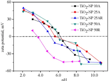
Figure 2. Changes of zeta potentials of the TiO2NPs against a
solution pH.
Results and Discussion
1. Characteristics of TiO2NPs
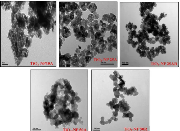
Selected properties of the TiO2NPs are listed in Table 1 with
their TEM images shown in Figure 1. Big NP aggregates present in the TEM images and the measured large hydrodynamic diameters indicate the aggregation of the NP suspensions even after the sonication. TiO2-NP 10A, having the smallest particle
size (TEM size of 11.063.4 nm) among the five TiO2NPs, owned
relative greater hydrodynamic diameter of 31468 nm. Changes in zeta potentials of the TiO2NPs against a solution pH are shown in
Figure 2. The calculated pHpzcof the NPs varied from pH 3.6 to
6.2, which could account for their negative zeta potentials (233.8–
25.48 mV) in the neutral toxicity test medium. Specific surface areas of the TiO2NPSranged from 30 m2g21(TiO2-NP 50R) to
324 m2g21(TiO2-NP 10A). Purities of the NPs determined by the
EDS were all above 98.0% (Figure 3). XRD patterns of the TiO2
NPS (Figure 3) confirmed the predominance of anatase phase of
TiO2-NP 10A, TiO2-NP 25A, and TiO2-NP 50A and the rutile
nature of TiO2-NP 50R; and TiO2-NP 25AR was a mixture of
anatase (93%) and rutile (7%).
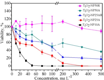
2. Cell viability assessment
The particle dose, size and phase dependent reductions in the cell viability ofE.coliupon exposure to the TiO2NPs for 3 h were
observed through the plate count assay (Figure 4). The four anatase NPs were more or less toxic toE.coliand the viability of the bacteria exhibited a pronounced concentration-dependent decrease. The calculated 3 h LC50of the four anatase NPs had an
order of TiO2-NP 10A (17.0 mg L21),TiO2-NP 25A (59.2 mg
L21),TiO2-NP 25AR (163 mg L2 1
),TiO2-NP 50A (304 mg
L21). The enhancement of bactericidal effect of the NPs with decreasing particle size was observed throughout the various particle concentrations. TiO2-NP 10A in the anatase phase with
the minimum particle size and the largest BET surface area was determined to be the most toxic toE.coli. The presence of rutile phase in the NPs lowered the bactericidal activity in comparison to the pure anatase NPs. As shown in Figure 4, although similar in particle size, the toxicity of TiO2-NP 25AR was much lower than
that of TiO2-NP 25A; the pure rutile TiO2-NP 50R was nontoxic
to the bacteria with concentration up to 500 mg L21, while the anatase TiO2-NP 50A could inactivate half of the bacteria at
304 mg L21.
3. TEM observations of the direct NP-cell interactions
Nanoparticle-type-dependent bacterial cell membrane localiza-tions of the TiO2 NPs as well as morphological changes of the
NPs-exposed cells were captured by the TEM images (Figure 5). The stronger NP-cell interaction was observed for the TiO2NPs
with anatase crystal structure and smaller particle size. Numerous TiO2-NP 10A aggregates with various sizes were observed tightly
attached to the bacterial cell surfaces (Figure 5B). The big and tight NP-cell aggregate in Figure 5C indicates the strong
Figure 3. EDS (left column) and XRD (right column) figures of the as-received TiO2NPs.
doi:10.1371/journal.pone.0110247.g003
Figure 4. Variations of the bacteria viability with concentra-tions of the TiO2NPs. The viability was the ratio of bacterial cell number under the NP treatment to the blank control.
interaction between TiO2-NP 25A and the cells. Some of the
TiO2-NP 25AR aggregates were also observed attaching to the
bacterial cells but some present away from the cells (Figure 5D), implying the relatively weaker NP-cell interaction as compared with the pure anatase TiO2-NP 25A of the same size. A few big
aggregates of TiO2-NP 50A were observed loosely attached to the
bacterial cell (Figure 5E), which suggests the much weaker interaction of TiO2-NP50A than the smaller sized TiO2-NP 25A
and TiO2-NP 10A with the cells. No obvious attachment between
the TiO2-NP 50R aggregates and the bacterial cells was observed
(Figure 5F).
Figures 5G to 5L show TEM images of the sliced bacterial cells untreated or treated with 50 mg L21 of the TiO2 NPs. The
untreated (Figure 5G) and TiO2-NP 50R-treated (Figure 5L) cells
remained intact with unimpaired cell morphology and structure, indicating the nontoxicity of TiO2-NP 50R. However, the NPs
with smaller size and anatase phase were observed sticking to the
cell surfaces (Figure 5H to 5K), which apparently induced cell distortion, plasmolysis and cell wall and membrane damage; penetration and internalization of the nanoparticles into the bacterial cells were also observed (Fig. 5H and 5I).
From the above TEM observations, it can be concluded that anatase TiO2NPs are more prone to attaching on the bacterial
surfaces than rutile NPs, and the larger NPs interact weaker with cells compared to the smaller NPs. As particle size decreases, the ratio of surface area to mass increases and changes in the physicochemical properties (e.g., surface atom reactivity, electronic and optical properties) of the nanoparticles occur, consequently, the smaller particles tend to agglomerate to a greater extent, which can further influence their reactivity and binding characteristics [40]. The NP-cell attachment may inhibit the movement of substances in and out of bacterial cells, thereby causing homeostatic imbalance, cellular metabolic disturbance and even cell death [41]. Moreover, the NP-cell attachment would facilitate
Figure 5. Selected TEM images of the unsliced (A to F) and sliced (G to L)E.colicells without (A and G) and with the treatments of TiO2-NP 10A (B and H), TiO2-NP 25A (C and I), TiO2-NP 25AR (D and J), TO2-NP 50A (E and K) and TiO2-NP 50R (F and L).The blue arrows point to the cells and the red arrows direct to the NP aggregates.
the cell internalization of NPs and the intracellular ROS production [12]. If TiO2 NPs are sufficiently small, they can
penetrate in the cells, and then induce the potential photocatalytic process inside and adsorb and deactivate biomolecules such as proteins [22,42]. Therefore, the physical NP-cell attachment and interaction could substantially contribute to the observed nano-toxicity. Many studies [13,43–44] suggest that the antibacterial mechanism of NPs includes the disruption of bacterial cellular membrane. However, we do not know for sure yet why the anatase NPs had higher affinity to the cell surfaces than the rutile NPs, which could be possibly due to their different surface properties. It is indicated that the coordination and surface properties allow anatase but not rutile NPs after dispersion induce the generation of ROS [24].
4. Oxidative stress and lipid peroxidation induced by the NPs
Relative intracellular ROS productions following the exposures to the TiO2 NPs (50 mg L21) are shown in Figure 6A. The
produced ROS in the bacterial cells exposed to the anatase TiO2
NPs was significantly (p,0.05) higher than that in the blank control cells and increased with decreasing particle size; whereas the pure rutile TiO2-NP 50R had insignificant effect on the
intracellular ROS production and TiO2-NP 25AR containing 7%
rutile induced significantly lower intracellular ROS production
compared with the pure anatase TiO2-NP 25A of the same
particle size. The enhanced intracellular ROS would affect protein expression and function in the bacteria by interrupting translation and post-translational modification [45]. It has been indicated that TiO2NPs in anatase phase are capable of inducing generation of
more ROS than that in the rutile phase [24,46] and thereby may cause higher cytotoxicity [47] including towardE.coli[13,48–52]. The increased ROS generation inE.coliexposed to the smaller and anatase TiO2NPs coincided with their enhanced bactericidal
effects as shown in Figure 4, which suggests that the size and crystal phase of TiO2NPs played a critical role in the nanotoxicity
and the nanotoxicity could be caused mainly by the elevated oxidative stress.
MDA productions in theE colicells upon the exposures to the 50 mg L21TiO2NPs are shown in Figure 6B. Significantly higher
MDA contents were observed in the NPs-treated cells compared with the blank control, indicating the cell membrane lipid peroxidation induced by the NPs. The MDA content was the highest in the TiO2-NP 10A treated cells and overall decreased
with decreasing size of the anatase NPs, which was in the same order of the ROS production by anatase NPs. This implies that the lipid peroxidation could be mainly caused by the increased ROS.
Figure 6. Relative contents of intracellular ROS (A) and MDA (B) in the bacterial cells after 3 h exposure to the TiO2NPs (50 mg L21). a–e stand for TiO2-NP 10A, TiO2-NP 25A, TiO2-NP 25AR, TiO2-NP 50A, and TiO2-NP 50R, respectively. Asterisk indicates a significant difference relative
to the control (*,p,0.05; **,p,0.01) based on the Student’sttest. Error bars represent standard deviation (n = 3). doi:10.1371/journal.pone.0110247.g006
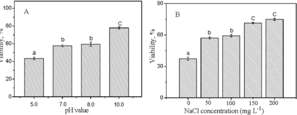
Figure 7. The effects of pH (A) and NaCl (B) on the relative viability ofE.coliexposed to 10 mg L21TiO
5. Effects of pH and ionic strength on the nanotoxicity
The bactericidal effect of the 10 mg L21 TiO2-NP 10A
exhibited a significant dependence on the solution chemistry (Figure 7). No significant difference in the nanotoxicity was observed between pH 7.0 and 8.0, while the exposed bacteria presented significantly lower and higher viability at pH 5.0 and 10.0 as compared with that at pH 7.0, respectively (Figure 7A). Increasing the suspension pH from 5.0 to 10.0, the bacterial viability increased from 43.3% to 77.9%, indicating the decreasing nanotoxicity with increasing pH. It is generally considered that direct contact and adherence of NPs with the organism cell surfaces plays a critical role in the nanotoxicity [12]. Zeta potentials of the bacterial cells were all negative at the four pHs, being258.4,256.7,256.7 and 252.5 mV at pH 5.0, 7.0, 8.0 and 10.0, respectively; whereas zeta potential of TiO2-NP 10A
decreased from about 20 mV at pH 5.0 to lower than240 mV at pH 10.0 (Figure 2). The positively-charged NPs at pH 5.0 could have a higher potential of contact and hetero-agglomeration with the negatively-charged bacterial cells through the electrostatic attraction and therefore had a higher antibacterial effect compared with the negatively-charged NPs at the three higher pHs.
It has been indicated that the antibacterial effect of TiO2NPs
(25 nm, P25) onE.coliwas stronger at pH 5.5 versus 7.0 and 9.5 and the stronger antibacterial effect at the lower pH was attributed to the stronger accumulation of the NPs on the cell surfaces [53,54]. However, contradictory research result has also been reported. Planchon et al. (2013) found a stronger adsorption of TiO2NPs (25 nm, P25) onE.colibut a slightly lower toxicity at
pH 5.0 versus 8.0, which was attributed to a better physiological state of E.coli bacteria at pH 5.0 (artificial water) versus 8.0 (surface water sample) [15]. Rincon and Pulgarin (2004) did not observe any difference in theE.colideactivation rate by the TiO2
NPs (25 nm, P25) in the pH range of 4.0–9.0 [55]. The contradictory results on the pH effect may partly come from the difference in the crystal structure of the used TiO2NPs, but the
exact mechanisms remain to be studied.
It is observed that the addition of NaCl (50,200 mg L21) reduced the toxicity of TiO2-NP 10A toward E.coli to an
extremely significant extent (Figure 7B). Some studies suggest that NaCl introduced to the medium can decrease the toxicity effect on the bacteria by providing a barrier of steric hindrance between NPs and cells [15]. Li et al. (2013) found that saline ions promoted NP aggregation and reduced surface charge, and then inhibited the adsorption of NPs on bacterial surfaces, so higher saline ions could lead to higher cell viability [38]. Furthermore, ionic strength can also influence the tolerance of bacteria to toxicants [37,38]. The ionic strength of physiology salt-water (8.5 g L21 NaCl) is isotonic and favorable for the bacterium survival. Hence, the bacteria were more tolerant to the NP suspensions at the higher ionic strengths (closer to the physiology salt-water).
Conclusions
The present study investigated the antibacterial effect of five types of TiO2NPs with various crystal phase and particle size. A
marked particle size and crystal phase dependent nanotoxicity was observed. Water chemistry, i.e. pH and ionic strength, could also significantly influence the bactericidal activity of the anatase TiO2
NPs. In conclusion, the TiO2NPs with anatase phase and smaller
particle size had higher affinity to the cell surfaces and induced heavier oxidative damage and toxicity to the bacterial cells, and the toxicity decreased with increasing pH (5.0–10.0) and ionic strength (50–200 mg L21 NaCl). These findings substantiate the need to correlate the NP characterization and behavior in environmental matrices with the toxicological endpoints and to develop a common test strategy for the eco-toxicity study of NPs taking into consideration of various confounding factors relating to the NPs, bacterial cells, and the test environment in the near future.
Author Contributions
Conceived and designed the experiments: DHL XCL. Performed the experiments: XCL JYL SM GSL. Analyzed the data: XCL. Contributed reagents/materials/analysis tools: DHL KY MPT. Contributed to the writing of the manuscript: XCL DHL.
References
1. Chen XB, Mao SS (2007) Titanium dioxide nanomaterials: synthesis, properties, modifications, and applications. Chem Rev 107: 2891–2959.
2. Robichaud CO, Uyar AE, Darby MR, Zucker LG, Wiesner MR (2009) Estimates of upper bounds and trends in nano-TiO2production as a basis for
exposure assessment. Environ Sci Technol 43: 4227–4233.
3. Kaegi R, Ulrich A, Sinnet B, Vonbank R, Wichser A, et al. (2008) Synthetic TiO2nanoparticle emission from exterior facades into the aquatic environment.
Environ Pollut 156: 233–239.
4. Lin DH, Tian XL, Wu FC, Xing BS (2010) Fate and transport of engineered nanomaterials in the environment. J Environ Qual 39: 1896–1908. 5. Johnson AC, Bowes MJ, Crossley A, Jarviea HP, Jurkschat K, et al. (2011) An
assessment of the fate, behaviour and environmental risk associated with sunscreen TiO2nanoparticles in UK field scenarios. Sci Total Environ 409:
2503–2510.
6. Batley GE, Kirby JK, Mclaughlin MJ (2013) Fate and risks of nanomaterials in aquatic and terrestrial environments. Accounts Chem Res 46: 854–862. 7. Ma S, Lin DH (2013) The biophysicochemical interactions at the interfaces
between nanoparticles and aquatic organisms: adsorption and internalization. Environ Sci: Processes Impacts 15: 145–160.
8. Menard A, Drobne D, Jemec A (2011) Ecotoxicity of nanosized TiO2: review of
in vivo data. Environ Pollut 159: 677–684.
9. Griffitt RJ, Luo J, Gao J, Bonzongo JC, Barber DS (2008) Effects of particle composition and species on toxicity of metallic nanomaterials in aquatic organisms. Environ Toxic Chem 27: 1972–1978.
10. Battin TJ, Kammer FVD, Weilhartner A, Ottofuelling S, Hofmann T (2009) Nanostructured TiO2: transport behavior and effects on aquatic microbial
communities under environmental conditions. Environ Sci Technol 43: 8098– 8104.
11. Ji J, Long ZF, Lin DH (2011) Toxicity of oxide nanoparticles to the green algae Chlorellasp. Chem Eng J 170: 525–530.
12. Lin DH, Ji J, Long ZF, Yang K, Wu FC (2012) The influence of dissolved and surface-bound humic acid on the toxicity of TiO2nanoparticles toChlorellasp.
Water Res 46: 4477–4487.
13. Dalai S, Pakrashi S, Kumar RSS, Chandrasekaran N, Mukherjee A (2012) A comparative cytotoxicity study of TiO2nanoparticles under light and dark
conditions at low exposure concentrations. Toxicol Res 1: 116–130. 14. Cle´ment L, Hurel C, Marmier N (2013) Toxicity of TiO2nanoparticles to
cladocerans, algae, rotifers and plants – effects of size and crystalline structure. Chemosphere 90: 1083–1090.
15. Planchon M, Ferrari R, Guyot F, Ge´labertb A, Menguy N, et al. (2013) Interaction betweenEscherichia coliand TiO2nanoparticles in natural and
artificial waters. Colloid Surface B 102: 158–164.
16. Kim J, Lee S, Kim C, Seo J, Park Y, et al. (2014) Non-monotonic concentration–response relationship of TiO2 nanoparticles in freshwater
cladocerans under environmentally relevant UV-A light. Ecotox Environ Safe 101: 240–247.
17. Jiang JK, Oberdo˝rster G, Biswas P (2009) Characterization of size, surface charge, and agglomeration state of nanoparticle dispersions for toxicological studies. J Nanopart Res 11: 77–89.
18. Kim DS, Kwak SY (2009) Photocatalytic inactivation of E.coli with a mesoporous TiO2coated film using the film adhesion method. Environ Sci
Technol 43: 148–151.
19. Deckers AS, Loo S, L’hermite MM, Boime NH, Menguy N, et al. (2009) Size-, composition- and shape-dependent toxicological impact of metal oxide nanoparticles and carbon nanotubes toward bacteria. Environ Sci Technol 43: 8423–8429.
21. Tong TZ, Binh CTT, Kelly JJ, Gaillard JF, Gray KA (2013) Cytotoxicity of commercial nano-TiO2 to Escherichia coli assessed by high-throughput
screening: effects of environmental factors. Water Res 47: 2352–2362. 22. Xiong SJ, George SJ, Ji ZX, Lin SJ, Yu HY, et al. (2013) Size of TiO2
nanoparticles influences their phototoxicity: an in vitro investigation. Arch Toxicol 87: 99–109.
23. Nel A, Xia T, Ma¨dler L, Li N (2006) Toxic potential of materials at the nanolevel. Science 311: 622–627.
24. Jin C, Tang Y, Yang FG, Li XL, Xu S, et al. (2011) Cellular Toxicity of TiO2
nanoparticles in anatase and rutile crystal phase. Biol Trace Elem Res 141: 3– 15.
25. Whirter MJM, Quillan AJM, Bremer PJ (2002) Influence of ionic strength and pH on the first 60 min ofPseudomonas aeruginosaattachment to ZnSe and to TiO2monitored by ATR-IR spectroscopy. Colloid Surface B 26: 365–372.
26. French RA, Jacobson AR, Kim B, Isley SL, Penn RL, et al. (2009) Influence of ionic strength, pH, and cation valence on aggregation kinetics of titanium dioxide nanoparticles. Environ Sci Technol 43: 1354–1359.
27. Chowdhury I, Cwiertny DM, Walker SL (2012) Combined factors influencing the aggregation and deposition of nano-TiO2in the presence of humic acid and
bacteria. Environ Sci Technol 46: 696826976.
28. Ng AMC, Chan CMN, Guo MY, Leung YH, Djurisˇic´ AB, et al. (2013) Antibacterial and photocatalytic activity of TiO2and ZnO nanomaterials in
phosphate buffer and saline solution. App Microbiol Biot 97: 5565–5573. 29. Brunet L, Lyon DY, Hotze EM, Alvarez PJJ, Wiesner MR, et al. (2009)
Comparative photoactivity and antibacterial properties of C60fullerenes and
titanium dioxide nanoparticles. Environ Sci Technol 43: 4355–4360. 30. Jiang W, Mashayekhi H, Xing BX (2009) Bacterial toxicity comparison between
nano- and micro-scaled oxide particles. Environ Pollut 157: 1619–1625. 31. Kim SW, An YJ (2012) Effect of ZnO and TiO2nanoparticles preilluminated
with UVA and UVB light on Escherichia coli and Bacillus subtilis. App Microbiol Biot 95: 243–253.
32. Bokare A, Sanap A, Pai M, Sabharwal S, Athawale AA (2013) Antibacterial activities of Nd doped and Ag coated TiO2nanoparticles under solar light
irradiation. Colloid Surface B 102: 273–280.
33. Li S, Wallis LK, Ma H, Diamond SA (2014) Phototoxicity of TiO2nanoparticles
to a freshwater benthic amphipod: Are benthic systems at risk? Sci Total Environ 466–467: 800–808.
34. Li S, Wallis LK, Diamond SA, Ma H, Hoff DJ (2014) Species sensitivity and dependence on exposure conditions impacting phototoxicity of TiO2
nanopar-ticles to benthic organisms. Environ Toxicol Chem 33: 1563–1569. 35. Li S, Pan X, Wallis LK, Fan ZY, Chen ZL, et al. (2014) Comparison of TiO2
nanoparticle and graphene-TiO2 nanoparticle composite phototoxicity to
Daphnia magnaandOryzias latipes. Chemosphere 112: 62–69.
36. Adams LK, Lyon DY, Alvarez PJJ (2006) Comparative eco-toxicity of nanoscale TiO2, SiO2, and ZnO water suspensions. Water Res 40: 3527–3532.
37. Li M, Zhu LZ, Lin DH (2011) Toxicity of ZnO nanoparticles toEscherichia coli: Mechanism and the influence of medium components. Environ Sci Technol 45: 1977–1983.
38. Li M, Lin DH, Zhu LZ (2013) Effects of water chemistry on the dissolution of ZnO nanoparticles and their toxicity toEscherichia coli. Environ Pollut 173: 97– 102.
39. Long ZF, Ji J, Yang K, Lin DH, Wu FC (2012) Systematic and quantitative investigation of the mechanism of carbon nanotubes’ toxicity toward algae. Environ Sci Technol 46: 8458–8466.
40. Suresh AK, Pelletier DA, Doktycz MJ (2013) Relating nanomaterial properties and microbial toxicity. Nanoscale 5: 463–474.
41. Wang Z, Lee YH, Wu B, Horst A, Kang Y, et al. (2010) Anti-microbial activities of aerosolized transition metal oxide nanoparticles. Chemosphere 80: 525–529. 42. Szczupak AM, Ulfig K, Morawski AW (2011) The application of titanium dioxide for deactivation of bioparticulates: An overview. Catalysis Today 169: 249–257.
43. Adams CP, Walker KA, Obare SO, Docherty KM (2014) Size-dependent antimicrobial effects of novel palladium nanoparticles. Plos One 9 (1), e89581: 1–12.
44. Musee N, Thwalaa M, Nota N (2011) The antibacterial effects of engineered nanomaterials: implications for wastewater treatment plants. J Environ Monitor 13: 1164–1183.
45. Jiang GX, Shen ZY, Niu JF, Bao YP, Chen J, et al. (2011) Toxicological assessment of TiO2nanoparticles by recombinant Escherichia coli bacteria.
J Environ Monitor 13: 42–48.
46. Linsebigler AL, Lu GQ, Yates JT (1995) Photocatalysis on TiO2 surfaces:
Principles, mechanisms, and selected results. Chem Rev 95: 735–758. 47. Kelly K, Havrilla C, Brady T, Abramo K, Levin E (1998) Oxidative stress in
toxicology: established mammalian and emerging piscine model systems. Environ Health Persp 106: 375–384.
48. Neal AL (2008) What can be inferred from bacteria–nanoparticle interactions about the potential consequences of environmental exposure to nanoparticles? Ecotoxicology 17: 362–371.
49. Dastjerdi R, Montazer M (2010) A review on the applications of inorganic nano-structured materials in the modification of textiles: focus on anti-microbial properties. Colloid Surface B 79: 5–18.
50. Foster HA, Ditta IB, Varghese S, Steele A (2011) Photocatalytic disinfection using titanium dioxide: spectrum and mechanism of antimicrobial activity. App Microbiol Biot 90: 1847–1868.
51. Kumar A, Pandey AK, Singh SS, Shanker R, Dhawan A (2011) Engineered ZnO and TiO2nanoparticles induce oxidative stress and DNA damage leading
to reduced viability ofEscherichia coli.Free Radical Bio Med 51: 1872–1881. 52. Barnes RJ, Molina R, Xu JB, Dobson PJ, Thompson IP (2013) Comparison of
TiO2and ZnO nanoparticles for photocatalytic degradation of methylene blue
and the correlated inactivation of gram-positive and gram-negative bacteria. J Nanopart Res 15: 1432.
53. Pagnout C, Jomini S, Dadhwal M, Caillet C, Thomasc F, et al. (2012) Role of electrostatic interactions in the toxicity of titanium dioxide nanoparticles toward Escherichia coli.Colloid Surface B 92: 315–321.
54. Schwegmann H, Ruppert J, Frimmel FH (2013) Influence of the pH-value on the photocatalytic disinfection of bacteria with TiO2- explanation by DLVO
and XDLVO theory. Water Res 47: 1503–1511.
55. Rincon AG, Pulgarin C (2004) Effect of pH, inorganic ions, organic matter and H2O2onE.coliK12 photocatalytic inactivation by TiO2implications in solar