Haloperidol Derivatives as Novel Potent Calcium
Channel Blockers with Vasodilator Activity
Yicun Chen1, Jinhong Zheng2, Fuchun Zheng3, Jinzhi Wang2, Yanmei Zhang1, Fenfei Gao1, Zhanqin Huang1, Ganggang Shi1,4*
1Department of Pharmacology, Shantou University Medical College, Shantou, Guangdong, China,2Department of Chemistry, Shantou University Medical College, Shantou, Guangdong, China,3Department of Pharmacy, First Affiliated Hospital, Shantou University Medical College, Shantou, Guangdong, China,4Department of Cardiovascular Diseases, First Affiliated Hospital, Shantou University Medical College, Shantou, Guangdong, China
Abstract
Several haloperidol derivatives with a piperidine scaffold that was decorated at the nitrogen atom with different alkyl, benzyl, or substituted benzyl moieties were synthesized at our laboratory to establish a library of compounds with vasodilator activity. Compounds were screened for vasodilatory activity on isolated thoracic aorta rings from rats, and their quantitative structure–activity relationships (QSAR) were examined. Based on the result of QSAR, N-4-tert-butyl benzyl haloperidol chloride (16c) was synthesized and showed the most potent vasodilatory activity of all designed compounds. 16cdose-dependently inhibited the contraction caused by the influx of extracellular Ca2+in isolated thoracic aorta rings from rats. It concentration-dependently attenuated the calcium channel current and extracellular Ca2+ influx, without affecting the intracellular Ca2+mobilization, in vascular smooth muscle cells from rats.16c, possessing theN-4-tert-butyl benzyl piperidine structure, as a novel calcium antagonist, may be effective as a calcium channel blocker in cardiovascular disease.
Citation:Chen Y, Zheng J, Zheng F, Wang J, Zhang Y, et al. (2011) Design, Synthesis, and Pharmacological Evaluation of Haloperidol Derivatives as Novel Potent Calcium Channel Blockers with Vasodilator Activity. PLoS ONE 6(11): e27673. doi:10.1371/journal.pone.0027673
Editor:Zhe Zhang, Virginia Commonwealth University, United States of America
ReceivedAugust 14, 2011;AcceptedOctober 21, 2011;PublishedNovember 16, 2011
Copyright:ß2011 Chen et al. This is an open-access article distributed under the terms of the Creative Commons Attribution License, which permits unrestricted use, distribution, and reproduction in any medium, provided the original author and source are credited.
Funding:This work was supported by a grant from the National Natural Science Foundation of China (No. 30973922, 30472028), and the United Fund of the National and GuangDong province Natural Science Foundation of China (No. U0932005). The funders had no role in study design, data collection and analysis, decision to publish, or preparation of the manuscript.
Competing Interests:The authors have declared that no competing interests exist.
* E-mail: [email protected]
Introduction
Calcium is essential for life and is the most common signal transduction element in cells [1,2]. The contraction of all types of muscle primarily depends on increased intracellular Ca2+
[3]. Calcium channel blockers constitute an important class of cardio-vascular drugs. Members of this class are clinically useful in the treatment of cardiovascular disorders in which calcium plays a regulatory role. However, most cannot be used to resolve clinical problems such as ischemia, reperfusion injury, and vascular structure remodeling [4]. Furthermore, some of them have serious side effects. Therefore, better calcium channel blockers are still needed.
Haloperidol is a widely used antipsychotic whose therapeutic properties have been associated with its D2antagonist activity on
the basis of the so-called dopamine hypothesis [5]. N-Benzyl haloperidol chloride (4) (Figure 1A), a derivative of haloperidol, was synthesized at our laboratory and was found to have a vasodilator effect [6]. Our previous study revealed that 4’s inhibitory effect on the influx of extracellular Ca2+
might be related to vasodilatory mechanisms [7]. Furthermore, theN-benzyl piperidine structure of 4is completely different from that of the clinically used calcium channel blockers, which belong to one of the following three chemical classes: the dihydropyridines, the phenylalkylamines, and the benzothiazepines [8]. Therefore, a
novel calcium channel antagonist may be obtained by molecular manipulation of the structure of4, which itself is not a sufficiently potent vasodilator.
With the aim of exploring the structure–activity relationship of 4 and obtaining more efficient calcium channel blockers, we had cultured single crystals to determine the structure of 4. Furthermore we designed the series of compounds shown in Table S1. In addition, we revealed some haloperidol derivatives (compounds6a–6c,9a–9c in Table S1) had vasodilator activity in previous study [9]. The piperidine scaffold, which we consider a supporting structure for novel calcium channel antagonists, was decorated at the nitrogen atom of piperidine with different substituent groups. We screened these compounds for vasodilatory activity in isolated rat thoracic aorta rings held in a Ca2+
-containing high-K+
solution. A detailed quantitative structure– activity relationship (QSAR) analysis of this series of haloperidol derivatives was subsequently performed to study the physicochem-ical properties that were responsible for their activity. Base on the result of QSAR, we synthesizedN-4-tert-butyl benzyl haloperidol chloride (16c) which was found to posses a good vasodilative activity in KCl-constricted vessels in line with expectation.
Many studies have demonstrated that vasorelaxation in KCl-constricted vessels can be ascribed directly to the inactivation of voltage-dependent Ca2+
further investigated for its inhibitory action on the contraction caused by an influx of exogenous Ca2+
in thoracic aorta rings to prove that its vasodilator activity was related to Ca2+
. Then, the inhibitory effects of 16c on the activity of VDCCs, the extracellular Ca2+ influx, and the intracellular Ca2+ release in
vascular smooth muscle cells (VSMCs) were explored in detail to reveal the calcium antagonist mechanism.
Results
Synthesis
The reaction pathways used to synthesize the designed compounds (compounds 1–16) were described in Schemes S1, S2, and the chemical and physical characteristics of the compounds were reported in Table S1. The key intermediate haloperidol (4-[4-(4-chlorophenyl)-4-hydroxy-1- piperidyl]-1-(4-fluorophenyl)-butan-1-one) could be obtained via piperidyl alkylation reaction according to the literature [11]. Commercially available alkyl and benzyl halides were used. Some of benzyl halides (compoundsI–V) were synthesized from the corresponding benzaldehyde compounds (Scheme S1). The quaternary ammo-nium salt derivatives of haloperidol (compounds 1–16) were synthesized from haloperidol by alkylation or benzylation at the nitrogen atom using a suitable alkyl or benzyl halide (Scheme S2). In order to know about the structure of4as the leading compound clearly, we had cultured single crystals and determined the crystal structure by X-ray single crystal diffraction (Figure 1B, Table S2). The chemical structures of these compounds were characterized by 1H NMR spectra (Table S3). 16c was illustrated as a representation in Figure 2.
Determination of vasorelaxation
All synthesized compounds caused a concentration-dependent relaxation of aortic rings that had been precontracted to varying extents with a high level of K+
(80 mM). The vehicle (0.1% DMSO) used for all compounds had no effect on vascular tone. Concentration–response curves were determined for each com-pound, and the potency was expressed as the IC50 value in the
presence and absence of endothelium (Table S4). From Table S4, we can see no significant IC50difference between aortic rings with
intact endothelium or denuded endothelium. Concentration-response curves showed no significant shift in the absence of endothelium, than in the presence of endothelium, which suggested no nitric oxide involvement in the vasodilatation (data not shown).
QSAR study of some derivatives. A detailed QSAR
analysis of a series of haloperidol derivatives (compounds 4–15, splitting into a testing set of 3 compounds which are randomly
selected and training set of 26 compounds) had been performed to determine the physicochemical properties responsible for their activity. The physicochemical parameters used were listed in Table 1, namely the hydrophobic constantp, Hammetts, molar refractivity (MR), and were compiled from the literature [12–14]. The IC50values were transformed to pIC50(negative logarithm of
IC50). The QSAR results indicated that activity was best modeled
by molecular steric parameters represented by MR, hydrophobic parameters represented byp. The best QSAR obtained was:
pIC50~0:238MRpz0:181p-0:195 MRoz5:061
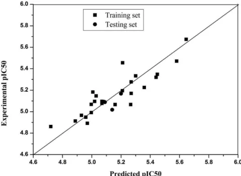
The subscripts in the parameter relate to the position of the substituent considered. A determinants of coefficient R2is 0.787 for 26 compounds in training set, and the residuals of compounds 9b,11b,14c as testing set are 0.03176; 0.1224, 0.00163. It confirmed that the equation was of some value in predicting activity (Table 1, Figure 3).
The inhibitory action of 16c on the contraction caused by KCl and the influx of extracellular Ca2+
The results indicated 16c showed the most significant vasorelaxation effect on KCl-dependent contraction compared with compounds 1–15, and the IC50 value of16cwas 0.95mM (Table 1). And 16c was chosen for further research.
KCl-dependent contraction is due to the influx of extracellular Ca2+
through VDCCs [10]. We first investigated the ability of16c
to inhibit the contraction induced by the exogenous application of Ca2+in a Ca2+-free high-K+solution. As Figure 4 showed,16c
at different concentrations (0.1—10mM) significantly impaired the response to Ca2+
(3 mM) to 87.49%—11.02% of the initial response. The IC50 value was 0.76mM, similar to the result obtained from the determination of vasorelaxation (Table S1, Table 1). The results indicated that16cinhibited the responses to Ca2+
in a concentration-dependent manner. So we deduced that
16csignificantly inhibited the response to Ca2+
influx from the extracellular space.
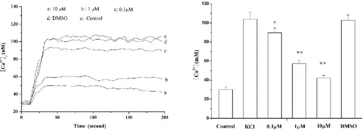
Effects of 16c on [Ca2+
]i in arterial VSMCs. The Ca
2+
responses of arterial VSMCs to KCl (80 mM) were studied after incubation with16c.16cdid not affect basal [Ca2+]
i(29.861.2vs
30.761.4 nM for 16cand control, respectively,n= 25,p.0.05). The response of the control cells to KCl was characterized by a sharp increase in [Ca2+
]i(from 30.1862.82 to 103.3467.26 nM,
mean 6S.E. n= 24) followed by a sustained plateau (106.646 6.24 nM at 200 s). This response was totally inhibited by16cat 0.1, 1, 10mM (both KClpeakand KCl200 s= 89.7664.96, 57.2463.64, 42.3262.65 nM, respectively n= 24, p,0.01 vs control values; Figure 5).
Effect of 16c on L-type Ca2+
channel activity. We then
applied the patch-clamp technique to measure the effect of16con the activity of VDCCs on the primary cells. In our study,IBawas
elicited by depolarization from the depolarizing potential of 230 mV to +50 mV. The peakIBawas elicited at the potential
of 0 mV. Barium currents were reversibly inhibited by 1mM nimodipine, and augmented by 50 nM Bay K 8644 (data not shown). 16c (10, 1, 0.1mM) reduced the peak current to 21.365.1% (p,0.01, n = 5), 39.466.2% (p,0.01, n = 5), 89.8615.1% (p,0.01, n = 5) of control values, respectively (Figure 6A). The VSMCs showed current/voltage relationships typical of high voltage-activated L-type Ca2+
channels, with apparent threshold and reversal potentials of approximately230 and+50 mV, respectively (Figure 6B). No change in leak current Figure 1. The chemical structure of 4(A), The molecular
structure of 4 (B).
doi:10.1371/journal.pone.0027673.g001
was observed during the application of the compounds, which suggests that at these concentrations, the compounds had no ‘‘detergent-like’’ effect on cell plasma membrane. Half-maximal inhibition of the Ca2+
channel current was observed at about 1mM.
Discussion
Calcium channel blockers play an important role in cardiovas-cular diseases, but better drugs are still needed for some clinical problem. In our study, we found some Haloperidol derivatives showed vasorelaxation activity to varying degree. We synthesized a series of haloperidol derivatives and we used the test of vasodilator effect on the rat isolated thoracic aorta rings with a high level of K+(80 mM) to screen molecules for further studies, and examined
the structure-activity relationship of compounds. Studies have demonstrated that the contraction of vascular smooth muscle is
initiated by an increased intracellular calcium level [15–17], which may be achieved in two ways: extracellular Ca2+
influx from VDCCs evoked by depolarization with high potassium concen-tration, and intracellular Ca2+
release from the intracellular stores [18,19]. From the result, we can deduce these compounds’ inhibition on the contraction of the vascular smooth muscle might relate to the extracellular Ca2+
influx from VDCCs evoked by depolarization with high K+.
Furthermore, most published QSARs show the importance of particular physicochemical parameters in describing activity [20]. We observed a correlation between the pharmacological activities and structures in this study. The best QSAR obtained was: pIC50= 0.238 MRp+0.181 p- 0.195 MRo+5.061. This result
showed that the main factors governing activity were the MR term of the specific position of the substituent which will determine the fit at the receptor site and the hydrophobic parameters which determine the ability of the compounds to transport the cellular Figure 2. The chemical structure of 16c (A); 400 MHz1H NMR spectrum of 16c in CDCl3(B); Infrared Spectroscopy of 16c (C).
membrane and to bind in the hydrophobic space of the proteins in the cellular membrane. In addition, compound 4 displayed higher vasodilation activity than compounds 1–3 (from Table S1). These results also indicated that for this series of derivatives, the MR term of a particular molecule was the significant element of the substituent. The importance of both the substituent steric parameters and their position appears to be in their ability to maintain the molecule in an orientation that is conducive to receptor binding. This was inferred from the fact that MR term, as a steric parameter was consistently expressed in a regression analysis. The equation also highlighted the importance of the substituent for vasodilator activity in the ortho- and para-positions. The MR parameter needs some discussion at this point. MR has been viewed as a measure of bulk and as a ‘‘rough and ready’’ steric parameter. It is used to model the intermolecular effects between a ligand and receptor. From the x-ray structure of 4 (Figure 1B), it is easy to understand that the negative ortho effect may be due to a conformational preference of 4-phenyl piperidine
ring for the substituted benzyl group. It is, therefore, most probably due to direct steric hindrance interfering with drug receptor binding [21]. Although a more complex problem involving, interpretation of a positive coefficient with MR revealed that the steric effect at the para-position of the aryl ring was advantageous to the activity. In summary, the result showed activity may be further improved if substituents with greater hydrophobic parameters in the para-position are considered (such as p-C (CH3)3, p-(CH2)4N(CH3)3+, p-(CH2)4C6H5). The
com-pound with p-(CH3)3had a lower molecular weight than the others
(such as p-(CH2)4N(CH3)3+, p-(CH2)4C6H5). So this compound
(16c) was designed and synthesized, and 16c showed the most potent vasorelaxation effect agreeing with the predicted effect (experimental IC50: 0.95mM, predicted IC50: 1.36mM, Table 1).
As KCl-dependent contraction is due to the influx of extracellular Ca2+
through VDCCs [10], it is likely that the haloperidol derivatives, such as 16c, may have effects on the inhibition of the influx extracellular Ca2+
, and the blockade of calcium channels. The ability Table 1.Physicochemical properties and lgIC50of the substituent.
Compound p s MRo# MRm# MRp#
lgIC50(M)*
Experimental lgIC50(M)*Predicted
4 0 0 0.1 0.1 0.1 24.9965 25.06832
5a 20.67 20.2 0.28 0.1 0.1 24.8877 24.91286
5b 20.67 0.12 0.1 0.28 0.1 24.9597 24.94857
5c 20.67 20.37 0.1 0.1 0.28 24.9974 24.99122
6a 0.71 0.40 0.6 0.1 0.1 25.07 25.09602
6b 0.71 0.37 0.1 0.6 0.1 25.2097 25.19522
6c 0.71 0.23 0 0.1 0.6 25.3001 25.33352
7b 0.64 0.39 0.1 1.25 0.1 25.0074 25.18271
7c 0.64 0.31 0.1 0.1 1.25 25.2111 25.45515
8a 0.88 0.97 0.5 0.1 0.1 25.0301 25.14624
8b 0.88 0.64 0.1 0.5 0.1 25.3595 25.22561
8c 0.88 0.56 0.1 0.1 0.5 25.44 25.32037
9a 0.56 0.93 0.56 0.1 0.1 25.07 25.07715
9b& 0.56 0.8 0.1 0.56 0.1
25.1999 25.16814
9c 0.56 0.73 0.1 0.1 0.56 25.27 25.27739
10a 0.14 0.98 0.09 0.1 0.1 25.02 25.09533
10b 0.14 0.92 0.1 0.09 0.1 25.0798 25.09335
10c 0.14 0.93 0.1 0.1 0.09 25.07 25.09098
11a 20.28 1.03 0.74 0.1 0.1 24.9698 24.8913
11b&
20.28 0.86 0.1 0.74 0.1 25.1402 25.01778
11c 20.28 0.73 0.1 0.1 0.74 25.27 25.1699
12c 1.53 20.15 0.1 0.1 1.50 25.6458 25.67345
13b 20.01 0.36 0.1 1.29 0.1 25.266 25.06654
13c 20.01 0.45 0.1 0.1 1.29 25.4485 25.34845
14a 20.57 1.28 0.63 0.1 0.1 24.7201 24.86129
14b 20.57 1.07 0.1 0.63 0.1 24.9299 24.96587
14c&
20.57 0.91 0.1 0.1 0.63 25.0899 25.08827
15a 1.02 20.17 1.03 0.1 0.1 25.1598 25.06611
15c 1.02 20.15 0.1 0.1 1.03 25.58 25.47095
16c 1.98 20.02 0.1 0.1 1.96 26.0222 25.86636
*, IC50: The half maximal inhibitory concentration, #, MR
o: molar refractivity in ortho-position, MRm: molar refractivity in meta-position, MRp: molar refractivity in para-position. &, compounds as testing set and their predicted IC
50were calculated by QSARs model. doi:10.1371/journal.pone.0027673.t001
of 16c to inhibit the contraction induced by the exogenous application of Ca2+
then was confirmed in a Ca2+
-free high-K+
solution. This observation suggests that C3might further inhibit the influx of Ca2+
through VDCCs. An elevated [Ca2+
]i level is the main trigger for VSMC
contraction [22], and we showed that 16c had an inhibitory effect on the contraction induced by exogenous Ca2+
. To reveal the
molecular mechanism by which 16c inhibited vasoconstriction and to obtain more information about the effect of 16c on calcium ions, we studied the effect of 16c on Ca2+
signaling in VSMC by LSCM. The increase in [Ca2+
]i is believed to result from
Ca2+
influx, corresponding to the opening of the voltage-sensitive plasma membrane Ca2+
channels [23]. This response was inhibited by 16c, which is consistent with the results in intact Figure 3. Correlation between predicted activities (IC50) by QSARs models and the experimental activities (IC50) (for compounds of
training set, R2= 0.787; for randomly selected compounds 9b,11b,14c as testing set, Residue are 0.03176; 0.1224, 0.00163. Correlation is significant at the 0.01 level.).
doi:10.1371/journal.pone.0027673.g003
Figure 4. Cumulative concentration-response curves for Ca2+in the presence of the vehicle and increasing concentrations of 16c (0.1–10mM) in isolated endothelium-intact aortic rings (n = 5).Results are the mean6S.E. from 5 independent experiments.
arteries (Figure 4). The results of the effect on KCl-dependent Ca2+
signaling indicated that 16c decreased the influx of extracellular Ca2+
caused by depolarization in VSMCs.
Based on these findings, we therefore postulate that the compounds targeted the plasma membrane, especially the VDCCs present in VSMCs. The patch-clamp technique was applied to measure the effect of 16c on the activity of VDCCs on the primary cells. 16c obviously suppressed the calcium channel current and the I-V relation ofIBaindicated that the blocking effect of 16c on calcium channels was voltage-independent. Studies indicate two types (L and T) of calcium channel exist in VSMCs [24]. Under the condition of individual cell depolarization from a holding potential of240 mV, the L-type calcium channel was activated, and the T-type Ca2+
and Na+
channels were inactivated. Moreover, TEA, a non-specific K channel blocker, was admin-istered in the extracellular solution. The pipette solution was also filled with 4-AP (a K+
channel blocker). So the outward K+
currents were completely blocked [25]. In addition, Barium
currents were reversibly inhibited by 1mM nimodipine, and augmented by 50 nM Bay K 8644, an activator of L-type Ca2+
channels [26] in our study. Therefore, the inward current we recorded under these conditions was an L-type Ca2+
current. Obviously, 16c suppressed the L-type calcium channel current. In addition, we can infer the mechanism of 16c inhibition of L type Ca2+
channels, from the structure of 4-phenyl piperidine which exists in the haloperidol derivatives. In our study, the structure-activity relationship analysis indicated the 16c and other haloperidol derivatives, which contain 4-phenyl piperidine ring possess calcium channel blocking (CCB) activity and the conformation of the 4-phenyl piperidine ring correlates with the CCB activity. The piperidine ring is the hydride 1, 4-DHP ring. Studies on the structure of nifedipine demonstrated the 1, 4-dihydropyridine (1, 4-DHP) ring system which was perpendicular to the C-4 substituted-phenyl ring, was the determinant of calcium channel antagonist activity [27]. Therefore we suppose the mechanism of 16c in the block of L-type Ca2+
channel is probably Figure 5. Effect of 16c (0.1,10mM) and the vehicle (0.1% DMSO) on KCl (80 mM)-induced changes of Ca2+ concentration in vascular smooth muscle cells.A, representative Ca2+
concentration response to KCl ; B, dose-dependent inhibitory effect of16con the Ca2+
concentration response to KCl. Results are the means6S.E. (n = 24). *,p,0.01 compared with normal. **,p,0.01 compared with model (KCl). doi:10.1371/journal.pone.0027673.g005
Figure 6. Effect of 16c on L-type Ca2+channel currents in vascular smooth muscle cells (A) and onI–Vrelation ofBa2+currents in vascular smooth muscle cells (B).The step protocol of recording was described in methods section.
doi:10.1371/journal.pone.0027673.g006
similar to the nifedipine. The heterologous expressing such as Xenopus oocytes, will be performed to explore the exact mechanism in our future studies.
Ca2+
is considered to be an intracellular signal implicated in both vasoconstriction and proliferation [28]. However, it is well known that some L-type Ca2+
channel antagonists(verapamil, diltiazem, and nifedipine)show little inhibitory effect on the proliferation of vascular smooth cells, which is related to the development of vascular diseases [29]. While, it was reported that 16c, a novel channel blocker, had anti-proliferation activity [30]. Ca2+
, as a second messenger, stimulates nuclear transcription factors Egr-1 expression which involves cell proliferation [30]. We previously showed that Egr-1 overexpression induced cell proliferation by using antisense Egr-1 ODNs, and demonstrated that 16c attenuated the Ang II-induced extracellular Ca2+
influx and inhibited Egr-1 overexpression. In this study, we demonstrat-ed further that 16c had vasodilatory activity, although the vasodilator effect of 16c was not more potent than other L-type Ca2+antagonists (nifedipine, 1–10 nM; verapamil, 0.1–1
mM; and diltiazem, 0.5–1mM; concentrations for 50% inhibition of vascular contractions in rat or rabbit aorta) [31].
However, 16c, as a novel channel blocker, had vasodilatory activity and anti-proliferation activity that provides a basis for further studies of novel calcium channel blockers on intervention of vascular remodeling and atherosclerosis [30].
Methods
Chemistry
General methods for preparation of the haloperidol
derivatives. The preparation of the title compounds was very
simple. Haloperidol was dissolved in chloroform and was then reacted with benzyl halide under reflux conditions, or halohydrocarbon (iodomethane, ethyliodide, iodopropane) were used as a solvent and were reacted with haloperidol directly. All melting points were taken on a Bu¨chi apparatus and were uncorrected. IR spectra were recorded with a Nicolet-Impact 410 system as KBr pellets. 1H NMR spectra were recorded on a
Bruker AM-400 spectrometer (400 MHz). The1H NMR internal standard used was tetramethylsilane (TMS). Chromatographic separations were performed on a silica gel column by gravity chromatography (Kieselgel 40, 0.01–0.04 mm; Merck). Yields are given after purification, unless otherwise stated. Where analyses are indicated by symbols, the analytical results are within61% of the theoretical values. All spectral and elemental analysis data (shown in the Supporting Information) were in agreement with the assigned structures. All compounds were estimated to be.95% pure according to analytical reversed-phase high performance liquid chromatography (RP-HPLC) and detection with MS (see Supporting Information for details) for details. No UV-active impurities were observed with analytical RP-HPLC and UV detection at 245 nm. The data for 16c was displayed as represented in Figure 2.
Synthesis of compounds 1–3. Haloperidol (10 mM) was
dissolved in the corresponding halohydrocarbon (4 ml). The mixture was heated in an oil bath for 24 h to maintain reflux and was then cooled to room temperature. The filtered solid was recrystallized from an ethanol-water mixed solvent to a crystalline product.
Synthesis of some benzyl halide (compounds I–V). The
corresponding benzaldehyde compounds (100 mM) were dissolved in methanol (20 ml)(for I–III) or THF (20 ml) (for IV–V). NaBH4
(200 mM) was then added gradually in ice-bath. The mixtures were elevated to room temperature and stirred for 5 min(for I–III)
or 1 h (for IV–V). Then water (20 ml) was added to end the reaction and the mixtures were evaporated to dryness. The products were again dissolved in ethylacetate (40 mL), washed with water (20 ml) and ethylacetate extractions were concentrated to dryness. The products of benzyl alcohol derivatives were obtained in this step. These products were then dissolved in THF (20 ml without water, contained DMF 1–2 d), SOCl2(3 ml) was
added gradually in ice-bath. After the mixtures were stirred at 0uC for 20 min, reactions were terminated and the pH value of the mixture was adjusted to about 7 with a 1 N NaOH solution. The oil layers were separated from the mixture with a separatory funnel and the products (benzyl halide) were obtained by distillation. The 1HNMR data of compounds I–V were compared with literature [32,33], and reported in supporting information (Table S5).
Others benzyl halide were purchased fromAlfa Aesar,A Jhnson Matthey Companyand some Chinese Companies. These compounds have been confirmed with literature data in HNMR.
Synthesis of compounds 4–16. Haloperidol (10 mM) was
dissolved in chloroform (4 mL) and the corresponding benzyl halide (30 mM) was then added. The mixture was heated to maintain reflux for 18–24 h, and was then cooled to room temperature. The white solid was filtered off, and was washed with chloroform. The crude product was recrystallized in an ethanol– water mixed solvent to provide a crystalline product.
Pharmacology
Vasoactivity determination. The vasodilation activity of
compounds1–16was evaluated in isolated rat thoracic aortic rings according to the methods of Towart and Polster et al [34,35]. Thoracic aortas were isolated from male Wistar rats weighing 200–250 g. The vessels were cut into ring segments, 2–3 mm wide, and 5 rings were obtained from each aorta. Rings were then mounted in standard 10-mL organ baths filled with Krebs bicarbonate solution ((PSS), containing (in mM): 118.0 NaCl, 4.7 KCl, 1.2 KH2PO4, 1.2 MgSO4.7H2O, 5.0 glucose, 25.0
NaHCO3, and 2.5 CaCl2.7H2O) at 37uC and were gassed with
95% O2–5% CO2. (pH = 7.4). The aortic rings were allowed to
equilibrate for 90 min at a testing tension of 1 g, and were then depolarized by the addition of a solution of KCl to a final concentration of 80 mM. The preparations were then extensively washed with PSS, and a second contraction was evoked by K+
depolarization (80 mM). When the amplitude of the contraction reached a plateau, cumulative concentrations of the tested compounds in a vehicle of water and DMSO (1000:1) were added every 45 min. Development of antagonism occurred so slowly that doses had to be increased at established times, without waiting for complete equilibrium to be reached. Cumulative concentration-response cures to all compounds were determined in the absence and presence of endothelium. According to experimental protocol, when required, the endothelium was mechanically removed from some rings by gently rubbing the lumen with the tips of a forceps. Acetylcholine was used to confirm that the endothelium had effectively been removed from the aortic rings [36].
Inhibitory action of active compound (16c) on the
contraction caused by influx of extracellular Ca2+
. To
determine the effect of 16c on the response to the influx of extracellular Ca2+
, the contractile response to the exogenous application of Ca2+
was examined in the presence of16cin Ca2+
-free high-K+
solution (80 mM, K+
-HBSS) as previously described [37]. Aortic rings were initially equilibrated at a resting tension of 1 g in normal Ca2+
-free HBSS for 60 min. The bath medium was then replaced with Ca2+
-free high-K+
determine a control response to Ca2+
(3 mM) and this was considered to be 100% contraction. After a 30 min equilibration with Ca2+-free HBSS, the cumulative contractile response to Ca2+
(0.01–3 mM) was determined in K+
-HBSS in the presence of increasing concentrations (0.1–10mM) of 16c. The aortic rings were incubated with 16c for 10 min before examining the responses to Ca2+
.
Electrophysiology. Inward barium (IBa) currents were
studied in single cells by the patch-clamp technique in the perforated-attached configuration [38]. The recordings were made using a Biologic RK-300 amplifier, filtered (23 dB, 5-pole Tchebicheff filter) at 1 kHz and sampled at 5 kHz. Currents were recorded from holding potential 240 mV during linear voltage ramps from240 to+60 mV, with 10 mV increments. The bath solution contained (in mM) NaCl 125, BaCl210.8, MgCl21,
CsCl 5.4, glucose 10, and Hepes 10 (pH = 7.4 at 24uC). Patch pipettes were pulled from glass capillaries (Warner Instruments) and were coated with wax to minimize capacitative transients and noise; the electrode resistance ranged from 2–5 MVwhen filled with the pipette solution. The patch pipette was filled with a solution containing (in mM) CsOH 75, CsCl 55, MgCl25, aspartic
acid 75, Hepes 10 (pH = 7.4 at 22uC). Nystatin (Biochrom KG, Berlin, Germany) was dissolved in Me2SO and diluted into the
pipette solution to give a final concentration ranging from 50 to 100 mg/mL. Drugs were applied by changing the bath solution. The final concentration of ethanol, the diluent, was 0.1% (v/v). The sampling and data analysis involved the use of pClamp 8.1 software (Axon Instruments). Experiments were carried out at room temperature (25uC).
Isolation of rat primary VSMCs. Primary cell cultures of
VSMCs were established with cells removed from abdominal aortas excised from 4-week-old Wistar rats. Isolation of aortic VSMCs followed the method of Campbell et al [39]. The cells were suspended in a growth medium (DMEM supplemented with 10% (v/v) fetal bovine serum [FBS] (JRH Biosciences, Lenexa, KS, USA), 2.2 g/L NaHCO3, 100 U/mL penicillin G, and
100mg/mL streptomycin) and seeded into 24-mm glass coverslips in 6-well plates (for LSCM) or a 0.5-mL chamber (for patch-clamp). Cells were cultured at 37uC in a humidified atmosphere of 5% CO2in air. The experimental protocol was approved by the
University Research Committee, and the animal protocol was in full compliance with the Guidelines for Animal Experimentation developed by the university.
Determination of intracellular free [Ca2+
]i. [Ca
2+
]i was
determined in adherent cells after 60 min of loading with 20mM Fluo3-AM in HBSS, as described previously [40]. Fluorometric data were obtained by means of LSCM (Zeiss LSM 510 META). The Fluo-3 dye was excited with a 488-nm wavelength argon laser beam and the emission fluorescence was monitored at 525 nm. [Ca2+]
iwas calculated according to the method of Grynkiewicz, et
al [41]. At the end of the experiment, Triton X-100 and, subsequently, EGTA were added at final concentrations of 0.1% and 1 mM, respectively, to obtain the fluorescence maximum
(Fmax) and the fluorescence minimum (Fmin). [Ca2+]i was
determined from the equilibrium equation, [Ca2+
]i= Kd
(F2Fmin)/(Fmax2F), where F is the experimental value of
fluorescence and K was defined as 0.4mM. [Ca2+
]i was
measured at base line first. After the cells were treated with16c
for 10 min, the agonists, KCl and TG, were added and [Ca2+
]i
was determined.
Analysis of statistics. Data were presented as mean6S.E.
Group differences were analyzed by Student’sttest. The effect of
16con currents was evaluated by Student’s pairedt-test.P,0.05 was considered statistically significant.
Associated content
Ethics Statement. All animal work was performed according
to the international animal welfare guidelines, and protocols were approved by Shantou University Medical College Institutional Animal Care and Use Committee (permit numbers: SUMC2009-010).
Supporting Information
Table S1 Designed compounds 1–16 and their
sensitiv-ity (IC50) in endothelium-intact thoracic aorta rings from rats.IC50: The half maximal inhibitory concentration.
(DOC)
Table S2 Crystal data and structure refinement for
compound 4.
(DOC)
Table S3 1HNMR data, EI-MS data of Compounds 1–16.
(DOC)
Table S4 A comparison of the sensitivity (IC50) to
different compounds in endothelium-intact and -denud-ed thoracic aorta rings from rats.
(DOC)
Table S5 1HNMR data of Compounds I–V.
(DOC)
Scheme S1 Reagents and conditions.(a) NaBH4, MeOH,
room temperature 5 min, or NaBH4, THF, room temperature
1 hour. (b) SOCl2,THF/DMF, ice-bath 20 min.
(TIF)
Scheme S2 Reagents and conditions. (a) CH3I or
CH3CH2I or CH3(CH2)2I, reflux, 24 h. (b) The benzyl halidel,
CHCl3, reflux, 18–24 h.
(TIF)
Author Contributions
Conceived and designed the experiments: GS YC. Performed the experiments: YC JZ FZ JW YZ FG ZH. Analyzed the data: YC GS JZ FG. Wrote the paper: YC GS FZ.
References
1. Akizuki O, Inayoshi A, Kitayama T, Yao K, Shirakura S, et al. (2008) Blockade of T-type voltage-dependent Ca2+
channels by benidipine, a dihydropyridine calcium channel blocker, inhibits aldosterone production in human adrenocor-tical cell line NCI-H295R. Eur J Pharmacol 584: 424–434.
2. Betzenhauser MJ, Wagner LE, 2nd, Iwai M, Michikawa T, Mikoshiba K, et al. (2008) ATP modulation of Ca2+release by type2 and type3 inositol (1, 4, 5) -triphosphate receptors. Differing ATP sensitivities and molecular determinants of action. J Biol Chem 283: 21579–21587.
3. Escobales N, Crespo MJ, Altieri PI, Furilla RA (1996) Inhibition of smooth muscle cell calcium mobilization and aortic ring contraction by lactone vastatins. J Hypertens 14: 115–121.
4. Wajima T, Beguier B, Yaguchi M (2004) Effects of cariporide (HOE642) on myocardial infarct size and ventricular arrhythmias in a rat ischemia/ reperfusion model: comparison with other drugs. Pharmacology 70: 68–73. 5. Sibley DR, Monsma FJ, Jr. (1992) Molecular biology of dopamine receptors.
Trends Pharmacol Sci 13: 61–69.
6. Shi GG, Zheng JH, Li CC, Chen JX, Zhuang XX, et al. (1998) The effect of quaternary ammonium salt derivation of haloperidol on coronary artery. Chin Pharm J 9: 529–531.
7. Shi GG, Fang H, Zheng JH (2001) Quaternary ammonium salt derivative of haloperidol inhibits KCl-induced calcium increase in rat aortic smooth muscle cells. Acta Pharmacol Sin 22: 837–840.
8. Spedding M, Paoletti R (1992) Classification of calcium channels and calcium antagonists: progress report. Cardiovasc Drugs Ther 6: 35–39.
9. Zheng JH, Wang JZ, Shi GG (2005) Studies on synthesis of quaternary ammonium salt derivatives of haloperidol and their vasodilative activities. Chinese Pharmaceutical Journal 40: 947–951.
10. Izumi H, Tanaka Y, Okada Y, Ogawa N, Izawa T (1996) Structure-activity relationship of a novel K+
channel opener, KRN4884, and related compounds in porcine coronary artery. Gen Pharmacol 27: 985–989.
11. Lyles-Eggleston M, Altundas R, Xia J, Sikazwe DM, Fan P, et al. (2004) Design, Synthesis, and Evaluation of Metabolism-Based Analogues of Haloperidol Incapable of Forming MPP+-like Species. J Med Chem 47: 497–508. 12. Hansch C, Leo A, Hoekman D (1995) Exploring QSAR: Hydrophobic,
electronic, and steric constants. ACS: Washington.
13. Hansch C, Leo A (1979) Substituent constants for correlation analysis in chemistry and biology. Wiley & Sons: New York.
14. Hansch C, Leo A (1995) Exploring QSAR: Fundamentals and applications in chemistry and biology. ACS: Washington.
15. Kiselev KV, Gorpenchenko T, Chernoded GK, Dubrovina AS, Grishchenko OV, et al. (2008) Calcium-dependent mechanism of somatic embryogenesis in oncogene rolC expressing cell cultures of Panax ginseng. Mol Biol (Mosk) 42: 275–285.
16. Saida K, Van Breemen C (1983) Mechanism of Ca2+
antagonist induced vasodilation intracellular actions. Circ Res 52: 137–142.
17. Unno T, Kwon SC, Okamoto H, Irie Y, Kato Y, et al. (2003) Receptor signaling mechanisms underlying muscarinic agonist-evoked contraction in guinea-pig ileal longitudinal smooth muscle. Br J Pharmacol 139: 337–350.
18. Xiong Z, Sperelakis N (1995) Regulation of L-type calcium channels of vascular smooth muscle cells. J Mol Cell Cardiol 27: 75–91.
19. Orallo F (1996) Regulation of cytosolic calcium levels in vascular smooth muscle. Pharmacol Ther 69: 153–171.
20. Verma RP, Hansch C (2008) Investigation of DNA-binding properties of organic molecules using quantitative structure-activity relationship (QSAR) models. J Pharm Sci 97: 88–110.
21. Dearden JC, George E (1997) Anti-inflammatory potencies of some aspirin derivatives: a quantitative structure-activity study [proceedings]. J Pharm Pharmacol 31, Suppl: 45.
22. Block LH, Emmons LR, Vogt E, Sachinidis A, Vetter W, et al. (1989) Ca2+ -channel blockers inhibit the action of recombinant platelet-derived growth factor in vascular smooth muscle cells. Proc Natl Acad Sci U S A 86: 2388–2392. 23. Ng LL, Davies JE, Wojcikiewicz RJ (1994) 3-Hydroxy-3-methyl glutaryl
coenzyme A reductase inhibition modulates vasopressin-stimulated Ca2+ responses in rat A10 vascular smooth muscle cells. Circ Res 74: 173–181. 24. Kuga T, Kobayashi S, Hirakama Y, Kanaide H, Takeshita A (1996) Cell
cycle-dependent expression. of L-and T-type Ca2+currents in rat aortic smooth muscle cells in primary culture. Cir Res 79: 14–19.
25. Gebremedhin D, Lange AR, Campbell WB, Hillard CJ, Harder DR (1999) Cannabinoid CB1 receptor of cat cerebral arterial muscle functions to inhibit
L-type Ca2+
channel current. Am J Physiol Heart Circ Physiol 276: H2085–H2093.
26. Best JM, Kamp TJ (2010) A sympathetic model of L-type Ca2+ channel-triggered arrhythmias. Am J Physiol Heart Circ Physiol 298: H3–H4. 27. Janis RA, Triggle DJ (1983) New developments in Ca2+
channel antagonists. J Med Chem 26: 775–785.
28. Berridge MJ (1995) Calcium signaling and cell proliferation. Bio Essays 17: 491–500.
29. Yoshida J, Ishibashi T, Nishio M (2003) Antiproliferative effect of Ca2+channel blockers on human epidermoid carcinoma A431 cells. Eur J Pharmacol 472: 23–31.
30. Chen Y, Zheng J, Zhang Y, Wang J, Liu Q, et al. (2009) N-4-Tert-Butyl Benzyl Haloperidol Chloride Suppresses Ca2+-dependent Egr-1 Expression and Subsequently Inhibits Vascular Smooth Muscle Cell Proliferation Induced by Angiotensin II. Cell Physiol Biochem 23: 295–304.
31. Opie LH (1987) Calcium channel antagonists, Part I: Fundamental properties: mechanisms, classification, sites of action Cardiovasc. Drugs Ther 1: 411–430. 32. Breschi MC, Calderone V, Digiacomo M, Macchia M, Martelli A, et al. (2006)
New NO-Releasing Pharmacodynamic Hybrids of Losartan and Its Active Metabolite: Design,Synthesis, and Biopharmacological Properties. J Med Chem 49: 2628–2639.
33. Hulsman N, Medema JP, Bos C, Jongejan A, Leurs R, et al. (2007) Chemical insights in the concept of hybrid drugs: the anti-tumor effect of nitric oxide-donating aspirin involves a quinone methide but not nitric oxide nor aspirin. J Med Chem 50: 2424–2431.
34. Towart R (1982) A new method for investigating the isometric contractile responses of small blood vessels. Br J Pharmacol 75: 150.
35. Polster P, Christophe B, Van Damme M, Houlliche A, Chatelain P (1990) SR 33557, a novel calcium entry blocker. I. In vitro isolated tissue studies. J Pharmacol Exp Ther 255: 593–599.
36. Furchgott RF (1999) Endothelium-Derived Relaxing Factor: Discovery, Early Studies, and Identification as Nitric Oxide. Bioscience Reports 19: 235–251. 37. Chan EC, Pannangpetch P, Woodman OL (2000) Relaxation to flavones and
flavonols in rat isolated thoracic aorta: mechanism of action and structure-activity relationships. J Cardiovasc Pharmacol 35: 326–333.
38. Gollasch M, Haller H, Schultz G, Hescheler J (1991) Thyrotropin-releasing hormone induces opposite effects on Ca2+
channel currents in pituitary cells by two pathways. Proc Natl Acad Sci U S A 88: 10262–10266.
39. Campbell JH, Kocher O, Skalli O, Gabbiani G, Campbell GR (1989) Cytodifferentiation and expression of alpha-smooth muscle actin mRNA and protein during primary culture of aortic smooth muscle cells. Correlation with cell density and proliferative state. Arteriosclerosis 9: 633–643.
40. Minta A, Kao JP, Tsien RY (1989) Fluorescent indicators for cytosolic calcium based on rhodamine and fluorescein chromophores. J Biol Chem 264: 8171–8178.