Bond strength of adhesive systems to
human tooth enamel
Resistência adesiva de sistemas adesivos ao
esmalte dentário humano
Abstract: The purpose of this study was to evaluate in vitro three adhesive systems: a total etching single-component system (G1 Prime & Bond 2.1), a self-etching primer (G2 Clearil SE Bond), and a self-etching adhesive (G3 One Up Bond F), through shear bond strength to enamel of human teeth, evaluating the type of fracture through stereomicros-copy, following the ISO guidance on adhesive testing. Thirty sound premolars were bi-sected mesiodistally and the buccal and lingual surfaces were embedded in acrylic resin, polished up to 600-grit sandpapers, and randomly assigned to three experimental groups (n = 20). Composite resin cylinders were added to the tested surfaces. The specimens were kept in distilled water (37°C/24 h), thermocycled for 500 cycles (5°C-55°C) and submitted to shear testing at a crosshead speed of 0.5 mm/min. The type of fracture was analyzed under stereomicroscopy and the data were submitted to Anova, Tukey and Chi-squared (5%) statistical analyses. The mean adhesive strengths were G1: 18.13 ± 6.49 MPa, (55% of resin cohesive fractures); G2: 17.12 ± 5.80 MPa (90% of adhesive fractures); and G3: 10.47 ± 3.14 MPa (85% of adhesive fractures). In terms of bond strength, there were no signiicant differences between G1 and G2, and G3 was signiicantly different from the other groups. G1 presented a different type of fracture from that of G2 and G3. In con-clusion, although the total etching and self-etching systems presented similar shear bond strength values, the types of fracture presented by them were different, which can have clinical implications.
Descriptors: Shear strength; Dentin-bonding agents; Dental enamel.
Resumo: O objetivo deste estudo foi avaliar in vitro três sistemas adesivos: um monocom-ponente com condicionamento ácido total (G1 Prime & Bond 2.1), um “primer” autocon-dicionante (G2 Clearil SE Bond) e um adesivo autoconautocon-dicionante (G3 One Up Bond F), através de resistência ao cisalhamento ao esmalte de dentes humanos, avaliando o tipo de fratura por estereomicroscopia, seguindo as normas ISO para testes adesivos. Trinta pré-molares hígidos foram seccionados ao meio em sentido mésio-distal, incluídos em resina acrílica, polidos até lixa d’água de granulação 600 e aleatoriamente divididos em três grupos (n = 20). Cilindros de resina composta foram adicionados às superfícies de teste. Os espécimes foram armazenados em água destilada (37°C/24 h), termociclados por 500 ciclos (5°C-55°C) e submetidos ao teste de cisalhamento com velocidade de 0,5 mm/min, sendo o tipo de fratura analisado sob estereomicroscopia e os dados submetidos à análise estatística Anova, Tukey e Qui-quadrado (5%). As médias de resistência adesiva foram: G1: 18,13 ± 6,49 MPa, (55% de fraturas coesivas em resina); G2: 17,12 ± 5,80 MPa (90% de fraturas adesivas) e G3 10,47 ± 3,14 MPa (85% de fraturas adesivas). Em termos de resistência adesiva, não houve diferenças signiicantes entre G1 e G2, tendo G3 apresen-tado diferença signiicante em relação aos demais grupos. G1 apresentou tipo de fratura diferente de G2 e G3. Em conclusão, apesar de os sistemas adesivos com condicionamento ácido total e “primer” autocondicionante terem apresentado valores de resistência adesiva similares, o tipo de fratura apresentado por eles foi diferente, o que pode ter implicações clínicas.
Descritores: Resistência ao cisalhamento; Adesivos dentinários; Esmalte dentário. Thaís Cachuté Paradella(a)
Marcelo Fava(b)
(a)Doctorate Student, Department of
Bioscience and Oral Diagnosis;(b)Pediatric
Dentistry Professor, Department of Social Dentistry and Children’s Clinic – School of Dentistry of São José dos Campos, São Paulo State University.
Corresponding author: Thaís Cachuté Paradella Rua Padre Rodolfo, 71- Vila Ema São José dos Campos - SP - Brazil CEP: 12243-080
E-mail: [email protected]
Introduction
Modern dentistry treatments are based on mini-mal tooth wear due to adhesive techniques and cur-rent aesthetic restorative materials. The enamel etch-ing concept has been improved through the years, and new adhesive systems have been released and researched.2,3,4,12,13,19
Resin adhesion to acid etched enamel is mainly due to the formation of resin tags. Acid etching re-moves nearly 10 µm of enamel surface and creates a 5 to 50 µm deep porous layer. This irregular surface is the result of hydroxyapatite crystals dissolution. Therefore, when a low-viscosity resin is applied, it penetrates into the microporosities and polymerizes to form a micromechanical bond with enamel. Hy-drophilic adhesives have been marketed in the form of multiple-bottle or single-component systems, but both with separate conditioner. Nonetheless, self-etching systems, which include self-self-etching primers and self-etching adhesives, were developed in order to simplify and eliminate clinical steps. However, it is important that these simpliications do not affect enamel adhesion.7
The bond strength provided by adhesive sys-tems is the force per unit of area required to break a bonded assembly with failure occurring in or near the adhesive surface. The purpose of either a ten-sile, microtensile or shear bond strength test is to establish a numeric value in order to determine how strong that bond was.3,18,22 Shear testing is impor-tant since it presents reliable results and because shear stress is more representative in a clinical situ-ation.3,18 In 1994, the International Organization for Standardization (ISO)11 presented guidance on adhesive testing in order to standardize adhesion tests so that in vitro studies could provide similar, relevant and reproducible results that would support
in vivo testing.20
The purpose of this study was to evaluate in vitro
three adhesive systems: a total etching adhesive sys-tem, a self-etching primer and a self-etching adhe-sive, through shear bond strength to enamel of hu-man teeth, evaluating the type of fracture through stereomicroscopy, following the ISO11 guidance on adhesive testing. The working hypothesis was that there would be no signiicant differences between
total etching and self-etching adhesives regard-ing shear bond strength and type of fracture after debonding.
Material and Methods
The methodology applied in this study was ap-proved by the Ethics Committee, State University of São Paulo (Protocol # 067/2003-PH/CEP). Thirty premolars, extracted due to orthodontic reasons, from 14-16 year-old patients, were kept in a 0.5% chloramine solution, for no longer than a week. Af-ter this period of time, the teeth were kept in dis-tilled water at 4°C, for no longer than 6 months, with a weekly change of water.11 The teeth’s crowns were analyzed under stereomicroscopy (Zeiss/Stemi 2000C-MC-80-DX, Berlin, Germany) and the teeth with visible cracks or enamel alterations were elimi-nated from the study.
The samples were bisected mesiodistally with a high-speed diamond saw (KG Sorensen, Reference 7020, São Paulo, SP, Brazil), under air/water cooling, and the buccal and lingual surfaces were embedded in acrylic resin, polished up to 600-grit sandpapers, until a lat enamel area of 5 mm in diameter was exposed.4,5,9,14,17. The specimens were randomly as-signed to three experimental groups (n = 20): G1: Prime & Bond 2.1 (Dentsply Ind., Rio de Janeiro, RJ, Brazil) total etching single-component system, G2: Clearil SE Bond (Kuraray Medical Inc., To-kyo, Japan) self-etching primer, and G3: One-Up Bond F (Tokuyama Dental Corp., Tokyo, Japan) self-etching adhesive system. The adhesive system’s testing area was delimited using a circular adhesive tape with a 4-mm diameter central oriice.13,14 The adhesive systems were used according to the manu-facturers’ instructions described in Table 1. A telon mould (4 mm x 5 mm) was used to build resin cyl-inders (Z-250 3M ESPE Dental Products Division, St. Paul, MN, USA) on the test surface. The mate-rial was light-cured for 40 seconds using a XL3000 light-curing device (3M ESPE Dental Products, St. Paul, MN, USA), with 400 mW/cm²of light inten-sity.
testing machine (Instron, model 411, Chicago, IL, USA) set to operate at a 0.5 mm/min crosshead speed until breakdown. The specimens were posi-tioned in a stainless steel mould in order to maintain a 90° angle in relation to the applied force.10,11,18
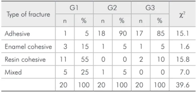
The obtained data (kgf/cm²) was transformed in MPa and submitted to parametric statistical analy-sis, ANOVA and Tukey (5%). The null hypothesis (H0) was that there would be no difference between the tested adhesives. After shear bond strength test-ing, the specimens had their types of fractures ana-lyzed under stereomicroscopy. Fractures were classi-ied as either adhesive, cohesive (resin or enamel) or mixed fractures12, and the data were submitted to Chi-squared (χ2) statistical analysis.
Results
The results and statistical indings for the three studied groups regarding shear bond strength are presented in Table 2. According to the values ob-tained in this study, it was possible to notice that the standard deviations for the three groups were close and the variation coeficients were inferior to 50%, which justiies a parametric statistical analysis. Ac-cording to ANOVA testing, the mean values differed statistically (p = 0.001 at a conidence level of 95%),
so H0 was rejected. With post-hoc Tukey’s multiple comparison test, the formation of two groups with same bond strength values was possible, as shown in Graph 1. Statistical analysis showed no signiicant differences between G1 and G2. Only G3 showed signiicant statistical difference in relation to both G1 and G2.
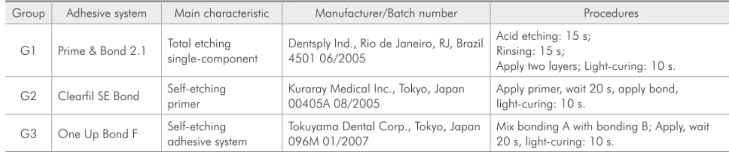
Regarding type of fracture, the results are pre-sented in Table 3, which shows that G1 prepre-sented a higher number of resin cohesive fractures, whereas G2 and G3 presented a higher number of adhesive fractures. These data were submitted to Chi-squared (χ2) statistical analysis, at a 5% level of conidence, which evaluated the equality or similarity of mutu-ally exclusive discrete categories. This means that this statistical analysis would either conirm or not the similarity of the type of fracture presented by the studied groups. The results of the Chi-squared Table 1 - Description of the adhesive systems.
Group Adhesive system Main characteristic Manufacturer/Batch number Procedures
G1 Prime & Bond 2.1 Total etching single-component
Dentsply Ind., Rio de Janeiro, RJ, Brazil 4501 06/2005
Acid etching: 15 s; Rinsing: 15 s;
Apply two layers; Light-curing: 10 s.
G2 Clearfil SE Bond Self-etching primer
Kuraray Medical Inc., Tokyo, Japan 00405A 08/2005
Apply primer, wait 20 s, apply bond, light-curing: 10 s.
G3 One Up Bond F Self-etching adhesive system
Tokuyama Dental Corp., Tokyo, Japan 096M 01/2007
Mix bonding A with bonding B; Apply, wait 20 s, light-curing: 10 s.
Table 2 - Description of the results regarding shear bond strength values (MPa).
Statistics G1 G2 G3
Mean 18.13 17.12 10.47
Standard Deviation 6.49 5.80 3.14
Variation coefficient 35.80 33.89 30.07
Minimum value 10.71 8.03 5.68
Maximum value 34.75 26.48 16.12
Graph 1 - Box chart display of the results after Tukey’s test. Similar letters (a) indicate same statistical group. Different letters (b), different statistical findings.
G1 G2 G3
Mean Mean – SD Mean +SD 30
25
20
15
10
5
0
(MP
a)
Shear bond strength
a
a
b 18.13
17.12
statistical analysis are presented in Table 3 and Ta-ble 4.
Analyzing the data in Table 3, when the three ex-perimental groups are considered, the Chi-squared value (χ2 = 39.6) is higher than the Chi-squared crit-ical value (12.5). Therefore, it was concluded that there was signiicant statistical difference between the types of fracture observed in G1, G2 and G3. In a second analysis, presented in Table 4, there is no signiicant statistical difference between the self-etching systems (G2 and G3), since χ2 = 3.0, which is lower than the χ2 critical value (7.8).
Discussion
Researches involving adhesive systems have be-come frequent in dentistry, since in vitro adhesive testing is simple and fast. However, adhesive testing must be performed in a standard manner, so that its results can be trustworthy.3,11,18,20. ISO11 guidance on adhesive testing standardizes adhesion testing, detailing how tests should be performed. Although
in vitro tests do not completely predict how dental
materials will behave in the oral cavity, these tests are valuable. Due to the quick development and re-lease of new bonding agents, it has become neces-sary to ind simple and fast methods to evaluate the effectiveness of these new agents, since clinical tri-als are time- and money-consuming. In vitro testing represents an effective and fast way to evaluate den-tal materials.14,20,23
In this study, the ISO11 guidance on adhesive test-ing was used for storage of the test specimens, stain rate for bond breaking, treatment of results and ac-celerated ageing through thermocycling, which is also important in bond testing since its effects are valuable parameters in determining the stability of adhesive bonding agents, because it simulates in-traoral conditions.14,16. The purpose of these proce-dures was not only to standardize the study, but also to maximally simulate oral conditions.
The shear bond strength values of acid etched enamel in association with single-component adhe-sive systems are in the range of 20 MPa.1 The results in this study regarding G1 are lower than those pre-sented by Gordan et al.6 (1998), which found bond strength values of 27.2 (± 6.22) MPa. However, no thermocycling was performed. Thermocycling can lower bonding values, since hot water accelerates hydrolysis,14 although this is a matter of discussion in literature. A recent study,1 which evaluated bond strength after 10,000 thermal cycles with various adhesive systems, concluded that the mean bond strengths remained unchanged after thermal stress. On the other hand, another recent study8 using hu-man teeth found higher mean bond strength values for Clearil SE Bond after 24 hours (23.4 MPa) than Table - Chi-squared (χ2) statistical analysis of the type of
fracture by groups individually.
Type of fracture G1 G2 G3 χ2
n % n % n %
Adhesive 1 5 18 90 17 85 15.1
Enamel cohesive 3 15 1 5 1 5 1.6
Resin cohesive 11 55 0 0 2 10 15.8
Mixed 5 25 1 5 0 0 7.0
20 100 20 100 20 100 39.6
χ2 critical value = 12.5.
Table 4 - Chi-squared statistical analysis (χ2) of the type of fracture considering every two groups.
Type of fracture G1/G2 G1/G3 G2/G3
n(G1) n(G2) χ2 n(G1) n(G3) χ2 n(G2) n(G3) χ2
Adhesive 1 18 15.2 1 17 14.2 18 17 0.0
Enamel cohesive 3 1 1.0 3 1 1.0 1 1 0.0
Resin cohesive 11 0 11.0 11 2 6.2 0 2 2.0
Mixed 5 1 2.6 5 0 5.0 1 0 1.0
20 20 29.8 20 20 26.4 20 20 3.0
in the present study, and thermocycling was not per-formed. Further studies should be performed in or-der to establish the effect of thermocycling on bond strength.
Torii et al.22 (2002) found similar bond strength values with the same self-etching adhesive system used in G2 of the present study. However, bovine teeth were used, and although many studies regard-ing bond strength include bovine teeth as substitutes for human teeth, with no statistical differences be-tween bovine and human enamel,13 the enamel sur-face of bovine teeth presents differences when com-pared to that of human teeth and, therefore, human teeth should be preferable when available.11 Moura et al.15 (2006), using micro-tensile bond strength testing, evaluated different adhesive systems and their results corroborate the indings of the present study as they found a similar mean bond strength (18.7 MPa) for Clearil SE. The authors also stat-ed that an overall increase in porosity was evident along the entire enamel surface treated with the self-etching primers, concluding that no selective de-mineralization similar to that produced with 35% phosphoric acid was observed, and that the highest bond strength means and the more retentive etch-ing pattern were observed for the total etchetch-ing ad-hesives. Regarding the self-etching systems tested, the authors stated that Clearil SE Bond should be preferred. Although in the present study Clearil SE Bond presented higher mean bond strengths than One Up Bond F, no statements can be made regard-ing the demineralization pattern produced by the adhesive systems since scanning electron
microsco-py (SEM) was not performed for all the samples. In the present study, the self-etching systems pre-sented a greater number of adhesive fractures when compared to the total etching single-component sys-tems. There is not a consensus in the literature regard-ing the type of fracture after bondregard-ing tests. Toledano
et al.21 (2001) found 50% of adhesive fractures and 50% of mixed fractures with the same adhesive sys-tem used in G2. However, Fritz et al.5 (2001) found 60% of enamel-cohesive fractures, and, in Miyazaki
et al.14 (2002), a tendency of mixed and cohesive frac-tures was observed when this system was used.
It has been reported that when an adhesive fracture occurs, enamel bonding has not been well-established.7 Thus, in orthodontics, the use of self-etching systems is being encouraged since it promotes adhesive fractures when the bracket is removed, preventing enamel loss.2,24,25 Therefore, the type of fracture after debonding can have clini-cal implications. However, parameters need to be taken into consideration for interstudy comparison of in vitro results2,10 and further standardized stud-ies should be performed in order to corroborate the indings of the present study.
Conclusion
The total etching adhesive system Prime & Bond 2.1 and self-etching primer Clearil SE Bond both presented acceptable shear bond strength values. However, the types of fracture presented by the self-etching systems were different from those presented by the total etching system, since the self-etching systems presented mostly adhesive fractures.
References
1. Asaka Y, Yamaguchi K, Inage H, Takamizawa T, Kurokawa H, Rikuta A et al. Effect of thermal cycling on bond strengths of single-step self-etch adhesives to bovine dentin. J Oral Sci. 2006;48(3):63-9.
2. Bishara SE, Soliman M, Lafoon J, Warren JJ. Effect of chang-ing a test parameter on the shear bond strength of orthodontic brackets. Angle Orthod. 2005;75(5):832-5.
3. Cardoso PEC, Braga RR, Carrilho MRO. Evaluation of micro-tensile, shear and tensile tests determining the bond strength of three adhesive systems. Dent Mater. 1998;14(6):394-8.
4. Di Hipolito V, de Goes MF, Carrilho MR, Chan DC, Daronch M, Sinhoreti MA. SEM evaluation of contemporary self-etch-ing primers applied to ground and unground enamel. J Adhes Dent. 2005;7(3):203-11.
5. Fritz UB, Diedrich P, Finger WJ. Self-etching primers – an alternative to the conventional acid etch technique? J Orofac Orthop. 2001;62(3):238-45.
7. Hara AT, Amaral CM, Pimenta LAF, Sinhoreti MAC. Shear bond strength of hydrophilic adhesive systems to enamel. Am J Dent. 1999;12(4):181-4.
8. Kanehira M, Finger WJ, Hoffmann M, Endo T, Komatsu M. Relationship between degree of polymerization and enamel bonding strength with self-etching adhesives. J Adhes Dent. 2006;8(4):211-6.
9. Kanemura N, Sano H, Tagami J. Tensile bond strength to and SEM evaluation of ground and intact enamel surfaces. J Dent. 1999;27(7):523-30.
10. Klocke A, Kahl-Nieke B. Influence of force location in orthodontic shear bond strength testing. Dent Mater. 2005;21(5):391-6.
11. International Organization for Standardization. TR 11405. Dental Materials – Guidance on testing of adhesion to tooth structure. 1994;1-15.
12. Jain P, Stewart GP. Effect of dentin primer on shear bond strength of composite resin to moist and dry enamel. Oper Dent. 2000;25(1):51-8.
13. Lopes MB, Sinhoreti MAC, Sobrinho LC, Consani S. Compar-ative study of the dental substrate used in shear bond strength tests. Pesqui Odontol Bras. 2003;17(2):171-5.
14. Miyazaki M, Kinoura K, Honjo G, Onose H. Effect of self-etching primer application method on enamel bond strength. Am J Dent. 2002;15(2):412-6.
15. Moura SK, Pelizzaro A, Dal Bianco K, de Goes MF, Loguercio AD, Reis A et al. Does the acidity of self-etching primers affect bond strength and surface morphology of enamel? J Adhes Dent. 2006;8(2):75-83.
16. Nikaido T, Kuzelmann K-H, Chen H, Ogata M, Harada N, Yamaguchi S et al. Evaluation of thermal cycling and
mechani-cal loading on bond strength of a self-etching primer system to dentin. Dent Mater. 2002;18(6):269-75.
17. Oberländer H, Friedl K-H, Schmalz G. Bond strength of poly-acid-modified resins using a new one-step adhesive system. Oper Dent. 2001;26(4):127-33.
18. Oilo G. Bond strength testing – what does it mean? Int Dent J. 1993;43(5):492-8.
19. Salz U, Zimmermann J, Zeuner F, Moszner N. Hydrolitic stability of self-etching adhesive systems. J Adhes Dent. 2005;7(2):107-16.
20. Stanley HR. Guest editorial: an urgent plea for standardized bonding adhesion test. J Dent Res. 1993;72(1):1362-3. 21. Toledano M, Osorio R, Leonardi G, Rosales-Leal JI,
Ce-ballos L, Cabrerizo-Vilchez MA. Influence of self-etching primer on the resin adhesion to enamel and dentin. Am J Dent. 2001;14(5):205-10.
22. Torii Y, Itou K, Hikasa R, Iwata S, Nishitani Y. Enamel ten-sile bond strength and morphology of resin-enamel interface created by acid etching system with or without moisture and self-etching priming system. J Oral Rehabil. 2002;29(6):528-33.
23. Torii Y, Itou K, Nishitani Y, Yoshiyama M, Ishikawa K, Suzuki K. Effect of self-etching primer containing N-acryloyl aspartic acid on enamel adhesion. Dent Mater. 2003;19(4):253-8. 24. Vicente A, Bravo LA, Romero M, Ortiz AJ, Canteras M. Shear
bond strength of orthodontic brackets bonded with self-etch-ing primers. Am J Dent. 2005;18(4):256-60.
25. Yamada R, Hayakawa T, Kasai K. Effect of using self-etch-ing primer for bondself-etch-ing orthodontic brackets. Angle Orthod. 2002;72(4):558-64.
ERRATUM
In the article “In vitro evaluation of the antimicrobial activity of endodontic sealers”, by authors Daniela Cristina Miyagak, Elaine Manso Oliveira Franco de Carvalho, Carlos Roberto Colombo Robazza, Jorge Kleber Chavasco and Gustavo Labegalline Levorato, published in Brazilian Oral Research, volume 20, number 4, oct/dec, 2006, pages 303-6, references 19 and 20 were published with errors. The correct data of these references are as follows:
19. Tronstad L, Andreasen JO, Hasselgren G, Kristerson L, Riis I. pH changes in dental tissues after root canal filling with calcium hydroxide. J Endod. 1980;7:17-21.