PB 23
Influence of carbamide peroxide-based bleaching agents on the
bond strength of resin-enamel/dentin interfaces
Influência de agentes clareadores à base de peróxido de
carbamida na resistência de união entre resina-esmalte/dentina
Vanessa Cavalli*
Ricardo Marins de Carvalho** Marcelo Giannini***
ABSTRACT: In this bond strength study, a bleaching agent containing 10% carbamide peroxide was applied over composite-teeth bonded interfaces of two adhesive systems applied to enamel and dentin. Sixteen human third molars were used for bonding procedures. Single Bond (SB) and Clearfil SE Bond (CB) were applied to enamel and dentin according to the manufacturers’ instructions. A resin composite cube-like structure was incrementally built on the bonded surfaces. The restored teeth were sectioned into 0.7 mm thick slices that were trimmed at enamel or dentin bonded interfaces to an hourglass shape with a cross-sectional area of approximately 0.5 mm2. Specimens
were assigned to 8 groups (n = 10) according to the following factors under study: dental substrate (enamel and dentin); adhesive system (SB and CB) and treatment (10% carbamide peroxide and not bleached/control). The bleaching gel (Opalescence) was applied at the bonded interfaces for 6 hours during 14 days and after daily treat-ment specimens were stored in artificial saliva. Unbleached specimens were stored in artificial saliva for 14 days. Specimens were tested for tension and the data were analyzed by three-way ANOVA and Tukey’s test (p < 0.05). Enamel tensile bond strength of CB was reduced after carbamide peroxide application. The bleaching treatment did not alter dentin bond strength of both adhesives. The results suggest that bleaching significantly affects bond strength of CB to enamel, but no influence on bond strength to dentin was noted for both adhesive systems. DESCRIPTORS: Peroxides; Dentin-bonding agents; Dentin; Enamel; Tensile strength.
RESUMO: Este estudo avaliou a resistência de união de dois sistemas adesivos ao esmalte e à dentina após a apli-cação de agente clareador sobre a união compósito-dente. Dezesseis terceiros molares humanos foram usados nos procedimentos restauradores. Single Bond (SB) e Clearfil SE Bond (CB) foram aplicados no esmalte e na dentina de acordo com as instruções dos fabricantes. Um bloco de compósito foi construído nas superfícies tratadas com os adesivos. Os dentes restaurados foram seccionados em fatias com espessura de 0,7 mm, que receberam constrição na interface de união num formato de ampulheta, com área de secção transversal de ± 0,5 mm2. Os espécimes
foram distribuídos em 8 grupos (n = 10) de acordo com os fatores em estudo: substrato dental (esmalte e dentina); sistema adesivo (SB e CB) e tratamento (peróxido de carbamida a 10% e controle). O agente clareador (Opalescen-ce) foi aplicado na interface de união por 6 horas durante 14 dias e, após o tratamento diário, os espécimes foram armazenados em saliva artificial. Os espécimes não clareados foram mantidos em saliva artificial por 14 dias. Os espécimes foram testados e os dados foram analisados pela ANOVA (três fatores) e pelo teste Tukey (p < 0,05). A resistência à tração do esmalte tratado com o adesivo CB foi reduzida após aplicação do peróxido de carbamida, entretanto, a resistência de união em dentina para ambos os adesivos não foi modificada. Os resultados sugerem que o clareamento afeta a resistência de união do CB ao esmalte, mas nenhuma influência foi observada em den-tina.
DESCRITORES: Peróxidos; Adesivos dentinários; Dentina; Esmalte; Resistência à tração.
INTRODUCTION
Dental bleaching using carbamide peroxide gel has been reported as a conservative and effective technique to treat anterior discolored and stained
teeth. Ten percent carbamide peroxide degrades into approximately 7% urea and 3% hydrogen per-oxide, which is considered the most commonly
* Graduate Student; ***Associate Professor – Department of Restorative Dentistry, School of Dentistry of Piracicaba, State Univer-sity of Campinas.
24 25
24 25
by patients during the bleaching treatment are tooth sensitivity and gingival irritation. These symptoms have been described as causing slight discomfort and being transient8. In vitro studies indicate surface morphology changes in enamel and dentin after peroxide bleaching9,23.
Regarding the bond strength of dental restor-ative composites after whitening procedures, tooth bleaching with carbamide peroxide can affect the immediate bond strength of adhesive systems to bleached enamel2. However, the effect of the bleaching solution on placed restoration bonded interfaces has not been determined yet. Bleaching gels can contain solvents and other components, which might contribute to increase the solubility or degradation of the adhesive resin, compromising the restoration longevity11. Thus, the aim of this study was to evaluate the tensile bond strength of two adhesive systems to enamel and dentin substrates after a bleaching regimen with 10% carbamide peroxide, in an attempt to simulate the intraoral exposure of composite restorative bonded interfaces during the bleaching treatment.
MATERIALS AND METHODS
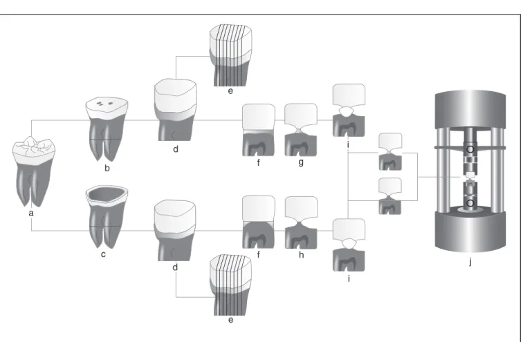
Sixteen sound human third molars (Figure 1a) that were refrigerated in a solution of 0.05% thy-mol (LabSynth Produtos para Laboratórios Ltda., Diadema, SP, Brazil) for no longer than two months after extraction were cleaned of gross debris and placed in distilled water (Permution Ltda., Curi-tiba, PR, Brazil) for twenty-four hours before begin-ning the experiment. The teeth used in this study were obtained under the protocol 106/2002, which was analyzed and approved by the Research Eth-ics Committee, School of Dentistry of Piracicaba, State University of Campinas.
Flat enamel bonding sites (Figure 1b) were prepared on the occlusal surfaces of eight teeth by wet grinding with a 600-grit silicon carbide paper (3M, Sumaré, SP, Brazil) in a polishing ma-chine (APL-4, Arotec, Cotia, SP, Brazil). Bonded enamel was prepared in a way that permitted the load testing to be applied parallel to its prismatic orientation. To prepare dentin bonding sites, the occlusal enamel of eight teeth was removed
us-Single Bond (3M ESPE, St. Paul, MN, USA) and Clearfil SE Bond (Kuraray Medical Inc., Okayama, Kurashiki, Japan) were applied to enamel and den-tin surfaces according to the manufacturers’ ins-tructions. Bonded surfaces received three layers of TPH Spectrum resin composite (Dentsply Caulk, Milford, DE, USA) to build up a cube-like crown of approximately 6.0 mm in height (Figure 1d). Each resin layer was light cured for 40 s with a XL 3000 light-curing unit (3M ESPE, St. Paul, MN, USA) and the bonded teeth were stored in water at 37°C.
After 24 h, the roots were removed and the crowns were vertically, serially sectioned into 0.7 mm thick slabs with a diamond saw (Buehler, Lake Bluff, IL, USA), under water lubrication (Fig-ure 1e). Five slabs were selected from each tooth (Figure 1f). The selected slabs were from the cen-ter of teeth and had directions of enamel prisms and dentin tubules that were perpendicular to the composite-tooth bonded interface. Slabs were pre-pared for microtensile testing and randomly as-signed to 8 experimental groups (n = 10). Bonded enamel specimens were obtained at the cusp ar-eas (Figure 1g), while bonded dentin specimens were prepared from the central area of dentin (Fig-ure 1h). Slabs were trimmed on both sides with a fine diamond bur (1040, KG Sorensen, Barueri, SP, Brazil) under water lubrication, reducing the bonded interface and making the specimen simi-lar in shape to an hourglass. The average cross-sectional bonded area at the “neck” was approxi-mately 0.5 mm2.
(Permu-24 25
24 25
tion Ltda., Curitiba, PR, Brazil) for 10 seconds and stored in 0.5 ml of artificial saliva at 37°C. After the end of the bleaching regimen (14th day) or stor-age in saliva, specimens were rinsed and placed in deionized water for 24 h at 37°C.
Afterwards, enamel and dentin tensile bond strengths were determined. Each specimen was fixed to the grips of a microtensile testing device (Cometa, Piracicaba, SP, Brazil) with cyanoacrylate glue (Zapit, DVA, Corona, CA, USA) and tested for tension in a universal testing machine (4411, Instron, Canton, MA, USA) at 0.5 mm/min until failure (Figure 1j). After testing, specimens were carefully removed from the device with a scalpel blade (Duflex, SS White, Rio de Janeiro, RJ, Brazil) and the cross-sectional area at the site of the frac-ture was measured to the nearest 0.01 mm with a digital caliper (727-6/150, Starret, SP, Brazil) to calculate ultimate tensile strength, expressed in MPa. Data were analyzed by three-way ANOVA and Tukey’s test at a 0.05 confidence level.
After testing, the enamel and dentin sides of the fractured specimens were mounted on alumi-num stubs (Procind Ltda., Piracicaba, SP, Brazil), gold-sputter coated (MED 010, Balzers Union, Balzers, Liechtenstein) and observed with a scan-ning electron microscope (Leo 435 VP, Cambridge, United Kingdom) for determination of the fracture mode.
RESULTS
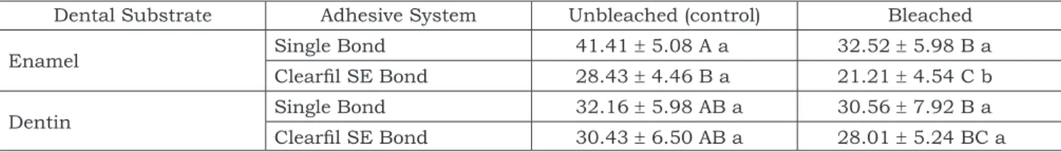
Table 1 displays the mean tensile bond strengths and standard deviations for the ex-perimental groups. Three-way ANOVA revealed significant difference among the tested groups (p = 0.0001) and for the interaction between the Adhesive System and Dental Substrate factors (p = 0.00005).
Tukey’s test showed that tensile bond strength to enamel using Single Bond always presented significantly higher mean values than that using
a
b
d
f
f
g
i
i
j h
e
e d
c
FIGURE 1 - Schematic representation of specimen preparation. From human third molars (a), enamel (b) and dentin
(c) bonding sites were prepared on occlusal surfaces. After building up a composite block (d), the teeth were
verti-cally sectioned (e) into 0.7 mm thick slabs (f), which were trimmed to an hourglass shape (g and h). Bonded
26 27
26 27
FIGURE 2a - SEM micrographs of the enamel side of fractured specimens that had been restored with Single Bond. Asterisks (*) indicate the etched enamel impregnated with adhesive resin.
FIGURE 2b - SEM micrographs of the enamel side of fractured specimens that had been restored with Single Bond. Cohesive failures were also observed on fractured enamel surfaces.
FIGURE 2c - SEM micrographs of the enamel side of fractured specimens that had been restored with Single Bond. Higher magnification shows porosities at the transversally fractured enamel prisms (arrows) of bleached specimens.
2b
*
*
2a 2c
bleached and unbleached groups (p > 0.05). SEM examination of fractured interfaces showed variations among groups. For Single
testing (Figure 2c). For Clearfil SE Bond applied to enamel not submitted to the bleaching treat-ment, fractures occurred predominantly between
TABLE 1 - Tensile Bond Strength (MPa) for both enamel and dentin.
Dental Substrate Adhesive System Unbleached (control) Bleached
Enamel Single Bond 41.41 ± 5.08 A a 32.52 ± 5.98 B a
Clearfil SE Bond 28.43 ± 4.46 B a 21.21 ± 4.54 C b
Dentin Single Bond 32.16 ± 5.98 AB a 30.56 ± 7.92 B a
Clearfil SE Bond 30.43 ± 6.50 AB a 28.01 ± 5.24 BC a
26 27
26 27
3b
FIGURE 3a - SEM micrographs of the enamel side of fractured specimens that had been restored with Clearfil SE
Bond. Unbleached specimens showed mixed failures between primed enamel and the adhesive layer (A) and
be-tween the adhesive layer and the resin composite (B).
FIGURE 3b - SEM micrographs of the enamel side of fractured specimens that had been restored with Clearfil SE Bond. Self-etched enamel can be seen in a fracture of the bleached group.
3a
A B
FIGURE 4a - SEM micrographs of the dentin side of fractured specimens restored with Single Bond. Cohesive fail-ures are observed in dentin.
FIGURE 4b - SEM micrographs of the dentin side of fractured specimens restored with Single Bond. Failures in the adhesive layer were prevalent.
4a 4b
the primed enamel and the adhesive layer (A), and between the adhesive layer and the resin composite (B) (Figure 3a). When the enamel-composite bond-ed interface (with Clearfil SE Bond) was subjectbond-ed to bleaching, adhesive fractures occurred and the enamel presented the etching pattern produced by the self-etching system application (Figure 3b).
SEM analysis of the dentin fractured sites showed similar results for bleached or unbleached specimens. Illustrative micrographs of specimen dentin sides restored with Single Bond
demon-strated cohesive failures in dentin (Figure 4a) or in the adhesive layer (Figure 4b). For the self-etching adhesive, the fracture mode comprised the primed dentin (D), the adhesive layer (AD) and the resin composite (RC) (Figure 5).
DISCUSSION
proper-28 29
28 29
ties4,6,21. Regarding the influence of bleaching on bonding, Crim3 (1992) reported increased micro-leakage at gingival margins, which compromised the marginal seal of class V composite resin res-torations.
The adverse effects of bleaching agents on resin-based materials, adhesive resins and com-posites have been described only for microfilled composite materials4. The use of oxidizing agents could cause chemical softening, erosion or degra-dation of resinous materials; however, no changes were observed at the fractured hybrid composite surfaces. Moreover, no damage on the adhesive resin structures of placed restorations that could affect bond strength has been reported.
Carbamide peroxide bleaching reduced the bond strength of the self-etching primer adhe-sive to enamel. Moreover, results showed that the bond strength of the totetch adhesive was al-ways higher than that of the self-etching primer on enamel surfaces. The bleaching treatment could promote an additional effect on the surface, leading to an unpredictable bonding to enamel with the self-etching adhesive7. Some studies have evalu-ated the bond strength of self-etching adhesive systems to enamel and the controversial results have shown that bonding to enamel should be improved15,22 or that it is similar to total-etch ad-hesives10,19. The bleaching regimen also reduced bond strengths for Single Bond, but no significant difference was detected. The interfacial failures were predominant in enamel-composite interfaces subjected to bleaching. Figure 3b shows that the
dental bleaching on dental hard tissues and res-ins. Clinically, the bleaching agents are applied to intact, mineralized external enamel surfaces, and not to a ground subsurface. However, in view of the high permeability of enamel to hydrogen perox-ide, it is likely that the entire thickness of enamel might be modified by such prolonged treatments, regardless of the surface used13. The mechanism of bonding to enamel with self-etching primers has been reported to be more superficial than that of total-etch adhesives and based on inter- and in-tra-crystallite hybridization of 0.6 to 0.7 µm into enamel7. Since deeper etching pattern and long resin tags in phosphoric acid-etched specimens can be obtained with Single Bond, little effect was produced on the bond strength of the total-etch adhesive.
Bleached specimens presented porosities on the surface of transversally fractured enamel prisms (Figure 2c), indicating the penetration and effects of oxidizing agents into the enamel internal structure. The bleaching agent possibly interacted with the dental tissues, since cohesive fractures in enamel were observed (Figure 2b). The effect of microstructural changes in the enamel mechani-cal properties have been investigated and the results suggest reduction of fracture toughness and tensile strength1,18, which could be related to enamel cohesive fracture of specimens. Rotstein et al.17 (1992) reported that bleaching treatments may alter the dentin chemical structure, due to a reduction in the Ca/P ratio after carbamide and hydrogen peroxide bleaching16. Moreover, dentin microhardness12, permeability5 and surface mor-phology23 can be changed following bleaching. Al-though whitening treatments can possibly alter dentin properties, the application of the bleaching gel at bonded dentin-composite interfaces did not affect the bond strength of both adhesives. Frac-ture patterns of dentin bonded specimens were in accordance with those found in previous stud-ies14,20.
Bleaching procedures are often indicated for patients with placed composite restorations. Den-tin-composite interfaces are not affected by bleach-ing, according to the microtensile test. However, special care must be taken at enamel-composite
5
AD
RC
FIGURE 5 - SEM micrograph of the dentin side of speci-mens restored with Clearfil SE Bond. Mixed failures
comprised the primed dentin (D), the adhesive layer
28 29
28 29
interfaces, because dentists are not always aware of which dental adhesive system has been used for restoration.
CONCLUSION
The results suggest that the bleaching treat-ment affected the tensile bond strength of the self-etching primer to enamel. For the total-etch one-bottle adhesive, there was no significant dif-ference in bond strength between bleached and
unbleached (controls) groups to both dental hard tissues.
ACKNOWLEDGMENTS
The authors are indebted to Prof. E. W. Kita-jima (ESALQ - USP) for the use of SEM. This study was supported by the Coordination for the Im-provement of Higher Education Personnel (CAPES) and the State of São Paulo Research Foundation (FAPESP) (01/2771-7).
REFERENCES
1. Cavalli V, Giannini M, Carvalho RM. Effect of carbamide peroxide bleaching agents on tensile strength of human enamel. Dent Mater 2004;20:733-9.
2. Cavalli V, Reis AF, Giannini M, Ambrosano GMB. The ef-fect of elapsed time following bleaching on enamel bond strength of resin composite. Oper Dent 2001;26:597-602. 3. Crim GA. Post-operative bleaching: effects on microleakage.
Am J Dent 1992;5:109-12.
4. Cullen DR, Nelson JA, Sandrik JL. Peroxide bleaches: effect on tensile strength of composite resins. J Prosthet Dent 1993;69:247-9.
5. Dezotti MSG, Silva e Souza Jr MH, Nishiyama CK. Evalu-ation of pH variEvalu-ation and cervical dentin permeability in teeth submitted to bleaching treatment. Braz Oral Res 2002;16:263-8.
6. Garcia-Godoy F, Garcia-Godoy A, Garcia-Godoy F. Effect of bleaching gels on the surface roughness, hardness, and mi-cromorphology of composites. Gen Dent 2002;50:247-50. 7. Hannig M, Bock H, Bott B, Hoth-Hannig W.
Inter-crystal-lite nanoretention of self-etching adhesives at enamel im-aged by transmission electron microscopy. Eur J Oral Sci 2002;110:464-70.
8. Haywood VB, Robinson FG. Vital tooth bleaching with Nightguard vital bleaching. Curr Opin Cosmet Dent 1997;4:45-52.
9. Hegedüs C, Bistey T, Flóra-Nagy E, Keszthelyi G, Jenei A. An atomic force microscopy study on the effect of bleaching agents on enamel surface. J Dent 1999;27:509-15. 10. Kanemura N, Sano H, Tagami J. Tensile bond strength
to and SEM evaluation of ground and intact enamel sur-faces. J Dent 1999;27:523-30.
11. Langsten RE, Dunn WJ, Hartup GR, Murchison DF. Higher-concentration carbamide peroxide effects on surface roughness of composites. J Esthet Restor Dent 2002,14:92-6. 12. Lewinstein I, Hirschfeld Z, Stabholz A, Rotstein I. Effect
of hydrogen and sodium perborate on the microhardness of human enamel and dentin. J Endod 1994;20:61-3.
13. Markovic M, Sieck BA, Takagi S, Chow LC, Majeti S. Diffusion of hydrogen peroxide through sound enamel
[abstract 1290]. J Dent Res 2000;79:305.
14. Montes MAJR, De Goes MF, Cunha MRB, Soares AB. A morphological and tensile bond strength evaluation of an unfilled adhesive with low-viscosity composites and a filled adhesive in one and two coats. J Dent 2001;29:435-41. 15. Perdigão J, Geraldeli S. Bonding characteristics of
self-etching adhesives to intact versus prepared enamel. J Esthet Restor Dent 2003;15:32-41.
16. Rotstein I, Danker E, Goldman A, Heling I, Stabholz A, Zalkind M. Histochemical analysis of dental hard tissues following bleaching. J Endod 1996;22:23-6.
17. Rotstein I, Lehr Z, Gedalia I. Effect of bleaching agents on inorganic components of human dentin and cementum. J Endod 1992;18:290-3.
18. Seghi RR, Denry I. Effects of external bleaching on indentation and abrasion characteristics of human enamel. J Oral Rehabil 1992;71:1340-4.
19. Shimada Y, Senawongse P, Harnirattisai C, Burrow MF, Nakaoki Y, Tagami J. Bond strength of two adhesive systems to primary and permanent enamel. Oper Dent 2002;27:403-9.
20. Tay FR, Carvalho R, Sano H, Pashley DH. Effect of smear layers on the bonding of a self-etching primer to dentin. J Adhes Dent 2000;2:99-116.
21. Yap AU, Wattanapayungkul P. Effects of in-office tooth whiteners on hardness of tooth-colored restoratives. Oper Dent 2002;27:137-41.
22. Yoshiyama M, Matsuo T, Ebisu S, Pashley DH. Re-gional bond strengths of self-etching/self-priming adhesive systems. J Dent 1998;26:609-16.
23. Zalkind M, Arwaz JR, Goldman A, Rotstein I. Surface morphology changes in human enamel, dentin and cemen-tum following bleaching: a scanning electron microscopy study. Endod Dent Traumatol 1996;12:82-8.