Received: September 7, 2011 Accepted: November 15, 2011
Conflict of Interests: The authors state that there are no financial and personal conflicts of interest that could have inappropriately influenced their work.
Copyright: © 2011 Cechin et al.; licensee EDIPUCRS. This is an Open Access article distributed under the terms of the Creative Commons Attribution-Noncommercial-No Derivative Works 3.0 Unported License.
Effect of endodontic irrigating solutions on the
adhesive bond strength to dentin
Efeito de soluções irrigadoras endodônticas na
resistência adesiva à dentina
Doglas Cecchin a
Ana Paula Farina a
João Vicente Baroni Barbizam b
Maria Paula Gandolfi Paranhos c
Bruno Carlini-Júnior a
a Department of Restorative Dentistry, School of Dentistry, University of Passo Fundo, Passo Fundo, RS, Brazil
b Department of Restorative Dentistry, School of Dentistry, University of Seatle, Seatle, Washington, USA
c Department of Restorative Dentistry, School of Dentistry, University of California, Los Angeles, California, USA
Correspondence:
Doglas Cecchin
Universidade de Passo Fundo, Campus I Faculdade de Odontologia
BR 285, Km 171 – Bairro São José Caixa Postal 611
Passo Fundo, RS – Brazil 99052-900
E-mail: [email protected]
Abstract
Purpose: To evaluate the bond strength of a total-etching adhesive system to dentin irrigated with 1% sodium hypochlorite (NaOCl) and ethylenediaminetetraacetic acid 17% (EDTA). Methods: Thirty human molars were sectioned 3mm below the oclusal surface and were randomly divided into 3 groups (n=10): G1, no treatment (control); G2, 1% NaOCl; G3, NaOCl followed by EDTA. The specimens received the total-etching adhesive system, restored with microhybrid composite resin, sectioned and trimmed to create 4 hourglass-shaped slabs of each tooth. The slabs were tested in microtensile bond strength in a universal testing machine (Emic DL 2000) at a crosshead speed of 0.5 mm/min until fracture. The results were analyzed statistically by ANOVA and Newman-Keuls test. G2 samples were not submitted to the test because they fractured just before testing, and its values were considered zero to the statistical analysis.
Results: The means and standard deviations (MPa) were: G1, 8.41 (±3.51); G2, 0.0 (±0.00); G3, 8.47 (±3.53).
Conclusion: It was concluded that the application of the NaOCl irrigating solution significantly decreased the bond strength values. The use of NaOCl followed by EDTA resulted in bond strength values not statistically different from control group.
Key words: Adhesives; dentin; root canal irrigants
Resumo
Objetivo: Avaliar a resistência de união de um sistema adesivo condicione-e-lave à dentina irrigada com hipoclorito de sódio 1% (NaOCl) e ácido etilenodiaminotetracético 17% (EDTA).
Metodologia: Trinta molares humanos foram seccionados 3mm abaixo da superficie oclusal e aleatoriamente divididos em 3 grupos (n=10): G1 (controle), nenhum tratamento; G2, NaOCl; G3, NaOCl seguido por EDTA. Os especimes foram hibridizados com um sistema adesivo condicione-e-lave, foram restaurados com resino composta microhibrida e seccionados de forma a originar 4 amplulhetas de cada dente. As fatias foram submetidas ao teste de resistência de união à microtraçnao em máquina universal de ensaios (EMIC DL 2000) à uma velocidade de 0,5 mm/min até o momento da fratura. Os resultados foram analisados estatisticamente por ANOVA seguido por Newman-Keuls. As amostras do G2 não foram submetidas aos testes de resistência de união pois fraturaram antes dos testes, e seus valores foram considerados zero para análise estatística.
Resultados: A média e desvios-padrão foram (MPa): G1 – 8,41 (±3,51); G2 – 0,0 (±0,00); e G3 – 8,47 (±3,53).
Conclusão: Pode-se concluir que a irrigação com NaOCl diminui significativamente os valores de resistência de união. O uso de NaOCl seguido por EDTA resultaram em valores de resistência de união sem diferença estatística com o grupo controle.
Introduction
The dentine substrate is a hydrated biologic complex,
with differential regional patterns that can be modiied
by physiological processes, age and disease (1). Bonding to dentin is a very complicated process, mainly due to its predominantly organic content, tubular structure, variety of
substrate mineralization degrees and to the external luids lux (2).
The actual adhesive systems contain substances that prepare the dentinal substrate for bonding. The acid etching
removes the mineral portion and exposes the collagen ibers
that must be kept moist. The forth generation adhesive systems bond to the demineralized and moist dentine tissue by means of acetone or alcohol-based primers that remove the water and maintain the collagen network, permitting resinous monomers penetration (3). This is a critical technique because of the subjectivity inherent to the ideal humidity and because
of the possibility of maintaining exposed collagen ibers
where the microleakage could be initiated (4). Therefore, one of the main reasons of the hybridization success is the
achievement of dentine ideal conditions (2).
In the teeth where endodontic treatment is indicated, the combined use of endodontic irrigating solutions that clean,
disinfect and shape the root canal system for posterior illing
are necessary and essential for the therapy success (5,6). Among the endodontic irrigating solutions, indicated as auxiliaries for endodontic therapy, sodium hypochlorite is the most used all over the world due to its anti-bacterial and organic tissue dissolving properties, which are directly proportional to its concentration (7).
EDTA is also a solution commonly used in Endondotics. It acts on the dentin mineral content and promotes smear layer removal (8). The use of sodium hypochlorite followed
by the inal irrigation with EDTA, for smear layer removal,
could enhance the contact between intra-canal irrigating
solutions and illing materials with dentine walls (9).
However, the use of irrigating solutions during endodontic treatment could modify the dentine structure (10) and interfere on dentin/resin bonding (11-13). Therefore, the purpose of this study was to evaluate the bond strength of a total-etching adhesive system to dentin irrigated with 1% sodium hypochlorite (NaOCl) and ethylenediaminetetraacetic acid 17% (EDTA). The null hypothesis that endodontic irrigants (NaOCl and EDTA) do not affect the bond strength of the self-etching adhesive system to dentin.
Material and Methods
Thirty sound freshly-extracted human third molars were used in this study. Teeth were stored 0.5% chloramine solution at 4ºC for 48 h for disinfection. Next, the teeth were cleaned with pumice/water slurry in Robinson brushes (Microdont; Socorro, SP, Brazil) and analyzed under x10 magnifying glass (Carl Zeiss; Jena, Germany) to exclude
those with fracture lines or issures. The teeth were stored
in distilled water at 4ºC.
Each tooth was vertically embedded in a PVC cylinder
(25 mm diameter and 20 mm high) using an auto-polymerizing
acrylic resin (Jet Clássico Ind.; São Paulo, Brazil). The oclusal third (3 mm) of the molar crowns was removed by means of a slow speed saw (400 rpm) with a diamond disk (Arotec; São Paulo, Brazil) in a cutting machine (Struers Miniton; Paraná, Brazil) under constant water-cooling. The
dentin was lattened with 600 and 1200-grit silicon carbide
abrasive papers.
The specimens were randomly distributed into the following groups (n=10): in group 1 (control), no irrigating
solution was applied; in group 2, 1% NaOCl (5 mL) was
applied to the dentin surface every 5 min for 1 h, simulating the time that NaOCl (based irrigants are usually left in the root canals during endodontic treatment under clinical conditions); in Group 3, 1% NaOCl was applied as described
for Group 2, followed by a 5-min inal rinse with 17% EDTA (5 mL), simulating the duration of the inal lush with this
chelating agent during endodontic treatment under clinical conditions. After dentin treatments, all specimens were washed with distilled water for 60 s.
Two coats of the adhesive system (Single Bond, 3M/ ESPE; St. Paul, MN, USA) were applied, with a break of
20s after each adhesive coat, and photo-polymerized (Radii, SDI; São Paulo, SP, Brazil) for 20s after the second coat. After hybridization, 3 increments (2 mm each) of composite resin (Z250, 3M/ESPE; St. Paul, MN, USA) were applied
over the dentine surface, using a spatula n. ½, completing 6mm high. Each increment was photo-polymerized for
20s. After bonding procedures specimens were stored in a sterilization oven in water for 24h, at 37ºC.
After storage in distilled water for 24 h, the specimens
were placed in a metallographic sectioning machine (Struers Miniton; Copenhagen, Denmark) and a water-cooled double-faced diamond disk was used to cut sequential longitudinal 1.0-mm-thick sections in a mesiodistal direction. Care was taken not to separate the slices. The specimens were then removed from the acrylic resin base through a transversal section, to obtain resin/dentin slabs measuring approximately 10-mm high, 5-mm wide and 1-mm thick. The slabs were
trimmed on both sides of resin-dentin interface with a #1093
FF bur (KG Sorensen; Barueri, SP, Brazil) at a high-speed handpiece (Kavo; Joinvile, SC, Brazil) to obtain a 1-mm thick central area and produce standard hourglass-shaped specimens.
The specimens were individually ixed in a metallic device with a cyanocrylate adhesive (Loctite Super Bonder;
São Paulo, SP, Brazil) so that the resin/dentin interface remained without any contact for the microtensile test. The metallic device coupled to a universal testing machine (Emic
DL 2000; São José of Pinhais, PR, Brazil) and the specimens
calculated in MPa using the following equation: Rt=F/A,
where Rt is the μTBS value, F is the force applied and A is
the bond area between the dentin and restorative system. The data obtained were subjected to ANOVA and Newman
Keuls tests (α=0.01).
For scanning electronic microscopy (SEM) analysis,
specimens were immersed in 2.5% glutaraldehyde for 12 hours at 4ºC for ixation, washed with 20mL buffer solution of sodium cacodylate 0.2M with pH 7.4 for 1 hour, and
washed in distilled water three times for 1 minute. For dehydration, the specimens were sequentially immersed in
ethyl alcohol (25% for 20 minutes, 50% for 20 minutes, 75% for 20 minutes, and 95% for 20 minutes), and dried at
37ºC for 48 hours with silica gel drying pearls. The prepared specimens were gold-sputtered at 10mA for 1 minute and observed by SEM.
Results
Table 1 shows the mean values and standard deviations (MPa) of groups tested in this study. There was no statistical difference between G1, where no treatment was performed and G3, where dentine surface was treated with NaOCl
followed by EDTA. However, between G2, where dentinal
surface was treated only with NaOCl, and the other groups,
there was statistical difference at a signiicance level of 1%. An important fact to report is that specimens of G2
were useless just before the test, since they fractured at a minimum touch, not allowing the microtensile testing. It
indicates that NaOCl had a strong negative inluence on the adhesive/dentin bond. The G2´s mean value was considered
as zero in the statistical analysis.
Table 1. Microtensile bond strength mean values (MPa) and the
respective standard deviations.
Groups Mean Standard deviation P
G1=Coontrol 8.41a ±3.51
<0.01
G2=NaOCl 0.00b ±0.00
G3=NaOCl+EDTA 8.47a ±3.53
* Mean followed by the same letters do not show statistical difference
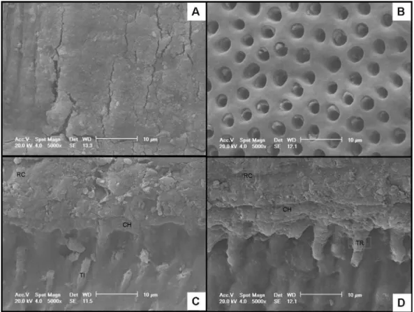
Fig. 1. SEM photomicrographs of dentin after: (A) NaOCl treatment for 1 h (5000x); (B) NaOCl treatment
for 1 h followed by EDTA treatment for 5 min (5000x); (C) NaOCl treatment for 1 h (RC – composite resin,
CH – hybrid layer, TI – incomplete resin tag (5000x); and (D) NaOCl treatment for 1 h followed by EDTA
treatment for 5 min (CR – composite resin, CH – hybrid layer, TR – resin tag (5000x).
Figure 1 shows by SEM a modiied smear layer (1A) that could dificult phosphoric acid etching. It can be observed
opened dentine tubules (Fig. 1B) with few pulp tissue
remnants and no smear layer. Less resin tags formation are observed in Figure 1C because of the dificulty of the
Discussion
The results showed that G2, treated only with 1%
NaOCl, had its bond strength strongly compromised when compared to the control group G1, and G3 that was treated with EDTA after NaOCl, rejecting the hypothesis under study. This could be caused by organic matrix damage, mainly collagen, after NaOCl application and could also
be attributed to the presence of a NaOCl modiied smear
layer after treatment (Figure 1A). This smear layer could
dificult phosphoric acid action and monomers iniltration
throughout the demineralized dentin because the sodium
chlorite already iniltrated in the smear layer could react and partially neutralize the phosphoric acid action (12).
Moreover, NaOCl releases oxygen, and this oxygen could cause a huge inhibition of the adhesive system penetration and polymerization, consequently decreasing the bond strength values (14-16).
The use of EDTA after NaOCl (G3) in dentinal surface resulted in an increase in bond strength, when compared to
the groups that received NaOCl treatment only (G2) and these results are in accordance to the study of Lay et al.
(15). These authors suggest that the EDTA increases bond strength due to its anti-oxidant power via redox reaction, allowing for free radicals polymerization without premature chains or failures. Moreover, it has the ability to remove the smear layer (Fig. 1B) and the residual chlorine ions, what
could beneit phosphoric acid etching and enhance resinous
monomer penetration through dentinal tubules (17). Our results are in agreement with the study of Morris et
al. (18) and Vongphan et al. (19) who evaluated the chemical
effect of irrigating solutions on microtensile bond strength of adhesive system after acid etching (Single Bond) to dentine
walls. They noticed that the use of NaOCl signiicantly
reduced microtensile bond strength. However, the use of
10% sodium ascorbate signiicantly increased dentin bond
strength after NaOCl treatment. In this case, the 10% sodium ascorbate had a similar effect of EDTA in our study.
Nikaido et al. (12) reported that the NaOCl used during endodontic treatment produces adverse effects on etch-and-rinse adhesive systems. Although the use of EDTA had recovered the adhesive resistance to the reference levels,
the use of NaOCl alone had signiicantly reduced bond
strength.
Until recently, endodontically treated teeth were usually
treated with a crown, core or dowel (20). However, when they have a suficient amount of complete coronal structure they
can be restored with an adhesive system and resin composite
(21,22). It is known that a better bond of restorative materials
to dentine walls increases the possibility of marginal sealing, mechanical resistance to mastication stress and durability
of restorations (23). The clinical signiicance of the present study is that the use of EDTA as inal irrigating solution
during endodontic treatment with NaOCl is fundamental for tooth restoration without compromising the bond strength when using etch-and-rinse adhesive systems.
However, several aspects of the bond strength of a resin composite in dentin need further research, including the effect of various chemicals used during root canal treatment
on canal walls. For example, Moreira et al. (24)observed that
NaOCl, whether or not in association with 17% EDTA, caused alterations to the dentin collagen, whereas chlorhexidine
did not. Furthermore, Cecchin et al. (25) showed that the
chlorhexidine pretreatment dentin were capable of preserving
the bond strength to root dentin for 12 months. Therefore,
different protocols of hybridization of root and coronary dentin must be evaluated to increase the bond strength and long-term adhesion of the resin composite.
In conclusion, the application of the NaOCl irrigating
solution signiicantly decreased the bond strength values. The
use of NaOCl followed by EDTA resulted in bond strength values not statistically different from control group.
1. Pashley DH, Sano H, Ciucchi B, Yoshiyama M, Carvalho R. Adhesion testing of dentin bonding agents: a review. Dent Mater 1995;11:117-25.
2. Meerbeek BV, Munck J, Yoshida SI, Vargas M, Vijay P, Landuyt KV, Lambrechts P, Vanherle G. Buonocore Memorial Lecture. Adhesion to Enamel and Dentin: Current Status and Future Challenges. Oper Dent 2003;28:215-35.
3. Perdigão J, Ramos JC, Lambrechts P. In vitro interfacial relationship between human dentin and one-bottle dental adhesives. Dent Mater 1997;13:218-27.
4. Frankenberger R, Krämer N, Oberschachtsiek H, Petschelt A. Dentin bond strength and marginal adaptation after NaOCl pre-treatment. Oper Dent 2000;25:40-5.
5. Yamazaki AK, Moura-Netto C, Salgado RJ, Kleine BM, Prokopowitsch I. Ex vivo analysis of root canal cleaning using Endo-PTC associated to NaOCl and different irrigant solutions. Braz Oral Res 2010;24:15-20.
6. Baumgartner JC, Hohal S, Marshall JG. Comparison of the antimicrobial efficacy of 1.3% NaOCl/BioPure MTAD to 5.25% NaOCl/15% EDTA for root canal irrigation. J Endod 2007;33:48-51.
7. Gomes BPFA, Ferraz CCR, Vianna ME, Berber VB, Teixeira FB, Souza-Filho FJ. In vitro antimicrobial acrivity of several concentrations of sodium hypoclorite and chlorhexidine gluconate in the elimination of Enterococcus faecalis. Int Endod J 2001;34:424-8. 8. Wachlarowicz AJ, Joyce AP, Roberts S, Pashley DH. Effect of endodontic irrigants on the
shear bond strength of epiphany sealer to dentin. J Endod 2007;33:152-5.
9. Çalt S, Serper A. Smear layer removal by EDTA. J Endod 2000;26:459-61.
10. Goldman LM, Goldman M, Kronman JH, Lin PS. The efficacy of several irrigating solutions for endodontics: a scanning electron microscopic study. Oral Surg 1981;52:197-204. 11. Eldeniz AU, Erdemir A, Belli S. Shear bond strength of three resin based sealers to dentin
with and without the smear layer. J Endod 2005;31:293-6.
12. Nikaido T, Takano Y, Sasafuchi Y, Burrow MF, Tagami J. Bond strength to endodontically-treated teeth. Am J Dent 1999;12:177-80.
13. Farina AP, Cecchin D, Barbizam JV, Carlini-Júnior B. Influence of endodontic irrigants on bond strength of a self-etching adhesive. Aust Endod J 2011;37:26-30
14. Ruegeberg FA, Margeson DH. The effect of oxygen inhibition on an unfilled/filled system. J Dent Res 1990;69:1652-8.
15. Lai SCN, Mak YF, Cheung GSP, Osorio R, Toledano M, Carvalho RM, et al. Reversal of compromised bonding to oxidized etched dentine. J Dent Res 2001;80:1919-24. 16. Nascimento JS, Carrilho MR, Goes MF, Zaia AA, Gomes BPFA, Souza-Filho FJ, Ferraz
CCR. Effect of Chemical Irrigants on the Bond Strength of a Self-Etching Adhesive to Pulp Chamber Dentin. J Endod 2006;32:1088-90.
17. Miyasak K, Nakabayashi N. Combination of EDTA conditioner and Phenyl-HEMA self-etching primer for bonding to dentin. Dent Mater 1999;15:153-7.
18. Morris MD, Lee KW, Agee KA, Bouillaquet S, Pashley DH. Effects of resin cement to endodontic surfaces. J Endod 2001;27:753-7.
19. Vongphan N, Senawongse P, Somsiri W, Harnirattisai C. Effects of sodium ascorbato on microtensile bond strength of total-etching adhesive system to NaOCl treated dentine. J Dent 2005;33:689-95.
20. Boone KJ, Murchison DF, Schindler WG, Walker WA. Post retention: the effect of post-space preparation, cementation time, and different sealers. J Endod 2001;27:768-71. 21. Hernandez R, Bader S, Boston D, Trope M. Resistance to fracture of endodontically
treated premolars restored with new generation dentine bonding systems. Int Endod J 1994;27:281-4.
22. Saupe WA, Gluskin AH, Radke RA Jr. A comparative study of fracture resistance between morphologic dowel and cores and a resin-reinforced dowel system in the intraradicular restoration of structurally compromised. Quintessence Int 1996;27:483-91.
23. Fortin D, Swift EJ, Denehy GR, Reinhardt JW. Bond strength and microleakage of current dentin adhesives. Dent Mater 1994;10:253–8.
24. Moreira DM, Almeida JF, Ferraz CC, Gomes BP, Line SR, Zaia AA. Structural analysis of bovine root dentin after use of different endodontics auxiliary chemical substances. J Endod 2009;35:1023-7.