Submitted 24 April 2015 Accepted 25 June 2015 Published6 August 2015
Corresponding author Akio Inui,
Academic editor Yoshinori Marunaka
Additional Information and Declarations can be found on page 12
DOI10.7717/peerj.1105
Copyright 2015 Amitani et al.
Distributed under
Creative Commons CC-BY 4.0
OPEN ACCESS
Allantoin ameliorates chemically-induced
pancreatic
β
-cell damage through
activation of the imidazoline I3 receptors
Marie Amitani1, Kai-Chun Cheng1, Akihiro Asakawa1, Haruka Amitani1, Timothy Sean Kairupan1, Nanami Sameshima1, Toshiaki Shimizu2, Teruto Hashiguchi2and Akio Inui1
1Department of Psychosomatic Internal Medicine, Kagoshima University Graduate School of
Medical and Dental Sciences, Kagoshima, Japan
2Department of Laboratory and Vascular Medicine, Kagoshima University Graduate School of
Medical and Dental Sciences, Kagoshima, Japan
ABSTRACT
Objective.Allantoin is the primary active compound in yams (Dioscorea spp.). Recently, allantoin has been demonstrated to activate imidazoline 3 (I3) receptors located in pancreatic tissues. Thus, the present study aimed to investigate the role of allantoin in the effect to improve damage induced in pancreaticβ-cells by streptozotocin (STZ) via the I3 receptors.
Research Design and Methods.The effect of allantoin on STZ-induced apoptosis in pancreaticβ-cells was examined using the ApoTox-Glo triplex assay, live/dead cell double staining assay, flow cytometric analysis, and Western blottings. The potential mechanism was investigated using KU14R: an I3 receptor antagonist, and U73122:
a phospholipase C (PLC) inhibitor. The effects of allantoin on serum glucose and
insulin secretion were measured in STZ-treated rats.
Results.Allantoin attenuated apoptosis and cytotoxicity and increased the viability
of STZ-inducedβ-cells in a dose-dependent manner; this effect was suppressed by
KU14R and U73112. Allantoin decreased the level of caspase-3 and increased the level of phosphorylated B-cell lymphoma 2 (Bcl-2) expression detected by Western
blotting. The improvement inβ-cells viability was confirmed using flow cytometry
analysis. Daily injection of allantoin for 8 days in STZ-treated rats significantly lowered plasma glucose and increased plasma insulin levels. This action was inhibited by treatment with KU14R.
Conclusion.Allantoin ameliorates the damage ofβ-cells induced by STZ. The blockade by pharmacological inhibitors indicated that allantoin can activate the I3 receptors through a PLC-related pathway to decrease this damage. Therefore, allantoin and related analogs may be effective in the therapy forβ-cell damage.
Subjects Cell Biology, Diabetes and Endocrinology, Nutrition, Oncology, Pharmacology
Keywords Allantoin, Imidazoline 3 receptor, Pancreaticβ-cell, PLC-related pathway, Streptozotocin
INTRODUCTION
because they contain ureides such as allantoin (Lee et al., 2010). Dioscoreaceae plants have also been shown to improve metabolic and diabetic disorders (Chang et al., 2005;Sato et al., 2009;Wang et al., 2012).
Several pathogenic processes are involved in the development of diabetes, including the destruction of pancreaticβ-cells that results in insulin resistance (Cnop et al.,
2005). Autoimmunity is one of the main causes of diabetes type 1 via damage of the
insulin-producingβ-cells in the pancreas (American Diabetes, 2010). In addition to insulin resistance, increased apoptosis and a significant reduction in the number ofβ-cells have been implicated in type 2 diabetes (Butler et al., 2003). Thus, prevention of pancreatic
damage and the development of therapeutic strategies to protectβ-cells have been
introduced as a major target for the management of diabetes (Mandrup-Poulsen, 2001).
The imidazoline receptor is an orphan receptor with three subtypes. The imidazoline 1 (I1) receptors act to lower blood pressure (Ernsberger et al., 1995), whereas the imidazoline 2 (I2) receptors serve as an allosteric binding site for monoamine oxidase and are known to be involved in pain modulation, neuroprotection and increased glucose uptake in muscle cells (Li & Zhang, 2011;Lui et al., 2010). The imidazoline 3 (I3) receptors play an important role in regulating insulin secretion fromβ-cells in the pancreas (Head & Mayorov, 2006).
Guanidine derivates can bind to the imidazoline receptors (Dardonville & Rozas, 2004). Allantoin, a guanidine derivative, has been shown to activate the I1 receptors to attenuate
hyperlipidemia, improve hepatic steatosis and act as an antihypertensive agent (Chen et
al., 2014a;Yang et al., 2012). Additionally, allantoin also increases glucose uptake in muscle cells via the I2 receptors (Chen et al., 2012;Lin et al., 2012). A recent study demonstrated that allantoin was able to bind to the I3 receptors, resulting in the lowering of blood glucose due to increased plasma insulin levels (Tsai et al., 2014). Moreover, insulinotropic agents such as glucagon-like peptide-1, an incretin derived from the transcription product of the proglucagon gene, can also protectβ-cells from apoptosis (Cernea, 2011;Liu et al., 2012). Thus, we speculated that allantoin may play a role in pancreaticβ-cell protection via the I3 receptor. The present study aimed to identify the role of allantoin in improving damage in pancreaticβ-cells induced by a low dose of streptozotocin (STZ).
MATERIAL AND METHODS
Animals
Male Wistar rats weighing 320–340 g obtained from Japan SLC, Inc (Shizuoka, Japan), were maintained in an environment under a 12 h light/12 h dark cycle with a controlled room temperature at the animal center of Kagoshima University (Kagoshima, Japan). Food and tap water were providedad libitumwith free access. All procedures in this study were approved by the Ethics Committee for Animal Care and Use of Kagoshima University (IRB approval number MD14059).
Islet isolation and primary culture
digested with 1 mg/mL collagenase (Roche, Basel, Switzerland) for 10 min. The digested samples were washed two times with RPMI 1640 medium (Sigma, St. Louis, Missouri, USA) containing 10% fetal bovine serum FBS (Thermo, Waltham, Massachusetts, USA) to inactivate the collagenase. The isolated pancreatic islets were cultured in RPMI 1640 supplemented with 1% penicillin and streptomycin (Life Technology, Carlsbad, California, USA), 1% amphotericin B (Sigma, St. Louis, Missouri, USA) and 10% FBS. The primary
culture was incubated (37◦C with 5% CO2) for 48 h. After the incubation period, the
primary culture was divided into 6-well plates for Live/Dead double staining assay, 96-well plates for the ApoTox-Glo Triplex assay, and 12-well plates for annexin and flow cytometry analysis.
Treatment of cells with reagents
The primary cultured cells were devided into 6-well plates. The medium was removed,
and the cells were washed once with phosphate-buffered saline (PBS). RPMI 1640 medium
containing 25 mM glucose was added to each well with 5 mM STZ (Sigma-Aldrich, St. Louis, Missouri, USA) and incubated for 6 h to induce cell apoptosis. To know the role of allantoin in the protection of pancreaticβ-cells against STZ, allantoin (Sigma-Aldrich, St. Louis, Missouri, USA) pretreatment at various doses was provided before 30 min prior to the addition of 5 mM STZ and incubated for 6 h. To identify the signaling pathway
of allantoin inβ-cells, 1µM KU14R (Santa Cruz Biotechnology, Santa Cruz, California,
USA): an I3 binding site antagonist, or 1µM U73122 (TOCRIS, Bristol, UK): the
phospho-lipase C (PLC) inhibitor were provided before 30 min prior to the addition of allantoin as previously described before (Yang et al., 2015). All the medium was removed, and the cells were washed three times with PBS prior to processing for the evaluation of morphology.
Live/dead double staining assay
Using the Live/dead assay kit (Life Technology, Carlsbad, California, USA), we stained
β-cells to distinguish the living cells from dead cells according to the manufacturer’s
in-struction. We added 100µl of Live/Dead solution to the samples and incubated them for 15
min at room temperature. Then, the staining solution was removed and the samples were viewed under a fluorescence microscope (LSM700)(Zeiss, Jena, Germany). Living cells were detected at green fluorescence, whereas dead cells were detected at red fluorescence.
ApoTox-Glo triplex assay
Theβ-cells were seeded into 96-well plates at a total density of 1×10⁴cells per well. Each
well contained 200µl RPMI 1640 medium and the test compound where appropriate.
Annexin V/PI staining and flow cytometry analysis
The primary culturedβ-cells were divided into 12-well plates and categorized into four
groups. Each group was treated with different reagents as follows: (1) 5 mM STZ; (2) 5 mM
STZ and 100µM allantoin; (3) 5 mM STZ, 1µM KU14R and 100µM allantoin; and (4)
control. The cells were incubated with the reagents for 48 h. Then, the cells were collected and the apoptotic cells in each group were quantified using Annexin V-PI staining (Life Technology, Carlsbad, California, USA) and analyzed using flow cytometer based on the previously described method (Luo et al., 2014).
Western blotting analysis
Western blotting analysis was performed to determine caspase-3 and Bcl-2 expression. The
β-cells were pre-cultured with 5 mM STZ for 6 h prior to the addition of 100µM allantoin
with or without 1µM KU14R or vehicle for 30 min. Theβ-cells were washed with ice-cold
PBS and incubated for 15 min to allow lysis to occur. The protein concentration was measured by BCA protein assay (Thermo Fisher Scientific Inc., Waltham, Massachusetts, USA). The protein samples were filtered and separated by SDS-PAGE (Polyacrylamide Gel Electrophoresis) (10% acrylamide gel) using the Bio-Rad Trans-Blot system and were transferred to polyvinylidene difluoride membranes. The membrane was blocked
with 5% non-fat milk in Tris-buffered saline containing 0.1% Tween 20 (TBS-T). The
membran was incubated for 2 h and washed with TBS-T and hybridized overnight with primary antibodies, caspase-3 (Merk Millpore, USA) and Bcl-2 (Cell signaling Technology, Danvers, Massachusetts, USA), diluted with a suitable concentration of TBS. Incubation with secondary antibodies and the detection of the antigen-antibody complex was performed using an ECL kit (Thermo Fisher Scientific Inc., Waltham, Massachusetts, USA). The bands densities were quantified using a laser densitometer.
Glucose and insulin levels in STZ-treated rats
The induction of pancreatic cell damage was accomplished by injecting 45 mg/kg STZ dis-solved in 10 mM Na-citrated buffer intraperitoneally. STZ-treated rats with blood glucose above 200 mg/dl at 7 days post-injection were included in the group. Total of 24 rats were divided into three groups as follows: Control (STZ) (n=8), STZ+allantoin (n=8), STZ
+KU14R+allantoin (n=8). The third group was treated with an intravenous injection
of 8 mg/kg/day KU14R; the first and second groups were treated with the same volume of vehicle injected intravenously. After 30 min of KU14R injection, the second and third groups received 10 mg/kg/day of allantoin intravenously. The first group was injected the same volume of vehicle intravenously. The experiments were performed for 8 days. The blood samples were obtained from tail vein everyday. The plasma glucose levels were measured everyday, and the plasma insulin levels were measured on day 0, 4, 6, 8.
Statistical analysis
Statistical analyses were performed using SPSS software (SPSS, Inc., Chicago, Illinois, USA). An analysis of variance (ANOVA) with Tukey’s test to determine significant
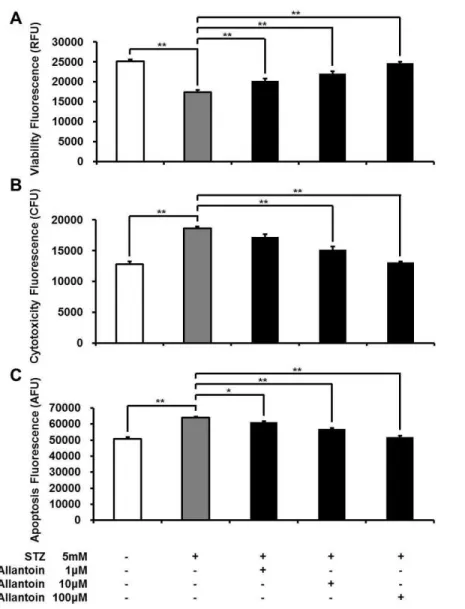
Figure 1 ApoTox-Glo triplex assay showing the viability (A), cytotoxicity (B), and apoptosis (C) of
β-cells treated with 5 mM streptozotocin (STZ), 5 mM STZ+1µM allantoin, 5 mM STZ+10µM allantoin, and 5 mM STZ+100µM allantoin (n=6 for each group).Data are presented as the mean
±SE.∗P<0.05,∗∗P<0.01.
mean±standard error (S.E.) based on the number (n) of samples in each group. Statistical
significance was set atp<0.05.
RESULTS
Allantoin decreased streptozocin-induced cytotoxicity and apoptosis inβ-cells
Viableβ-cells were significantly reduced in the STZ treated group, while cell toxicity and apoptosis were significantly increased compared to the control. In contrast, treatment with allantoin at various concentrations (1µM, 10µM, and 100µM) significantly increased cell
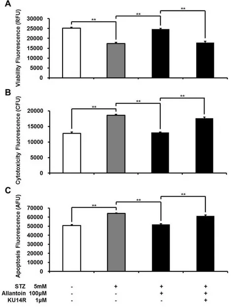
Figure 2 ApoTox-Glo Triplex assay showing the viability (A), cytotoxicity (B), and apoptosis (C) of
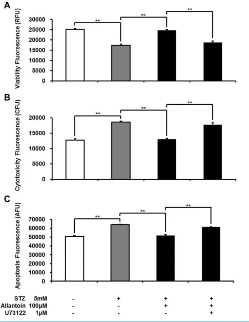
β-cells in rats treated with 5 mM streptozotocin (STZ), 5 mM STZ+100µM allantoin, 5 mM STZ
+1µM KU14R+100µM allantoin (n=6 for each group).Data are presented as the mean±SE.
∗∗P
<0.01.
Allantoin-induced increase inβ-cell viability was blocked by an I3 antagonist
I3 receptors located on pancreaticβ-cells are known to stimulate insulin secretion (Chen et al., 2014b;Dardonville & Rozas, 2004). Thus, the effect of allantoin on imidazoline I3 receptors was investigated using KU14R, an I3 specific antagonist, in the Apo Tox triplex assay. The effect of allantoin was inhibited by KU14R (Fig. 2).
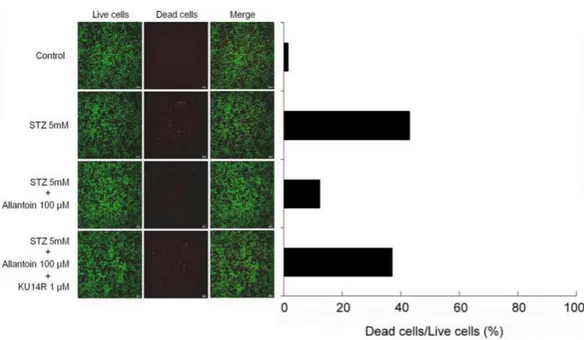
Allantoin increased the viability of STZ-treatedβ-cells
Exposure of theβ-cells to 5 mM STZ induced apoptosis based on the increase in red
Figure 3 Cell viability image ofβ-cell after treatment with 5 mM streptozotocin (STZ), 5 mM STZ
+100µM allantoin, 5 mM STZ+1µM KU14R+100µM allantoin.Allantoin greatly improved the viability of STZ-inducedβ-cells apoptosis.
addition of allantoin, shown by the marked decrease in red fluorescence emitted by EthD-1, which can bind to the DNA of the dead cells. We found that pretreatment with
1µM KU14R for 30 min resulted in the increased EthD-1 binding, thereby reducing the
action of allantoin (Fig. 3).
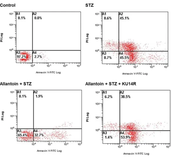
Allantoin decreased theβ-cell apoptosis percentage detected by flow cytometry
Viable cells are located in the lower left quadrant in the flow cytometric analysis. Treatment with 5 mM STZ for 6 h increased the percentage of apoptotic cells to 45.5%, indicated by the increased number of cells in the lower right quadrant. Allantoin reversed this effect and decreased the percentage of apoptotic cells to 32.7%, as shown by movement of the cell population from the lower right quadrant to the lower left quadrant. Co-treatment with KU14R blocked the action of allantoin and induce apoptosis to 53.9% (Fig. 4).
Allantoin-induced cell protective effect involved phospholipase C
To test whether the protective effect of allantoin involves the PLC pathway, we applied U73122: a PLC inhibitor. U73122 attenuated the protective effect of allantoin inβ-cells (Fig. 5).
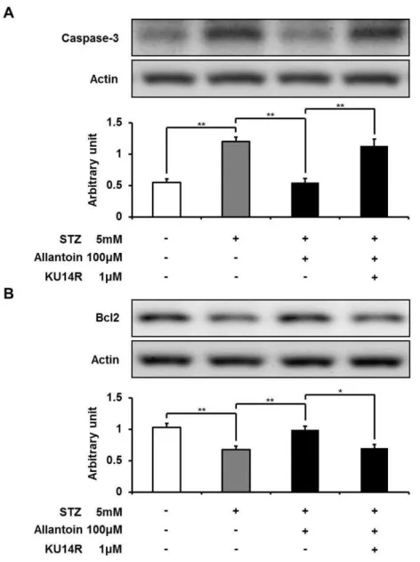
Allantoin decreased caspase-3 and increased Bcl-2 expression
Figure 4 Flow cytometry of apoptotic cells.Samples were incubated with FITC-labeled Annexin-V and propidium iodide. Number at the corner represent the percentage of cells found in each quadrant.
while the Bcl-2 level was significantly decreased. Allantoin significantly suppressed the expression of caspase-3 and significantly increased the expression of Bcl-2. In contrast, KU14R significantly inhibited these actions of allantoin (Fig. 6).
Plasma glucose levels in rats treated with STZ, allantoin, and KU14R
Plasma glucose levels were increased following intraperitoneal injection of STZ. Treatment with allantoin significantly lowered the blood glucose levels in STZ-treated rats. This effect
of allantoin was countered by combined treatment with KU14R (Fig. 7).
Plasma insulin levels in rats treated with STZ, allantoin, and KU14R
Figure 5 ApoTox-Glo Triplex assay showing the viability (A), cytotoxicity (B), and apoptosis (C) of
β-cells in rats treated with 5 mM streptozotocin (STZ), 5 mM STZ+100µM allantoin, 5 mM STZ
+1µM U73122+100µM allantoin (n=6 for each group).Data are presented as the mean±SE.
∗∗P
<0.01.
DISCUSSION
Allantoin is known to bind to the imidazoline receptors (Chung, Lee & Cheng, 2013;Tsai et al., 2014). In the present study, we found that allantoin could activate the I3 receptors to
protectβ-cells from the damage induced by STZ.
Figure 6 Western blotting analysis of the expression levels of caspase-3 and Bcl-2.The expression level of caspase-3 was reduced by allantoin (A), while Bcl2 expression was increased (B) (n=6 for each group). Data are presented as the mean±SE.∗P<0.05,∗∗P<0.01.
medium containing 25 mM glucose for 6 h. Moreover, high glucose is known to increase pancreatic cell vulnerability to toxic damage by increasing the expression of potential autoantigens on the cell membrane surface (Mellado-Gil & Aguilar-Diosdado, 2004). Therefore, we established a model that induced significant changes inβ-cells, including the induction of an apoptotic response.
In the present study, we found that allantoin attenuated the damage induced by STZ
in a dose-dependent manner, resulting in the reduction of STZ-inducedβ-cell apoptosis.
Figure 7 Effects of allantoin and KU14R on blood glucose levels in streptozotocin (STZ) -treated rats.STZ-treated rats were daily treated with 10 mg/kg allantoin and 8 mg/kg KU14R. Plasma glucose levels were measured daily for 8 days (n=8 for each group). Values are presented as the mean±SE.
∗P<0.05 and∗∗P<0.01 for the di
fference between STZ and STZ+Allantoin+KU14R. #<0.05 for the difference between STZ+Allatoin and STZ+Allantoin+KU14R.
Figure 8 Effects of allantoin and KU14R on plasma insulin levels in streptozotocin (STZ) -treated rats.STZ-treated rats were daily treated with 10 mg/kg allantoin and 8 mg/kg KU14R. Plasma insulin levels were measured on day 0, 4, 6, 8 (n=8 for each group). Values are presented as the mean±SE.
∗∗P
<0.01 for the difference between STZ and STZ+Allantoin or STZ+Allantoin+KU14R. #<0.05 and ##<0.01 for the difference between STZ+Allatoin and STZ+Allantoin+KU14R.
2006), while Bcl-2 is considered to act as an anti-apoptotic protein that promotes cell survival (Vaux, Cory & Adams, 1988). Thus, allantoin can increase the survival rate of
β-cells through improvement of apoptosis.
Imidazoline compounds have been suggested to induce insulin secretion from
(Tsai et al., 2014). In the presence of KU14R, I3 receptor antagonist, the protective effect induced by allantoin inβ-cells was partially blocked. Flow cytometric analysis also supported this findings. As shown inFig. 4, cell viabilities were improved after allantoin treatment, and this effect was also suppressed by the blockade of I3 receptors using KU14R. As previously described, rats injected with a low dose of STZ exhibited higher blood glucose and lower plasma insulin levels (Tsai et al., 2014). Allantoin improved the
damaged function ofβ-cells in this animal model, which resulted in an increase of plasma
insulin levels and a reduction of plasma glucose levels. Thisin vivoaction of allantoin was also inhibited by KU14R to block the I3 receptors. Thus, the action of allantoin via the activation of I3 receptors was shown bothin vivoandin vitro. Similar result was also observed in the action of canavanine (Yang et al., 2015).
Moreover, we found that the protective effect of allantoin was linked to the phospho-lipase C (PLC) pathway. In the presence of U73122, a well-known PLC inhibitor, the protective effect of allantoin was markedly reduced (Fig. 5). In theory, upon activation, PLC cleaves phosphatidylinositol 4,5-biphosphate into diacylglycerol and inositol 1,4,5-triphosphate, which may potentiate insulin secretion (Kahn et al., 2005). Whether this action is related to the protection ofβ-cells shall be investigated in future studies.
Additionally, several yam species (Dioscorea spp.) are also known to contain saponin and the aglycone portion of saponin called sapogenin, that are also proven to be beneficial in STZ induced diabetic rats (Omoruyi, 2008;Pessoa et al., 2015). Taken together, allantoin or yam (Dioscorea spp.) seems beneficial to treat and/or prevent the diabetes in the future.
Nevertheless, for the first time, we characterized the improvement of STZ-induced
β-cell damage by allantoin via the I3 receptors bothin vivoandin vitro.
CONCLUSION
Allantoin has the ability to increaseβ-cells viability and ameliorateβ-cell damage through activation of the I3 receptors. Thus, allantoin and related analogs supplied as nutrients may be useful for the improvement of early stage ofβ-cell damage.
ACKNOWLEDGEMENTS
We thank all the staffmembers of the Institute of Laboratory Animal Sciences, Kagoshima
University (Frontier Science Research Center) who kept the animals in good condition.
ADDITIONAL INFORMATION AND DECLARATIONS
Funding
The authors declare there was no funding for this work.
Competing Interests
The authors declare there are no competing interests.
Author Contributions
• Marie Amitani conceived and designed the experiments, performed the experiments,
• Kai-Chun Cheng conceived and designed the experiments, performed the experiments,
analyzed the data, contributed reagents/materials/analysis tools, prepared figures and/or tables.
• Akihiro Asakawa conceived and designed the experiments, wrote the paper, reviewed
drafts of the paper.
• Haruka Amitani performed the experiments, analyzed the data.
• Timothy Sean Kairupan wrote the paper, prepared figures and/or tables.
• Nanami Sameshima performed the experiments, analyzed the data, prepared figures
and/or tables.
• Toshiaki Shimizu contributed reagents/materials/analysis tools.
• Teruto Hashiguchi reviewed drafts of the paper.
• Akio Inui conceived and designed the experiments, reviewed drafts of the paper.
Animal Ethics
The following information was supplied relating to ethical approvals (i.e., approving body and any reference numbers):
The Ethics Committee for Animal Care and Use of Kagoshima University. IRB approval number MD14059.
Supplemental Information
Supplemental information for this article can be found online athttp://dx.doi.org/
10.7717/peerj.1105#supplemental-information.
REFERENCES
American Diabetes A. 2010.Diagnosis and classification of diabetes mellitus.Diabetes Care 33(Suppl 1):S62–S69DOI 10.2337/dc10-S062.
Butler AE, Janson J, Bonner-Weir S, Ritzel R, Rizza RA, Butler PC. 2003.Beta-cell deficit and increased beta-cell apoptosis in humans with type 2 diabetes.Diabetes52:102–110
DOI 10.2337/diabetes.52.1.102.
Cernea S. 2011.The role of incretin therapy at different stages of diabetes.The Review of Diabetic Studies8:323–338DOI 10.1900/RDS.2011.8.323.
Chang WC, Yu YM, Wu CH, Tseng YH, Wu KY. 2005. Reduction of oxidative stress and
atherosclerosis in hyperlipidemic rabbits by Dioscorea rhizome.Canadian Journal of Physiology and Pharmacology83:423–430DOI 10.1139/y05-028.
Chen MF, Tsai JT, Chen LJ, Wu TP, Yang JJ, Yin LT, Yang YL, Chiang TA, Lu HL, Wu MC. 2014a. Antihypertensive action of allantoin in animals.BioMed Research International2014:690135. Chen MF, Tsai JT, Chen LJ, Wu TP, Yang JJ, Yin LT, Yang YL, Chiang TA, Lu HL, Wu MC.
2014b.Characterization of imidazoline receptors in blood vessels for the development of antihypertensive agents.BioMed Research International2014:182846.
Chung HH, Lee KS, Cheng JT. 2013.Decrease of obesity by allantoin via imidazoline I1-receptor activation in high fat diet-fed mice.Evidence-based Complementary and Alternative Medicine 2013:589309.
Cnop M, Welsh N, Jonas JC, Jorns A, Lenzen S, Eizirik DL. 2005.Mechanisms of pancreatic beta-cell death in type 1 and type 2 diabetes: many differences, few similarities.Diabetes 54(Suppl 2):S97–S107DOI 10.2337/diabetes.54.suppl 2.S97.
Dardonville C, Rozas I. 2004.Imidazoline binding sites and their ligands: an overview of the different chemical structures.Medicinal Research Reviews24:639–661DOI 10.1002/med.20007. Eizirik DL, Mandrup-Poulsen T. 2001.A choice of death–the signal-transduction of
immune-mediated beta-cell apoptosis.Diabetologia44:2115–2133DOI 10.1007/s001250100021. Ernsberger P, Graves ME, GraffLM, Zakieh N, Nguyen P, Collins LA, Westbrooks KL,
Johnson GG. 1995.I1-imidazoline receptors. Definition, characterization, distribution, and transmembrane signaling.Annals of the New York Academy of Sciences763:22–42
DOI 10.1111/j.1749-6632.1995.tb32388.x.
Head GA, Mayorov DN. 2006. Imidazoline receptors, novel agents and therapeutic potential. Cardiovascular & Hematological Agents in Medicinal Chemistry 4:17–32
DOI 10.2174/187152506775268758.
Jin Z, El-Deiry WS. 2005.Overview of cell death signaling pathways.Cancer Biology & Therapy 4:139–163DOI 10.4161/cbt.4.2.1508.
Kahn CR, Weir GC, King GL, Jacobson AM, Moses AC, Smith RJ. 2005.Joslin’s diabetes mellitus. 14th edition. Boston: Lippincott Williams & Willkins.
Lee WK, Abouhamed M, Thevenod F. 2006.Caspase-dependent and -independent pathways for cadmium-induced apoptosis in cultured kidney proximal tubule cells.American Journal of Physiology. Renal Physiology291:F823–F832DOI 10.1152/ajprenal.00276.2005.
Lee MY, Lee NH, Jung D, Lee JA, Seo CS, Lee H, Kim JH, Shin HK. 2010.Protective effects of allantoin against ovalbumin (OVA)-induced lung inflammation in a murine model of asthma. International Immunopharmacology10:474–480DOI 10.1016/j.intimp.2010.01.008.
Lenzen S. 2008.The mechanisms of alloxan- and streptozotocin-induced diabetes.Diabetologia 51:216–226DOI 10.1007/s00125-007-0886-7.
Li JX, Zhang Y. 2011.Imidazoline I2 receptors: target for new analgesics?European Journal of Pharmacology658:49–56DOI 10.1016/j.ejphar.2011.02.038.
Lin KC, Yeh LR, Chen LJ, Wen YJ, Cheng KC, Cheng JT. 2012.Plasma glucose-lowering action of allantoin is induced by activation of imidazoline I-2 receptors in streptozotocin-induced diabetic rats.Hormone and Metabolic Research44:41–46DOI 10.1055/s-0031-1295439. Liu Z, Stanojevic V, Brindamour LJ, Habener JF. 2012. GLP1-derived nonapeptide
GLP1(28-36)amide protects pancreatic beta-cells from glucolipotoxicity. Journal of Endocrinology213:143–154DOI 10.1530/JOE-11-0328.
Lui TN, Tsao CW, Huang SY, Chang CH, Cheng JT. 2010.Activation of imidazoline I2B receptors is linked with AMP kinase pathway to increase glucose uptake in cultured C2C12 cells. Neuroscience Letters474:144–147DOI 10.1016/j.neulet.2010.03.024.
Luo J, Hu Y, Kong W, Yang M. 2014.Evaluation and structure–activity relationship analysis of a new series of arylnaphthalene lignans as potential anti-tumor agents.PLoS ONE9:e93516
DOI 10.1371/journal.pone.0093516.
Mellado-Gil JM, Aguilar-Diosdado M. 2004.High glucose potentiates cytokine- and
streptozotocin-induced apoptosis of rat islet cells: effect on apoptosis-related genes.Journal of Endocrinology183:155–162DOI 10.1677/joe.1.05542.
Omoruyi FO. 2008.Jamaican bitter yam sapogenin: potential mechanisms of action in diabetes. Plant Foods for Human Nutrition63:135–140DOI 10.1007/s11130-008-0082-z.
Pessoa LR, Rˆego TDS, Asht S, Cabral I, Fortunato RS, Barreto M, Correia-santos AM, Alberto C, Boaventura GT. 2015.Serum and liver lipids distributions in streptozotocin induced diabetic rat treated with diet containing Yam (Dioscorea bulbifera ) flour.Nutricion Hospitalaria 31:1647–1653DOI 10.3305/nh.2015.31.4.8438.
Rees DA, Alcolado JC. 2005.Animal models of diabetes mellitus.Diabetic Medicine22:359–370
DOI 10.1111/j.1464-5491.2005.01499.x.
Sagara K, Ojima M, Suto K, Yoshida T. 1989.Quantitative determination of allantoin in Dioscorea rhizome and an Oriental pharmaceutical preparation, hachimi-gan, by high-performance liquid chromatography.Planta Medica55:93DOI 10.1055/s-2006-961841.
Sato K, Iemitsu M, Aizawa K, Ajisaka R. 2009. DHEA improves impaired activation of Akt and PKC zeta/lambda-GLUT4 pathway in skeletal muscle and improves hyperglycaemia in streptozotocin-induced diabetes rats.Acta Physiologica197:217–225
DOI 10.1111/j.1748-1716.2009.02011.x.
Shewade YM, Umrani M, Bhonde RR. 1999.Large-scale isolation of islets by tissue culture of adult mouse pancreas.Transplantation Proceedings31:1721–1723DOI 10.1016/S0041-1345(99)00077-9.
Szkudelski T. 2001.The mechanism of alloxan and streptozotocin action in B cells of the rat pancreas.Physiological Research50:537–546.
Tsai C-C, Chen L-J, Niu H-S, Chung K-M, Cheng J-T, Lin K-C. 2014.Allantoin activates imidazoline I-3 receptors to enhance insulin secretion in pancreaticβ-cells.Nutrition & Metabolism11:41DOI 10.1186/1743-7075-11-41.
Vaux DL, Cory S, Adams JM. 1988.Bcl-2 gene promotes haemopoietic cell survival and cooperates with c-myc to immortalize pre-B cells.Nature335:440–442DOI 10.1038/335440a0.
Wang T, Choi RC, Li J, Bi CW, Ran W, Chen X, Dong TT, Bi K, Tsim KW. 2012. Trillin, a steroidal saponin isolated from the rhizomes of Dioscorea nipponica, exerts protective effects against hyperlipidemia and oxidative stress.Journal of Ethnopharmacology139:214–220
DOI 10.1016/j.jep.2011.11.001.
Yang TT, Chiu NH, Chung HH, Hsu CT, Lee WJ, Cheng JT. 2012.Stimulatory effect of allantoin on imidazoline I(1) receptors in animal and cell line.Hormone and Metabolic Research 44:879–884DOI 10.1055/s-0032-1312624.