Metabolites of Naphthalene by Mass Spectrometry
Nathalie T. Pham1*, William T. Jewell2, Dexter Morin1, A. Daniel Jones3, Alan R. Buckpitt1
1Department of Molecular Biosciences, School of Veterinary Medicine, University of California Davis, Davis, California, United States of America,2Molecular Structure Facility, University of California Davis, Davis, United States of America,3Department of Biochemistry and Molecular Biology and Department of Chemistry, Michigan State University, East Lansing, Michigan, United States of America
Abstract
Naphthalene is a volatile polycyclic aromatic hydrocarbon generated during combustion and is a ubiquitous chemical in the environment. Short term exposures of rodents to air concentrations less than the current OSHA standard yielded necrotic lesions in the airways and nasal epithelium of the mouse, and in the nasal epithelium of the rat. The cytotoxic effects of naphthalene have been correlated with the formation of covalent protein adducts after the generation of reactive metabolites, but there is little information about the specific sites of adduction or on the amino acid targets of these metabolites. To better understand the chemical species produced when naphthalene metabolites react with proteins and peptides, we studied the formation and structure of the resulting adducts from the incubation of model peptides with naphthalene epoxide, naphthalene diol epoxide, 1,2-naphthoquinone, and 1,4-naphthoquinone using high resolution mass spectrometry. Identification of the binding sites, relative rates of depletion of the unadducted peptide, and selectivity of binding to amino acid residues were determined. Adduction occurred on the cysteine, lysine, and histidine residues, and on the N-terminus. Monoadduct formation occurred in 39 of the 48 reactions. In reactions with the naphthoquinones, diadducts were observed, and in one case, a triadduct was detected. The results from this model peptide study will assist in data interpretation from ongoing work to detect peptide adductsin vivoas markers of biologic effect.
Citation:Pham NT, Jewell WT, Morin D, Jones AD, Buckpitt AR (2012) Characterization of Model Peptide Adducts with Reactive Metabolites of Naphthalene by Mass Spectrometry. PLoS ONE 7(8): e42053. doi:10.1371/journal.pone.0042053
Editor:Fernando Rodrigues-Lima, University Paris Diderot-Paris 7, France
ReceivedOctober 12, 2011;AcceptedJuly 2, 2012;PublishedAugust 3, 2012
Copyright:ß2012 Pham et al. This is an open-access article distributed under the terms of the Creative Commons Attribution License, which permits unrestricted use, distribution, and reproduction in any medium, provided the original author and source are credited.
Funding:This work was supported by the National Institutes of Environmental Health [Grant ES 04311, ES 04699]. NP was supported by a fellowship from the Floyd and Mary Schwall Foundation. The funders had no role in study design, data collection and analysis, decision to publish, or preparation of the manuscript.
Competing Interests:The authors have declared that no competing interests exist. * E-mail: [email protected]
Introduction
A large body of evidence supports the concept that electrophiles, generated intracellularly from chemically stable compounds, play a key role in chemical-induced cytotoxicity [1,2]. Despite positive correlations between total levels of reactive metabolite binding and the extent of toxicity for many chemicals, there are notable exceptions where high levels of bound metabolite do not lead to detrimental effects in the cell [3,4]. These inconsistencies suggest that simple measures of reactive metabolite formation cannot, a priori, be used to exclude a prospective therapeutic agent from development and that better methods are needed to discriminate those reactive metabolites with real potential for tissue injury from those which are unlikely to cause deleterious effects [5].
Considerable progress has been made in enhancing the ease and speed of studying proteins adducted by reactive metabolites and, owing largely to Hanzlik’s work, a database is now available which lists many of the cellular proteins adducted by reactive interme-diates [6]. In addition, biotin-tagged model chemicals that alkylate proteins via SN2 mechanisms have also allowed identification of several low abundance proteins which have been covalently modified [7,8]. However, although hypotheses have emerged on which proteins may be important in the pathways leading to cell death, the identification of many of the critical proteins remains elusive.
Naphthalene is a polycyclic aromatic hydrocarbon present in jet fuel, cigarette smoke, and fossil fuel combustion byproducts, and is used industrially in the production of phthalate plasticizers and dyes [9,10]. The recent listing of naphthalene as ‘reasonably anticipated to be a human carcinogen’ by the National Toxicology Program is based on dose-dependent tumors observed in rodents during cancer bioassays [11]. However, although naphthalene has been shown to produce species specific toxicity in the respiratory tract of rodents, epidemiological studies cannot discriminate contributions of naphthalene from other airborne toxicants to the incidence of diseases in the human respiratory system. Thus, further tools are needed to better establish the potential human health consequences from naphthalene exposure.
adduction sites were identified with thiobenzamide, there is relatively little information available about amino acid targets within whole proteinsin vivo[17].
Two of the six model peptide sequences in this study were from proteins previously identified as adducts of naphthalene. The other sequences were selected based on the presence of putative target residues (cysteine, lysine, and histidine) to aid in the technical evaluation of adduction sites in a controlled environment. Rates of modification of these nucleophilic sites would presumably help guide predictions of side chain reactivity for other candidate proteins.
High resolution tandem mass spectrometry (HR-MS/MS) was used to characterize the adducts. These analyses aimed to establish: 1) the binding site for each metabolite, 2) the reactivity of each peptide in different solution conditions, and 3) the affinity of an electrophilic metabolite for more than one nucleophilic site within the same peptide. As demonstrated recently, even with HR-MS, identifying specific adducted amino acid residues on proteins remains challenging [18]. The work described here is part of an ongoing effort to investigate adducted urinary peptides of naphthalene as a means to compare the formation of critical and non-critical protein adducts through signature MS character-istics. Specifically, this model peptide system has furthered our understanding of relevant MS/MS patterns and made possible identifications of adducting moieties and target amino acids in peptides isolated in urine of naphthalene-treated animals. Future comparisons between urinary peptide adduct patterns in suscep-tible and non suscepsuscep-tible species and exposed humans could be used to assess potential human health consequences of naphtha-lene exposure.
Materials and Methods
Reagents
Peptides GRGDSPC, DYKDDDDK, Leucokinin IV (DASFHSWG-NH2), as well as fragments froma-neo-endorphin (YGGFLRKR), protein disulfide isomerase (EFYAPWCG), and actin (EIVRDIKE) were purchased from the American Peptide Company, Inc (Sunnyvale, CA, USA), stored at220uC, and used
without further purification. All peptides yielded a single UV peak (215 nm) as evaluated by the manufacturer using reversed phase HPLC in two solvent systems. ESI/MS was used to demonstrate that peptides were intact. 1,2-Naphthoquinone (1,2-NQ) and 1,4-naphthoquinone (1,4-NQ) were purchased from Sigma-Aldrich (St. Louis, MO, USA), stored at280uC, and used without further
purification.
Synthesis of Naphthalene Epoxides
Naphthalene 1,2-epoxide (NO) and naphthalene 1,2-dihydro-1,2-dihydroxynaphthalene-3,4-epoxide (diol epoxide, NDO) were synthesized using methods previously published by Yagi and Jerina and Tsang et al., respectively [19,20]. NO was recrystallized from ethanol to yield white crystals. The concentration of the product in solution was determined by UV absorbance at 268 nm; absence of significant UV absorbance at 309 nm was evidence that the preparation was not contaminated by the primary rearrangement product, 1-naphthol. NDO was recrystallized from chloroform to yield white crystals. Products were redissolved in 99.5% ethanol/ 0.5% triethylamine (epoxide) or ethanol (diol epoxide) and stored at280uC under argon.
Preparation of Naphthalene Metabolite-peptide Adducts A solution of stock peptide (1 mg/ml, 1 ml) in 0.1M sodium phosphate buffer (pH 8.5 or 7.4) was incubated with the selected
metabolite (0.33M, 33ml) (epoxides in ethanol, naphthoquinones in dimethyl sulfoxide (DMSO)) in a sealed vial under argon. The final concentration of the metabolite was 9.90mM, which was an
approximate 10-fold molar excess of metabolite to peptide. The vial was stirred on a vortex mixer for 15 s and then mixed by continuous inversion for 1 h at room temperature. The pH was measured at the beginning and the end of each incubation and remained constant. For control incubations, the metabolite was omitted. The reaction was stopped by the addition of 10mL formic acid (FA). Products were stored at220uC until further analysis.
This procedure was performed with NO, 1,2-NQ, 1,4-NQ, and NDO separately with each of the peptides for a total of 48 incubations.
Rates of Adduct Formation and Preferential Binding To study the rate of peptide modification by metabolite, reactions were performed at pH 8.5 (optimal stability of the naphthalene epoxide toward hydrolysis) in 0.1M sodium phos-phate buffer. Products from incubations were analyzed by HR-MS/MS. Basic comparisons between epoxides and quinones were drawn using 1,2-naphthalene epoxide and 1,2-naphthoquinone.
Model peptide adducts were prepared as described above to assess adduct formation by measurement of unadducted peptide depletion. After metabolite addition, aliquots of the reaction were removed and quenched with 10ml FA at the following time points: 30 sec, 1 min, 2 min, 4 min, and 8 min; an aliquot for the positive control time point at 0 min was also taken. Peptide to metabolite ratios of 1:1 and 1:10 were investigated. Depletion of unadducted peptide was monitored by selective ion monitoring (SIM). These were conducted for GRGDSPC, DYKDDDDK, and DASFHSWG-NH2 with naphthalene epoxide and 1,2-naphthoquinone.
To examine preferential binding between the sites of adduction (Cys, Lys, and His) with epoxides and quinones, time course studies were also conducted with all three peptides incubated together with either NO or 1,2-NQ. Aliquots of the reaction were removed and quenched by the addition of FA at the time points: 0 min, 30 sec, 1 min, 2 min, 4 min, and 8 min; aliquots for the control (without metabolite) were taken at each time point.
High Resolution Mass Spectrometry Analysis
Samples were analyzed with a LTQ Orbitrap (Thermo Fisher, San Jose, CA) operated in positive mode with an IonMax electrospray ionization source. Compounds were separated by HPLC on a 1 x 50 mm Waters Symmetry C18column. Mobile phase A was 0.1% FA/H2O and mobile phase B was 0.1% FA acid/acetonitrile (ACN); initial HPLC gradient conditions were 95% A and 5% B at a flow rate 200mL/min. The gradient was
increased linearly from 5% B to 50% B over 25 min, increased to 100% B over the next 3 min, then returned to initial conditions over the last 3 min. The HPLC effluent was monitored in FT mode in them/z200 to 1500 range with a spray voltage of 5.00kV, a capillary temperature of 250uC, and a capillary voltage of 49V. Collision induced dissociation was utilized to fragment the [M+H]+ions. The MS/MS analysis of the peptide adducts was
performed with 35% normalized collision energy and the product ions were scanned in the Orbitrap. The resolving power was 30,000 for the full scan MS event and MS/MS events. Data were analyzed with Xcalibur Qual Browser software (Thermo Fisher) and spectra were validated against predicted fragment ions using Protein Prospector (http://prospector.ucsf.edu).
peptide adducts were chosen as ions for SIM detection. To look at competitive binding between the amino acid sites, the disappear-ance of the unadducted peptide was also measured using SIM. Steeper gradients consisting of 95% A and 5% B initially followed by a linear increase to 50% B over 12 min and a final increase to 100% B over the last 3 min were used. Integrations of peptide ion peaks were performed using Xcalibur Qual Browser after optimization of peak integration parameters. Peak detection was by the Genesis algorithm; LC profile trace was by base peak; smoothing was performed using a 15-point smooth, and baselines were subtracted. Although alignment software was not utilized, our samples were relatively pure and SIM was used to focus on target ions in the chromatographic profiles, and the identification and peak integration of peptide ions were unambiguous.
Results
MS and MS/MS Analysis of Naphthalene Metabolite-peptide Adducts
Six peptides (GRGDSPC, YGGFLRKR, DYKDDDDK, DASFHSWG-NH2, EFYAPWCG, EIVRDIKE) were incubated with the naphthalene metabolites NO, NDO, 1,2-NQ, and 1,4-NQ at pH 7.4 and 8.5. Metabolite structures are given in Figure 1. Adduct identification was achieved by comparing intact masses of the unadducted peptide with the corresponding adducted product and observing the predicted [M+H]+
(monoisotopic mass of unmodified peptide+exact mass of metabolite+H+); for NO, the
mass of the metabolite was 144.06 Da; for NDO, 178.06 Da; for both the NQs, 158.04 Da (hydroquinone form). Adduct formation was confirmed based on fragment ions in the MS/MS of each adduct. Mass spectral summaries of the b-ion and y-ion series for monoadducts are presented in Tables 1 and 2. The ion series for the diadducts are listed in Table 3. For peptides with adduction at both pH conditions, the product ion spectra were indistinguishable between the two conditions. Masses of the [M+H]+
ions of the adducted peptides were found to be in good agreement with the predicted masses (Table S1) and the MS/MS spectra from the reactions are shown in the supplemental data. Nomenclature of fragment ions modified with specific metabolite groups is indicated with the following symbols:#= NO adduct,y= NDO adduct,{= 1,2-NQ adduct, andu= 1,4-NQ adduct.
Naphthalene Epoxide (NO)
When incubations were conducted at pH 8.5, a NO mono-adduct was observed to form on GRGDSPC, DYKDDDDK, DASFHSWG-NH2, and EFYAPWCG (Table 1). Except for DYKDDDDK, adduction sites on the other three peptides were identified. The cysteine of GRGDSPC was believed to be the site of adduction (m/z 835, Figure S1). This is supported by the nonadducted b4and b5ions and the adducted y3
#
and y4
#
ions (# indicative of the NO adducted forms) observed in the MS/MS spectrum which provide evidence for adduct formation on one of the three C-terminal residues (SPC), with the cysteine anticipated to be the most reactive nucleophile. Based on the MS/MS, proline cannot be excluded as an adduction site, but because cysteine has been shown in the literature to be a highly nucleophilic site with epoxides, it is unlikely that proline was the preferred site in the monoadduct.
The dehydrated ion [M+H-H2O]+ at m/z 1139 suggested adduct formation for DYKDDDDK (Fig. S2). Several adducted b-ions (particularly b7# and dehydrated b4# – b6#) matching theoretical ions one would expect from the product ion spectrum of a NO adduct were observed. Unmodified y- ions were observed and argue for adduction at the N-terminal amino
group. Because [b4
#
– H2O] (m/z 648) was the smallest adducted b-ion observed, the site could not be unambiguously identified.
The ion at m/z 1049 was consistent with NO adduction on DASFHSWG-NH2 (Fig. S3). Adducted y
#
-ions including and greater than y4#as well as adducted b#-ions including and greater than b5# indicated that adduction was on the histidine. For EFYAPWCG (m/z1116, Fig. S4), evidence for modification was provided by adducted ions, y4
#
and y6
#
, which localized the adduct on one of the four C-terminal amino acids (PWCG) with cysteine anticipated to be the most likely site of adduction. For reactions conducted at pH 7.4, NO adducts were only observed for peptides DASFHSWG-NH2, DYKDDDDK, and EFYAPWCG.
Naphthalene Diol Epoxide (NDO)
At pH 8.5, the NDO adducted peptide masses were observed for all the peptides except for EIVRDIKE (Table 1). For GRGDSPC, the modified peptide was observed as [M+H]+
at m/z 869 and the MS/MS showed fragment ions (y5
y and y6
y ) consistent with adduction on cysteine (Figure 2); the lack of adducted b ions supports this assignment. The NDO adduct of YGGFLRKR was observed atm/z1175 (Fig. S5); the modified b6
y and b7
y
and unmodified y6and y7ions in the MS/MS of the modified parent ion suggested adduction on the N-terminus. Although the C-terminal Arg is a potential site of adduction, there were no modified y-ions in the spectrum. The DYKDDDDK adduct was observed atm/z1191 (Fig. S6). The lysine at position 3 was believed to be adducted based on the modified b7
y , y6
y , and y7
y
ions (m/z 1045, 913, and 1076, respectively). The adducted mass on DASFHSWG-NH2was observed atm/z1083 (Fig. S7); adduction on the histidine was supported by adducted ions: y4y–
Figure 1. Metabolite structures and peptide sequences. 1A shows the structures of the reactive metabolites of naphthalene. 1B shows the sequences of the model peptides. Sites of potential adduction by reactive metabolites are circled on the amino acid residues of the sequences.
y7 y
and b5 y
– b7 y
. For EFYAPWCG, the adducted [M+H]+was
observed atm/z1150 and supported by adducted ions y4 y
- y7 y
(Fig. S8). For incubations at pH 7.4, ions consistent with adduct formation were only observed for DASFHSWG-NH2, DYKDDDDK, and EFYAPWCG.
1,2-Naphthoquinone (1,2-NQ)
1,2-NQ formed adducts with all peptides in incubations conducted at pH 7.4 and 8.5 (Table 2). Diadducts were observed for all reactions conducted at both pH conditions except for YGGFLRKR and EIVRDIKE, which only formed monoadducts (Table 3). The [M+H]+
ion atm/z849 in the MS spectrum was consistent with adduct formation on GRGDSPC with support for adduction on one of the two C-terminal residues by adducted ion y2{(Fig. S9A). The diadduct of GRGDSPC was observed atm/z 1007 and confirmed with monoadducted ions: b4
{ , b5
{ , and y6
{
(m/z 544, 631, and 792) (Table 3, Fig. S9B), consistent with modification on the C-terminal cysteine and N-terminal amino group. A triadduct also was observed for this peptide atm/z1165. MS/MS fragmentation showed the dominant fragment corre-sponding to neutral loss of two NQ groups and two hydrogen atoms. Monoadducted b4
{
and b5 {
, and a diadducted y6 {{
suggested that the proline and N-terminal glycine were both adduction sites. For YGGFLRKR, the product ion spectrum of the adducted mass yielded N-terminal fragment ions matching theoretical b-ions as would be expected from a 1,2-NQ monoadduct (Fig. S10); adducted ions b4 and b6 confirmed adduction at the N-terminal tyrosine. For peptide DYKDDDDK, formation of a monoadduct was supported by [M+H]+
m/z1171 (Fig. S11), with adducted N-terminal ions b3{– b7{and C-terminal ions y6{and y7{, indicating adduction at the lysine at position 3. Fragmentation of the diadduct parent ion atm/z1329 produced Table 1.MS/MS Ion series of unmodified and adduct-modified model peptides by naphthalene epoxide (NO) and naphthalene diol epoxide (NDO) at pH 8.5.
Peptide Modification
Precursorm/z [M+H]+
b/y-ions detecteda SupplementalFigure Ref.b
GRGDSPC NOc 835.34 386.18 (b
4), 473.21 (b5),450.17 (y3#), 565.20 (y4#), 691.28 ([M+H]+- Xd),
817.33 ([M+H]+- H 2O)
1
GRGDSPC NDOe 869.31 271.15 (b
3), 386.17 (b4), 473.21 (b5), 570.26 (b6),656.21 (y5y), 812.32 (y6y),
691.28 ([M+H]+- X),851.32 ([M+H]+
- H2O)
Figure 2
GRGDSPC unmodified 691.28 214.13 (b2), 271.15 (b3), 386.18 (b4), 473.21 (b5), 570.26 (b6), 306.11 (y3),
634.26 (y6)
1
YGGFLRKR NO N/A Precursor mass was not detected. No fragmentation. N/A
YGGFLRKR NDO 1174.64 872.43 (b6
y
), 1000.53 (b7 y
),776.49 (y6), 833.51 (y7), 996.57 ([M+H]+- X), 1156.62 ([M+H]+
- H2O)
5
YGGFLRKR unmodified 996.57 538.26 (b5), 694.37 (b6), 712.38 (b6+H2O), 822.46 (b7), 840.47 (b7+H2O),
303.21 (y2)
5
DYKDDDDK NO 1139.45 ([M+H]+
-H2O)f
752.27 (b6),648.26 (b4#– H2O), 763.29 (b5#– H2O), 878.32 (b6#– H2O), 1011.36 (b7#),377.17 (y3), 492.19 (y4), 607.22 (y5)
2
DYKDDDDK NDO 1191.47 1045.36 (b7y), 492.19 (y4), 607.22 (y5),913.38 (y6y), 1076.44 (y7y),
1013.41 ([M+H]+- X),1173.45 ([M +H]+- H
2O)
6
DYKDDDDK unmodified 1013.41 407.19 (b3), 522.22 (b4), 637.25 (b5), 752.27 (b6), 867.30(b7), 377.17 (y3),
492.19 (y4), 607.22 (y5), 735.33 (y6), 898.38 (y7)
2,6
DASFHSWG-NH2 NO 1049.45 702.29 (b5
#
),789.32 (b6 #
), 975.40 (b7 #
),629.28 (y4 #
), 776.35 (y5#), 863.38 (y6#), 934.42 (y7#),905.39 ([M+H]+
- X),1031.43 ([M+H]+
- H2O)
3
DASFHSWG-NH2 NDO 1083.45 736.29 (b5
y
), 823.32 (b6 y
), 1009.40 (b7 y
), 663.29 (y4 y
), 810.35 (y5 y
), 897.39 (y6y), 968.42 (y7y),905.39 ([M+H]+
- X),1065.44 ([M+H]+
- H2O)
7
DASFHSWG-NH2 unmodified 905.39 558.23 (b5), 645.26 (b6), 831.34 (b7), 485.22 (y4), 719.33 (y6), 790.36 (y7) 3,7
EFYAPWCG NO 1116.50 606.26 (y4#),840.36 (y6#),972.39 ([M+H]+- X),1098.43([M+H]+
- H2O) 4
EFYAPWCG NDO 1150.46 794.35 (b6),640.24 (y4y),711.28 (y5y) 874.34 (y6y),1021.41 (y7y), 1132.43 ([M+H]+
- H2O)
8
EFYAPWCG unmodified 972.39 511.22(b4), 608.27 (b5), 794.35 (b6), 897.36 (b7), 462.18 (y4), 533.22 (y5),
696.28 (y6)
4,8
EIVRDIKE NO N/A Precursor mass was not detected. No fragmentation. N/A
EIVRDIKE NDO N/A Precursor mass was not detected. No fragmentation. N/A
EIVRDIKE unmodified 1001.56 498.30 (b4),613.33 (b5), 854.51 (b7), 983.55 ([M+H]+– H
2O – X) 14
Product ion spectra for adducted peptides were indistinguishable between both pH conditions; more products were observed at pH 8.5 therefore ions series for the products at this pH was given.
aObserved signals assigned as b- or y- ions are listed. The bold b- and y- ions represent ions modified by the naphthalene epoxides. bReference to detailed supplemental spectra corresponding to the appropriate ion series.
cThe modifications are naphthalene epoxide (mass
+144.06 Da).
dIn the fragment ion signals, X represents the mass of the adduct. eThe modifications are naphthalene diol epoxide (mass
+178.06 Da). f[DYKDDDDK
diadducted fragment ions b6{{ and b7{{, however the lack of abundant fragments prevented more specific adduct localization; the unmodified y5suggested adduct formation on one of the three N-terminal amino acids. The CID spectrum showed fragment ions consistent with loss of 2 NQ groups plus a hydrogen atom (m/z 1013) and the doubly adducted fragment atm/z1068 (b6
{{ - H). The monoadduct and diadduct masses for DASFHSWG-NH2 were observed atm/z 1063 andm/z1221, respectively (Fig. S12). Histidine was confirmed as the site of adduction based on the adducted b5{- b7{, y6{, and y7{ ions in the MS/MS of the monoadduct. A comparison of the CID spectra between the monoadduct and diadduct showed a mass shift of 158Da (mass of 1,2-NQ) on the b5{and b6{ions in the MS/MS of the diadduct, suggesting a second adduction at the N-terminus or diadduct formation on the histidine. Peptide EFYAPWCG formed
a monoadduct atm/z1130 and y4{– y6{(m/z620, 691, and 854, respectively) supported adduction on the cysteine (Fig. S13). No ions corresponding to unmodified N-terminal b-series were observed to provide further supporting evidence. The diadduct mass atm/z1288 was supported by a diadducted y4
{{
ion in the MS/MS. EIVRDIKE formed a monoadduct ([M+H]+
at m/z 1159) and MS/MS fragments suggested the adduct formed on one of the four amino acids closest to the N-terminus based on b4
{ , b5
{ , and b7{fragments (Fig. S14). An unmodified y7supports adduct formation at the N-terminal amino group.
1,4-Naphthoquinone (1,4-NQ)
Adducted masses were observed for all the peptides from reactions conducted at both pHs (Table 2). Diadducts were observed on all peptides except for YGGFLRKR and EIVRDIKE Table 2.MS/MS Ion series of unmodified and adduct-modified model peptides by 1,2-naphthoquinone (1,2-NQ) and 1,4-naphthoquinone (1,4-NQ) at pH 8.5.
Peptide Modification m/z[M+H]+
b/y-ions detecteda SupplementalFigure Ref.b
GRGDSPC 1,2-NQc 849.32d 386.18 (b
4), 473.21 (b5), 570.26 (b6),377.12 (y2{),792.30 (y6{),
691.28 ([M+H]+- Xe),831.29 ([M +H]+- H
2O), 849.32 ([M+H]+)
9
GRGDSPC 1,4-NQf 849.32 271.15(b
3), 386.18 (b4), 473.21 (b5), 570.26 (b6),377.11 (y2u), 464.15 (y3u), 831.30 ([M+H]+
- H2O)
15
GRGDSPC unmodified 691.28 214.13 (b2), 271.15 (b3), 386.18 (b4), 473.21 (b5), 570.26 (b6),
306.11 (y3), 634.26 (y6)
1g
YGGFLRKR 1,2-NQ 1154.61 583.22 (b4{), 852.40 (b6{), 980.50 (b7{), 719.47 (y5), 776.49 (y6),
833.51 (y7), 996.57 (M+H]+- X),1136.59 ([M+H]+- H2O)
10
YGGFLRKR 1,4-NQ 1154.61 538.26 (b5),583.22 (b4u), 852.41 (b6u), 980.50 (b7u),719.47 (y5),
776.49 (y6), 833.51 (y7), 996.58 ([M+H]+
- X),1136.60 ([M+H]+
- H2O)
16
YGGFLRKR unmodified 996.57 538.26 (b5), 694.37 (b6), 822.46 (b7), 303.21 (y2) 9,16
DYKDDDDK 1,2-NQ 1171.44 565.23 (b3{), 680.26 (b4{), 795.28 (b5{), 910.31 (b6{), 1025.34 (b7{),377.17 (y3), 492.19 (y4), 607.22 (y5),893.35 (y6{), 1056.40 (y7{), 1153.42 ([M+H]+
- H2O)
11
DYKDDDDK 1,4-NQ 1171.44 752.27 (b6),565.21 (b3u), 680.24 (b4u), 795.27 (b5u), 910.29 (b6u), 1025.32 (b7u), 377.17 (y3), 492.19 (y4), 607.22 (y5),893.35 (y6u), 1056.40 (y7u), 1153.40 ([M+H]+
- H2O)
Figure 3
DYKDDDDK unmodified 1013.41 407.19 (b3), 522.22 (b4), 637.25 (b5), 752.27 (b6), 867.30(b7),
377.17 (y3), 492.19 (y4), 607.22 (y5), 735.33 (y6), 898.38 (y7)
2,6h
DASFHSWG-NH2 1,2-NQ 1063.42 716.26 (b5{), 803.30 (b6{), 989.38 (b7{),877.36 (y6{), 948.40 (y7{), 1045.41 ([M+H]+- H2O)
12
DASFHSWG-NH2 1,4-NQ 1063.42 716.27 (b5u), 803.29 (b6u), 989.38 (b7u),877.36 (y6u), 948.39 (y7u), 1045.40 ([M+H]+
- H2O)
17
DASFHSWG-NH2 unmodified 905.39 558.23 (b5), 645.26 (b6), 831.34 (b7), 485.22 (y4), 719.33 (y6), 790.36 (y7) 3,7i
EFYAPWCG 1,2-NQ 1130.43 608.27 (b5),620.22 (y4{),691.25 (y5{), 854.32 (y6{), 1112.41 ([M+H]+
- H2O)
13
EFYAPWCG 1,4-NQ 1130.43 620.22 (y4u),691.25 (y5u), 854.32 (y6u),1055.40 (b7u) 18
EFYAPWCG unmodified 972.39 511.22(b4), 608.27 (b5), 794.35 (b6), 897.36 (b7), 462.18 (y4),
533.22 (y5), 696.28 (y6)
4,8
EIVRDIKE 1,2-NQ 1159.59 504.27 (y4),656.34 (b4{), 771.37 (b5{), 1012.55 (b7{), 1141.58 ([M+H]+- H
2O)
14
EIVRDIKE 1,4-NQ 1159.59 500.24 (b3u), 771.37 (b5u), 1141.58 ([M+H]+
- H2O) 19
EIVRDIKE unmodified 1001.56 498.30 (b4),613.33 (b5), 854.51 (b7), 983.55 ([M+H]+– H2O – X) 14, 19
aProduct ion spectra for adducted peptides were indistinguishable between both pH conditions; more products were observed at pH 8.5 therefore ions series for the
products at this pH was given. Observed signals assigned as b- or y- ions are listed. The bold b- and y- ions represent the adduct-modified ions by the naphthoquinones.
bReference to detailed supplemental data spectra corresponding to the appropriate ion series. cThe modifications are 1,2-naphthoquinone (mass
+158.04).
dThe adducted masses were observed from peptide-metabolite incubations performed at both pHs; spectra obtained from products formed at pH 8.5 are given. eIn the detected fragment ion signals, X represents the mass of the adduct.
fThe modifications are 1,4-naphthoquinone (mass +158.04).
g,h,iThe spectrum for these unmodified peptides are referenced to in the supplemental figures for the epoxide adducts, not the quinone adducts.
which lack the softer nucleophilic Cys and His side chains (Table 3). An adducted mass for GRGDSPC was observed atm/z 849 (Fig. S15). Adducted y-ions (y2uand y3u) and an absence of adducted b-ions established cysteine as the adduction site. A diadduct mass was observed atm/z1007, but with the exception of b6u, there was a lack of adducted ions which precludes precise assignment of the site of adduction for the 1,4-NQ. The YGGFLRKR adduct was observed atm/z 1155 (Fig. S16) and modified b-ions (b4u, b6u, and b7u) along with unmodified y-ions (y5–y7) supported adduction at the N-terminus. The adducted mass for DYKDDDDK atm/z1171 was supported by adducted ions b3u– b7uand y6uand y7u, indicating adduction on the lysine at position 3 (Figure 3). The mass of the diadduct was observed at m/z1327 (not shown). The mass of the DASFHSWG-NH2adduct was observed atm/z1063 (Fig. S17); based on the b5u-b7uand y6u and y7u ions, the adduction site was the histidine. An ion corresponding to a diadduct (m/z 1221) was observed and confirmed by monoadducted ions y4u and y5u, and diadducted ions b6uuand b7uu. Peptide EFYAPWCG yielded a monoadduct with 1,4-NQ at m/z 1130 and adduction onto the cysteine was confirmed by ions y4u– y6uand b7u(Fig. S18). The diadduct mass was observed atm/z1288 and confirmed by diadducted ions y4uu – y6uuand b7uu, suggestive of two adduct groups on Cys. The mass of EIVRDIKE adduct was observed at m/z 1160. The lack of detectable y-ions precluded definitive determination of the adduction site, but the CID spectrum revealed adducted ions b3u and b5u (Fig. S19), which argues for adduction at the N-terminus.
Rates of Adduct Formation with Naphthalene Epoxide and Naphthoquinone
Qualitative measurements of the rates of adduct formation were made by monitoring the change in peak area for each unadducted peptide ion over time. Peptides investigated were GRGDSPC,
DYKDDDDK, and DASFHSWG-NH2and these showed linear responses within the range of concentrations used in the incubations (Fig S20); standards were diluted to obtain on scale signals. Control incubations measured unadducted peptide con-centrations in the absence of metabolites and these data showed a slight loss of GRGDSPC over 8 min (,15% decrease), while
DASFHSWG-NH2 and DYKDDDDK remained relatively the same, with a decrease of about 3.6% from the original area during the 8 min incubation (Fig. S21).
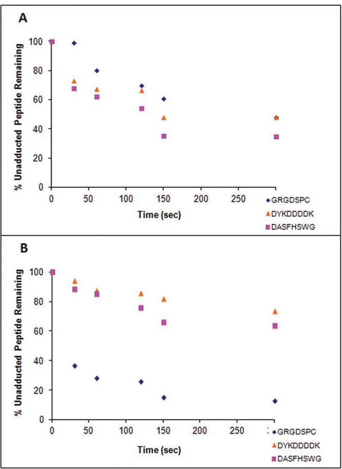
For GRGDSPC, where cysteine was the target, the 1:1 and 1:10 (peptide: metabolite) incubations with NO resulted in very similar losses of unadducted peptide after 8 min (Fig. 4A). The 1:10 incubation had a faster rate of peptide depletion overall, but by 8 min, both incubations had approximately 70% of unadducted peptide remaining. As stated above, approximately 15% of GRGDSPC was lost in control incubations. In comparison, there was a significant difference in signal loss from the unadducted peptide between the 1:1 and 1:10 peptide:metabolite incubations with NQ. After 8 minutes, while the unadducted peptide ion in the 1:1 incubation had decreased to about 40% of the original area, the ion in the 1:10 incubation had decreased to less than 20%. The hydrolysis of NQ may have accounted for this slowing in rate.
For DYKDDDDK, where lysine was the target residue, the 1:10 peptide:metabolite incubation with NO showed a steep initial decrease in peptide signal, but at the 8 min time point approximately the same percent peptide remained as in the 1:1 incubation, (30%, Fig. 4B). Approximately 60% of the unadducted peptide remained following 8 min incubations with NQ with both 1:1 and 1:10 peptide:metabolite ratios.
The loss of the unadducted peptide, DASFHSWG-NH2 (where histidine is the target), was similar to that observed with DYKDDDDK. At 8 min, the concentration of unadducted peptide remaining was similar in incubations conducted at 1:1 and 1:10 ratios of peptide to NO (Fig. 4B), even though the 1:10 Table 3.MS/MS Ion series of diadducted model peptides by naphthoquinones at pH 8.5.
Peptide Modification
Precursorm/z [M+H]+
Ions Detecteda
Supplemental Figure Ref.b
GRGDSPC 1,2-NQc 1007.35d 544.22 (b
4{)e,631.25 (b5{), 792.30 (y6{), 989.36 ([M+H]+
- H2O+Xg)
9
GRGDSPC 1,4-NQf 1007.35 728.29 (b
6)e,639.28 (y4), 989.35 ([M+H]+- H2O+X) 15
DYKDDDDK 1,2-NQ 1329.47 1068.35 (b6{{),1183.37 (b7{{), 607.33 (y5), 1013.41 ([M+H]+
- X),
1311.46 ([M+H]+
- H2O+X)
11
DYKDDDDK 1,4-NQ 1329.47 752.27 (b6), 867.30 (b7), 1013.40 ([M+H]+- X) Not shown
DASFHSWG-NH2 1,2-NQ 1221.46 874.31 (b5{{), 961.34 (b6{{), 643.26 (y4{), 790. 33 (y5{),
905.39 ([M+H]+
- X),1203.45 ([M+H]+
- H2O+X)
12
DASFHSWG-NH2 1,4-NQ 1221.46 961.34 (b6{{), 1147.42 (b7{{), 643.26 (y4{),790.36 (y5{),
905.39 ([M+H]+
- X),1203.45 ([M+H]+
- H2O+X)
17
EFYAPWCG 1,2-NQ 1288.47 620.22 (y4{),1270.45 ([M+H]+
- H2O+X) 13
EFYAPWCG 1,4-NQ 1288.47 794.35 (b6), 1213.4340 (b7{{), 778.26 (y4{{),849.29 (y5{{), 1012.36 (y6{{)
18
Product ion spectra for adducted peptides were indistinguishable between both pH conditions; more products were observed at pH 8.5 therefore ions series for the products at this pH was given.
a
Observed signals assigned as b- or y- ions for the diadducts are listed.
bReference to detailed spectra corresponding to the appropriate ion series. cThe modifications are 1,2-naphthoquinone (monoadduct mass
+158.04).
dThe adducted masses were observed in incubations performed at both pHs; data are from pH 8.5 incubations.
eIons in bold represent modification by a naphthoquinone. A bold b- or y- represents a monoadducted ion. Some of the diadduct MS/MS spectra had monoadduct
fragment ions present that were absent in the monoadduct MS/MS spectra.
fThe modifications are 1,4-naphthoquinone (monoadduct mass +158.04).
gIn the detected fragment ion signals, X represents the mass of the adduct.
incubation with NO showed a steeper initial decrease. When incubations were done with NQ at either 1:1 or 1:10 peptide:metabolite, peak areas for the unadducted peptide decreased rapidly leaving about 40% of the peptide after 8 minutes.
Adduct formation was also assessed for the incubations by monitoring the change in peak area of adducted ion over time, as shown in Fig. S22.
Preferential Binding to Different Nucleophilic Sites To assess whether naphthalene reactive metabolites would preferentially adduct different residues, metabolite was added to incubations containing equal concentrations of the model peptides containing a Cys, Lys, or His. Following 8 min incubation with NO, the amount remaining for each unadducted peptide was similar with 40–45% of the peptide remaining. The rate of loss of the lysine (DYKDDDDK) and histidine (DASFHSWG-NH2)
peptides was initially much faster than the decrease of the cysteine (GRGDSPC) peptide (Fig. 5A).
Adduction of the cysteine containing peptide was highly favored in the NQ incubation. While the histidine and lysine containing unadducted peptides were only depleted to about 70% of the initial level, the cysteine containing peptide was depleted to less than 20% over the same time period (Fig. 5B).
Discussion
Much of the current insight regarding the importance of electrophile adduction of proteins in cytotoxicity comes from
work on compounds where a single reactive metabolite is generated. In cases where multiple reactive metabolites are produced, our understanding of the relative rates of these processes is far more limited. This is the case with naphthalene where at least four reactive metabolites are generated, including the 1,2-epoxide, the diol epoxide, and both the 1,2- and 1,4-naphthoquinones, but little knowledge of the contribution of these metabolites to the levels of covalent adduct formation exists. If the differences in these metabolites’ reactivities with protein residues translate into disparate effects on whether a particular adduct alters the structural or functional properties of
Figure 3. MS/MS for adduction of peptide DYKDDDDK by 1,4-NQ (m/z1171.4366).3A shows the possible y- and b-ions associated with MS fragmentation of the peptide adducted on lysine. 3B shows the MS/MS spectrum of the adducted peptide acquired with labeled ions for designation. Adducted ions are in bold. Fragmentation patterns were the same when samples were incubated at both pH 7.4 and 8.5.
a protein critical to cellular homeostasis, then the overall metabolic disposition of this chemical and inter-individual differences in the enzymes responsible for controlling the
formation of these metabolites could substantially influence susceptibility to exposure. Thus, the ultimate goal is to assess the impact of electrophile-derived covalent adducts on protein
Figure 4. Depletion of unadducted peptides by metabolites.Depletion of GRGDSPC (A), DYKDDDDK (B), and DASFHSWG-NH2(C) by reaction
with either NO (naphthalene epoxide) or NQ (1,2-naphthoquinone) at two different ratios of peptide: metabolite, 1:1 and 1:10. Depletion of the unadducted peptide was monitored by measuring the peak area of the peptide at each time point.
Figure 5. Comparison of peptide binding by metabolite. Comparison of preferential binding between peptides containing different nucleophilic residues (Cys, Lys, and His) in incubations with NO (A) and NQ (B). Degree of preferential binding was monitored by measuring the peak area of the unadducted peptide ion over time.
structure and function. The first step toward addressing this larger question was to identify residues of reactive metabolite modification and to determine the preference for the epoxide and quinone metabolites on amino acid residues.
ESI-MS/MS was used to identify the predicted amino acid targets of naphthalene reactive intermediates (Fig. 6). All the peptides selected contained at least one nucleophilic amino acid shown previously to be adducted by epoxides and/or quinones [21,22,23]. The peptides were of high purity facilitating spectral interpretation. The truncated sequences containing the adduction sites (DSPC, LRKR, DYKD, DDDK, and FHSW) were found widely in the UniProt database(www.uniprot.org) within biolog-ical proteins; DSPC (i.e., P450 2B19, cell division kinase), LRKR (i.e., tyrosine protein kinase, myosin), DYKD (i.e., zinc phospho-diesterase, ubiquitin ligase), DDDK (i.e., serine phosphatase, EGF receptor substrate), and FHSW (i.e., kallikrein, cyclin-dependent kinase). The other two peptides (EFYAPWCG and EIVRDIKE) were from known naphthalene protein targets: actin and protein disulfide isomerase.
It is important to note that electrophilic metabolites do not react with all nucleophilic residues; rather, such interactions occur along a spectrum of reactivity [24]. The softness of a nucleophile is determined by the polarizability of its corresponding valence electrons: the higher the polarizability, the softer the nucleophile. Reactions between electrophiles and nucleophiles of similar softness are kinetically favored because the high energy transition state attained when sharing similar molecular orbitals requires less energy input than if they are dissimilar [25]. This was consistently observed between the naphthoquinones and amines and also contributes to the decreased likelihood of an epoxide to adduct onto the peptide a second time at the N-terminus.
In general, the thiolate of the cysteine is the softest nucleophile in proteins, which makes it the most likely target for the relatively soft naphthalene metabolite electrophiles [12]. Of the two peptides containing cysteine (GRGDSPC and EFYAPWCG), adduction occurred on both with all metabolites at pH 8.5 (Table 4). Whereas adduction was observed between the quinones and GRGDSPC at pH 7.4, no product was detected with the epoxides at pH 7.4. This is likely due to a combination of the reduced amounts of thiolate ion present, the relative reactivity of the quinones compared to the epoxides, and the higher instability of the epoxides at pH 7.4. At pH 7.4, the 1,2-epoxide more rapidly rearranges to 1-naphthol leading to a lower sustained concentra-tion of epoxide at physiological pH. A low pKaalso correlates to a higher availability of the thiolate anion at pH 7.4 and a lower overall nucleophilicity due to increased thiolate stabilization from electrostatic and polar interactions from the surrounding environ-ment [26]. Because naphthoquinones are more reactive, they compensate for the decreased nucleophilicity of the thiolate. From the rate of formation study at pH 8.5, there was a much faster depletion of GRGDSPC with NQ compared to NO (Fig. 4A). NQ also reacted more rapidly with the cysteine-containing peptide as compared to either the histidine or lysine containing peptides (Fig. 5B).
Adduction was observed on the histidine of DASFHSWG-NH2 at both pH 7.4 and 8.5 for all metabolites. The adduct stability is likely due to the imidazole moiety being a poor leaving group. The proximal aspartic acid at the N-terminus may have participated with the histidine in a catalytic triad making adduction more feasible [27]. Another explanation is the aspartate acting as an intramolecular base, enhancing removal of a proton from the His side chain.
Reactivity with the lysine-containing peptides varied. Whether the lack of NO adduct formation on EIVRDIKE and
YGGFLRKR was related to a low reaction rate of the epoxide with the peptides or whether the adducted peptides could not be readily detected in the mass spectrometer was unclear. The absence of NO adducts is partially supported by the direct influence of a protein’s microenvironment where it has been shown that pKa plays the dominant role in determining the reactivity of Lys side chains towards electrophiles [28]. In YGGFLRKR, the lysine was flanked by two adjacent arginines (pKa= 12.5), while in EIVRDIKE, the lysine residue was next to glutamic acid (pKa= 4.8). Charges from either highly acidic or highly basic neighbors can influence an adduction. With YGGFLRKR, the presence of the arginines may have decreased the reactivity of the Lys side chain. Likely because of this, a Lys adduction was not kinetically favored and binding occurred preferentially at the N-terminus for YGGLFRKR with the other three metabolites. With EIVRDIKE, adduction by the naphtho-quinones did not favor Lys either. With DYKDDDDK, adduction was observed under all conditions. Because the only major difference between this peptide and the others were flanking amino acids, this further validates the significant contribution of the microenvironment to the occurrence of an adduction.
Our studies suggest that the naphthoquinones are more reactive than the epoxides based on the range of detectable adducts as well as the presence of diadducts and a triadduct. Half of the peptides investigated did not react with either epoxide when reactions were done at physiologic pH; adducted products were observed with both naphthoquinones for all peptides when incubated at pH 7.4 and 8.5. The presence of the metabolites in large excess to the peptides was expected to enhance the likelihood of diadduct and triadduct formation, but multiple adductions on the same peptide were not detected with the epoxides, possibly due to a difference in the thermodynamic ‘‘softness’’ as mentioned previously [24]. The results of the current study are consistent with previously published work suggesting that the naphthoquinones are more reactive with proteins than the epoxide metabolites [29].
The time course disappearance of the unadducted peptide was measured in incubations containing 1:1 and 1:10 ratios of the peptide to metabolite to obtain an appreciation of the relative rates of reaction of epoxides and quinones with the various nucleophilic sites. LC-MS with SIM was utilized to provide high selectivity for the distinction of peptide peaks from background noise and neighboring peaks, which allowed direct comparison of peptide peak areas between different runs. The initial rates of reaction of NO with both DASFHSWG-NH2and DYKDDDDK had faster depletions in incubations containing a 1:10 ratio of peptide to metabolite than with those containing a 1:1 ratio. After 8 minutes, the unadducted peptides were almost completely depleted at both peptide: metabolite ratios, suggesting that the epoxide had saturated the available sites (Fig. 4B and C). No significant differences in the rates of unadducted peptide loss were observed in incubations of 1,2-NQ with these peptides at either the 1:1 or 1:10 peptide: metabolite ratios. Only about 50% of the peptides were lost during the 8 min incubation and this may reflect the relative instability of 1,2-NQ in aqueous solutions. Another explanation could be that the NQ formed initially are in the reduced form and can be oxidized by unreacted NQ, leading to depletion of free NQ and yielding quinonoid adducts.
specific activities of between 0.05 and 0.2 nmoles reactive metabolite bound/nmole protein, far lower than the ratios used here. However, our earlier work has shown that there is a reasonable degree of selectivity in adduct formation and so the specific activity of adducted proteins is likely to be much greater than the 0.05 to 0.2 nmoles/mg calculated above [31]. Moreover, as shown by the data in Figure 4, the ratios of reactants did not
strongly influence the rates of reaction when peptide: metabolite ratios were increased from 1:1 to 1:10. Finally, more recent studies monitoring adducted proteins in the urine [32] indicated that the specific activities of adducted proteins in the urine is 5–10 fold higher than observed in target tissues.
In experiments to evaluate possible competition which might occur when multiple nucleophilic amino acids were present in a
Figure 6. Structures of the predicted adducts.The products of adduct formation with epoxides were the result of SN2 reactions while the
mixture, model peptides which contained cysteine, lysine or histidine residues were reacted with NO or 1,2-NQ. Although some differences in rates of loss of unmodified peptide were noted particularly between 1,2-NQ and the cysteine-containing peptide compared to the lysine- and histidine-containing peptides, these experiments likely do not recapitulate the cellular milieu where the secondary and tertiary structure of proteins and proximity to hydrophobic amino acids and/or to basic amino acids such as arginine may dramatically alter reaction rates with electrophiles [26]. Indeed, recent work with protein disulfide isomerase and actin demonstrate relatively high selectivity for modification of specific cysteines, lysines and histidines by both epoxide and quinone metabolites of naphthalene [33].
The results of the current studies are consistent with previous findings showing epoxide binding to sulfur nucleophiles was minor relative to binding by the 1,2-naphthoquinone in Clara cell incubations with naphthalene [34]. In naphthalene-treated mice, 1,4-NQ adducts were more abundant than NO adducts in all tissue samples studied, with the lung carrying the highest NQ load [35]. In contrast, Waidyanatha and Rappaport showed NO-albumin and hemoglobin adducts were more abundant than NQ adducts in vivo [12,36]. Therefore, the relative amounts of metabolite bound covalently to proteins in vivo are dependent upon concentrations formed intracellularly, on the availability of cellular protein targets, as well as on the detoxification of reactive intermediates such as NO hydrolysis and GSH conjugation. Although the current work confirmed the target amino acids, additional studies are necessary to determine the relative proportions of epoxides, diol epoxides, and quinones bound to proteinsin vivo.
This current work characterized both epoxide and quinone adducts with model peptides and these data have been critical to the interpretation of adducted proteins and peptides eliminated in the urine of naphthalene-treated animals in ongoing studies in the laboratory. Understanding the reaction of naphthalene metabo-lites with biological molecules is critical to delineating the mechanisms by which this chemical leads to the loss of cellular homeostasis. However, rapid adduct formation with a given residue does not imply inherent toxicological significance. Rather, it is the role of the residue in protein structure or function and the resulting disruptive consequences of adduction that determine the relevance of an adduct. Accordingly, additional work which focuses on verifying the sites of adduction in vivo as well as investigation into the functional consequences of adduction is underway.
Supporting Information Available
MS/MS spectra of model peptide adduct products not already shown as figures are presented with corresponding b- and y- ions. Each figure presents either a spectral comparison between the nonadducted parent and the monoadduct, the monoadduct and the diadduct, or the nonadducted parent, the monoadduct, and the diadduct. Them/zof the parent peptide is given along with the corresponding MS/MS fragmentation. Adducted ions (modified by a metabolite) are labeled inbold, nonadducted ions are not. [M+H]+
is representative of the nonadducted parent peptide;
[M+H]+is representative of the monoadducted parent peptide.#
is representative of modification by naphthalene epoxide (NO).y is representative of modification by naphthalene diol epoxide (NDO).{is representative of modification of 1,2-naphthoqinone (1,2NQ).u is representative of modification by 1,4-naphthoqui-none (1,4NQ). Although spectra were acquired for both pHs, because spectra were closely similar, only spectra acquired at pH 8.5 are given.
Other supporting figures include standard curves and rate of formation. A table presenting mass errors (ppm) is also provided.
Supporting Information
Figure S1 MS/MS of peptide [GRGDSPC] at m/z 691.2826
(A)and adduct [GRGDSPC+NO] atm/z835.3408(B). (DOCX)
Figure S2 MS/MS of peptide [DYKDDDDK] at m/z 1013.4058(A)and adduct [DYKDDDDK+NO] – H2O atm/ z1139.4510(B).
(DOCX)
Figure S3 MS/MS of peptide [DASFHSWG-NH2] at m/z 905.3878 (A) and adduct [DASFHSWG-NH2+ NO] at m/z 1049.4487(B).
(DOCX)
Figure S4 MS/MS of peptide [EFYAPWCG] atm/z972.3920
(A)and adduct [EFYAPWCG+NO] atm/z1116.4995(B). (DOCX)
Figure S5 MS/MS of peptide [YGGFLRKR] atm/z996.5738
(A)and adduct [YGGFLRKR+NDO] atm/z1174.6365(B). (DOCX)
Figure S6 MS/MS of peptide [DYKDDDDK] at m/z 1013.4058 (A) and adduct [DYKDDDDK + NDO] at m/z 1191.4680(B).
(DOCX) Table 4.Sites of Adduction.
pH
Metabolite 7.4 8.5
NO DYKDDDDK*, DASFHSWG-NH2, EFYAPWCG GRGDSPC, DYKDDDDK*, DASFHSWG, EFYAPWC*G NDO DYKDDDDK, DASFHSWG-NH2, EFYAPWCG GRGDSPC,N-YGGFLRKR, DYKDDDDK, DASFHSWG-NH2,
EFYAPWCG
1,2NQ GRGDSPC,N-YGGFLRKR, DYKDDDDK, DASFHSWG-NH2,
EFYAPWCG,N-EIVRDIKE
GRGDSPC,N-YGGFLRKR, DYKDDDDK, DASFHSWG-NH2,
EFYAPWCG,N-EIVRDIKE
1,4NQ GRGDSPC,N-YGGFLRKR, DYKDDDDK, DASFHSWG-NH2,
EFYAPWCG,N-EIVRDIKE
GRGDSPC,N-YGGFLRKR, DYKDDDDK, DASFHSWG-NH2,
EFYAPWCG,N-EIVRDIKE
Sites of adduction deduced for each peptide.
Figure S7 MS/MS of peptide [DASFHSWG-NH2] at m/z 905.3900 (A) and adduct [DASFHSWG-NH2+ NDO] at m/z 1083.4537(B).
(DOCX)
Figure S8 MS/MS of peptide [EFYAPWCG] atm/z972.3920
(A)and adduct [EFYAPWCG+NDO] atm/z1150.4550(B). (DOCX)
Figure S9 MS/MS of peptide [GRGDSPC +1,2NQ] mono-adduct atm/z849.3181(A)and [GRGDSPC+1,2NQ] diadduct m/z 1007.3465 (B) and [GRGDSPC +1,2NQ] triadduct m/z 1165.3826(C).
(DOCX)
Figure S10 MS/MS of peptide [YGGFLRKR] atm/z996.5738
(A)and adduct [YGGFLRKR+1,2NQ] atm/z1154.6139(B). (DOCX)
Figure S11 MS/MS of peptide [DYKDDDDK +1,2NQ] monoadduct at m/z 1171.4382 (A) and [DYKDDDDK
+1,2NQ] diadduct atm/z1329.4729(B). (DOCX)
Figure S12 MS/MS of peptide [DASFHSWG-NH2+1,2NQ] monoadduct at m/z 1063.4248 (A) and [DASFHSWG-NH2+1,2NQ] diadduct atm/z1221.4593(B).
(DOCX)
Figure S13 MS/MS of peptide [EFYAPWCG+1,2NQ] mono-adduct at m/z 1130.4287 (A) and [EFYAPWCG +1,2NQ] diadduct atm/z1288.4655(B).
(DOCX)
Figure S14 MS/MS of peptide [EIVRDIKE] atm/z1001.5626
(A)and adduct [EIVRDIKE+1,2NQ] atm/z1159.5993(B). (DOCX)
Figure S15 MS/MS of peptide [GRGDSPC +1,4NQ] mono-adduct atm/z849.3167(A)and [GRGDSPC+1,4NQ] diadduct atm/z1007.3466(B).
(DOCX)
Figure S16 MS/MS of peptide [YGGFLRKR] atm/z996.5738
(A)and adduct [YGGFLRKR+1,4NQ] atm/z1154.6133(B). (DOCX)
Figure S17 MS/MS of peptide [DASFHSWG+1,4NQ] mono-adduct at m/z 1063.4237 (A) and [DASFHSWG +1,4NQ] diadduct atm/z1221.4593(B).
(DOCX)
Figure S18 MS/MS of peptide [EFYAPWCG+1,4NQ] mono-adduct at m/z 1130.4287 (A) and [EFYAPWCG +1,4NQ] diadduct atm/z1288.4655(B).
(DOCX)
Figure S19 MS/MS of peptide [EIVRDIKE] atm/z1001.5626
(A)and adduct [EIVRDIKE+1,4NQ] atm/z1159.5993(B). (DOCX)
Figure S20 Plots of areas for [M+ H]+
ions vs. concentration using selected ion monitoring for nonadducted parent peptide. Different dilutions of unadducted peptide were prepared as described in materials and methods.
(DOC)
Figure S21 Ion monitoring of depletion of unadducted control ([M+H]+) peptide over time; measurement of peak ion area.
(DOC)
Figure S22 Rate of adduct formation for individual incubations for GRGDSPC(A), DASFHSWG(B), and DYKDDDDK(C). (DOC)
Table S1 PPM errors of adduct ions observed. (DOC)
Acknowledgments
Mass spectrometric analysis was performed at the Campus Mass Spectrometry Facilities, University of California Davis.
Author Contributions
Conceived and designed the experiments: NP DM AB. Performed the experiments: NP WJ. Analyzed the data: NP WJ AJ AB. Contributed reagents/materials/analysis tools: NP DM WJ AB. Wrote the paper: NP AB.
References
1. Liebler DC (2008) Protein damage by reactive electrophiles: Targets and consequences. Chem Res Toxicol 21: 117–128.
2. Tang W, Lu AY (2010) Metabolic bioactivation and drug-related adverse effects: current status and future directions from a pharmaceutical research perspective. Drug Metab Rev 42: 225–249.
3. Roberts SA, Price VF, Jollow DJ (1990) Acetaminophen structure-toxicity studies: in vivo covalent binding of a nonhepatotoxic analog, 3-hydroxyaceta-nilide. Toxicol Appl Pharmacol 105: 195–208.
4. Obach RS, Kalgutkar AS, Soglia JR, Zhao SX (2008) Can in vitro metabolism-dependent covalent binding data in liver microsomes distinguish hepatotoxic from nonhepatotoxic drugs? An analysis of 18 drugs with consideration of intrinsic clearance and daily dose. Chem Res Toxicol 21: 1814–1822. 5. Humphreys WG (2011) Overview of strategies for addressing BRIs in drug
discovery: Impact on optimization and design. Chem Biol Interact 192: 56–59. 6. Hanzlik RP, Koen YM, Theertham B, Dong Y, Fang J (2007) The reactive metabolite target protein database (TPDB)–a web-accessible resource. BMC Bioinformatics 8: 95.
7. Liu J, Li Q, Yang X, van Breemen RB, Bolton JL, et al. (2005) Analysis of protein covalent modification by xenobiotics using a covert oxidatively activated tag: raloxifene proof-of-principle study. Chem Res Toxicol 18: 1485–1496. 8. Dennehy MK, Richards KA, Wernke GR, Shyr Y, Liebler DC (2006) Cytosolic
and nuclear protein targets of thiol-reactive electrophiles. Chem Res Toxicol 19: 20–29.
9. Preuss R, Angerer J, Drexler H (2003) Naphthalene–an environmental and occupational toxicant. Int Arch Occup Environ Health 76: 556–576.
10. Chao YC, Kupper LL, Serdar B, Egeghy PP, Rappaport SM, et al. (2006) Dermal exposure to jet fuel JP-8 significantly contributes to the production of urinary naphthols in fuel-cell maintenance workers. Environ Health Perspect 114: 182–185.
11. National Toxicology Program (2000) NTP Technical Report on the Toxicology and Carcinogenesis of Naphthalene in F344/N Rats Naphthalene, NTP TR 500.
12. Waidyanatha S, Rappaport SM (2008) Hemoglobin and albumin adducts of naphthalene-1,2-oxide, 1,2-naphthoquinone and 1,4-naphthoquinone in Swiss Webster mice. Chem Biol Interact 172: 105–114.
13. Miller MG, Rodgers A, Cohen GM (1986) Mechanisms of toxicity of naphthoquinones to isolated hepatocytes. Biochem Pharmacol 35: 1177–1184. 14. Cho M, Chichester C, Morin D, Plopper C, Buckpitt A (1994) Covalent
Interactions of Reactive Naphthalene Metabolites with Proteins. J Pharmacol Exp Ther 269: 881–889.
15. Hettick JM, Siegel PD (2011) Determination of the toluene diisocyanate binding sites on human serum albumin by tandem mass spectrometry. Anal Biochem 414: 232–238.
16. Kristiansson MH, Lindh CH, Jonsson BA (2003) Determination of hexahy-drophthalic anhydride adducts to human serum albumin. Biomarkers 8: 343– 359.
18. Stamper BD, Mohar I, Kavanagh TJ, Nelson SD (2011) Proteomic analysis of acetaminophen-induced changes in mitochondrial protein expression using spectral counting. Chem Res Toxicol 24: 549–558.
19. Yagi H, Jerina D (1975) A General Synthetic Method for Non-K-Region Arene Oxides. J Am Chem Soc 97: 3185–3192.
20. Tsang W, Griffin G, Horning M, Stillwell W (1981) Chemistry of anti- and syn-1,2:3,4-naphthalene dioxides and their potential relevance as metabolic intermediates. J Org Chem 47: 5339–5353.
21. Booth ED, Kilgour JD, Watson WP (2004) Dose responses for the formation of hemoglobin adducts and urinary metabolites in rats and mice exposed by inhalation to low concentrations of 1,3-[2,3-(14)C]-butadiene. Chem Biol Interact 147: 213–232.
22. Jagr M, Mraz J, Linhart I, Stransky V, Pospisil M (2007) Synthesis and characterization of styrene oxide adducts with cysteine, histidine, and lysine in human globin. Chem Res Toxicol 20: 1442–1452.
23. Bambal RB, Hanzlik RP (1995) Bromobenzene 3,4-oxide alkylates histidine and lysine side chains of rat liver proteins in vivo. Chem Res Toxicol 8: 729–735. 24. LoPachin RM, Gavin T, Petersen DR, Barber DS (2009) Molecular mechanisms
of 4-hydroxy-2-nonenal and acrolein toxicity: nucleophilic targets and adduct formation. Chem Res Toxicol 22: 1499–1508.
25. Coles B (1984) Effects of modifying structure on electrophilic reactions with biological nucleophiles. Drug Metab Rev 15: 1307–1334.
26. Ferrer-Sueta G, Manta B, Botti H, Radi R, Trujillo M, et al. (2011) Factors affecting protein thiol reactivity and specificity in peroxide reduction. Chem Res Toxicol 24 434–450.
27. Britto PJ, Knipling L, Wolff J (2002) The local electrostatic environment determines cysteine reactivity of tubulin. J Biol Chem 277: 29018–29027.
28. Zhang M, Vogel HJ (1993) Determination of the side chain pKa values of the lysine residues in calmodulin. J Biol Chem 268: 22420–22428.
29. Tsuruda LS, Lame MW, Jones AD (1995) Formation of epoxide and quinone protein adducts in B6C3F1 mice treated with naphthalene, sulfate conjugate of 1,4-dihydroxynaphthalene and 1,4-naphthoquinone. Arch Toxicol 69: 362–367. 30. DeStefano-Shields C, Morin D, Buckpitt A (2010) Formation of covalently bound protein adducts from the cytotoxicant naphthalene in nasal epithelium: species comparisons. Environ Health Perspect 118: 647–652.
31. Lin CY, Boland BC, Lee YJ, Salemi MR, Morin D, et al. (2006) Identification of proteins adducted by reactive metabolites of naphthalene and 1-nitronaphtha-lene in dissected airways of rhesus macaques. Proteomics 6: 972–982. 32. Pham N, Morin D, Jewell WT, Buckpitt AR (2011) Characterization of peptide
adducts of reactive naphthalene metabolites in the urine of male mice by mass spectrometry. The Toxicologist 120.
33. Pham N, Morin D, Jewell WT, Buckpitt AR (2012) Analysis of naphthalene adduct binding sites in model proteins by tandem mass spectrometry. Chemico-Biological Interactions http://dx.doi.org/10.1016/j.cbi.2012.05.009. 34. Zheng J, Cho M, Jones AD, Hammock BD (1997) Evidence of quinone
metabolites of naphthalene covalently bound to sulfur nucleophiles of proteins of murine Clara cells after exposure to naphthalene. Chem Res Toxicol 10: 1008– 1014.
![Figure S1 MS/MS of peptide [GRGDSPC] at m/z 691.2826 (A) and adduct [GRGDSPC + NO] at m/z 835.3408 (B).](https://thumb-eu.123doks.com/thumbv2/123dok_br/18158966.328544/13.918.92.833.114.304/figure-s-ms-ms-peptide-grgdspc-adduct-grgdspc.webp)
