Journal of Investigative Medicine High Impact Case Reports
October-December 2016: 1 –5 © 2016 American Federation for Medical Research
DOI: 10.1177/2324709616674316 hic.sagepub.com
Creative Commons CC-BY: This article is distributed under the terms of the Creative Commons Attribution 3.0 License (http://www.creativecommons.org/licenses/by/3.0/) which permits any use, reproduction and distribution of the work without further permission provided the original work is attributed as specified on the SAGE and Open Access pages (https://us.sagepub.com/en-us/nam/open-access-at-sage). Case Report
Introduction
Immunotherapy using interleukin-2 (IL-2) has long been a part of our historically small armamentarium against advanced melanoma. Treatment with IL-2 was largely lim-ited to patients who could tolerate the potentially serious adverse effects, and while only a minority of patients obtained benefit, those who did experienced prolonged response durations.1-3 Interferon has also been studied in the metastatic setting, and while it showed modest efficacy, durations of response were short and toxicities potentially prohibitive.4,5 Therapeutic options are now increasing with the emergence of checkpoint inhibitors, which are shifting the way that oncologists view a once universally lethal dis-ease. Although checkpoint inhibitors are relatively well tol-erated, some patients will experience immunologic side effects, known as immune-related adverse events (irAE). Additionally, those with preexisting autoimmune disorders may experience flares of their disease. Here we present 2 patients with metastatic melanoma treated with the PD-1 checkpoint inhibitor pembrolizumab who developed exacer-bations of preexisting autoimmune disease.
Case 1
I.M. is a 67-year-old Caucasian man with a history of noninva-sive low-grade (TaLG) bladder cancer diagnosed in 2013, treated with transurethral resection. Two years later, on a
surveillance cystoscopy, he was found to have lesions at the posterior bladder dome and within the prostatic fossa. Biopsies of the lesions revealed malignant melanoma, BRAF V600E positive. The patient denied any history of melanoma and had no skin lesions suspicious for a primary melanoma. A review of the bladder pathology from 2013 confirmed papillary bladder cancer with no evidence of melanoma. Staging positron emis-sion tomography/computed tomography (PET/CT) scan revealed scattered osseous lesions as well as right hilar and mediastinal lymphadenopathy. A brain magnetic resonance imaging (MRI) was negative for intracranial metastases. Other pertinent medical history included psoriasis and psoriatic arthritis, which had been well controlled on a weekly dose of
1
Department of Internal Medicine, Divison of Hematology/Oncology, University of Iowa Hospitals & Clinics, Iowa City, IA, USA
2
Department of Internal Medicine, University of Iowa Hospitals & Clinics, Iowa City, IA, USA
3
Department of Dermatology, University of Iowa Hospitals & Clinics, Iowa City, IA, USA
4
Department of Neurology, University of Iowa Hospitals & Clinics, Iowa City, IA, USA
Received July 2, 2016. Revised September 15, 2016. Accepted September 18, 2016.
Corresponding Author:
Sneha D. Phadke, DO, University of Iowa Hospitals & Clinics, 200 Hawkins Drive, C21GH, Iowa City, IA 52242, USA.
Email: [email protected]
Pembrolizumab Therapy Triggering an
Exacerbation of Preexisting Autoimmune
Disease: A Report of 2 Patient Cases
Sneha D. Phadke, DO
1, Ramez Ghabour, DO
2, Brian L. Swick, MD
3,
Andrea Swenson, MD
4, Mohammed Milhem, MD
1, and Yousef Zakharia, MD
1Abstract
Historically, metastatic melanoma was uniformly and rapidly lethal, and treatment options were limited. In recent years, however, checkpoint inhibitors have emerged as an accepted standard treatment for patients with advanced melanoma. In clinical trials, these agents have been largely well tolerated and have the potential to result in durable responses. Importantly though, one must recognize the unique side effect profile of these therapies, which can trigger or exacerbate underlying autoimmune disease. Whether this autoimmune activation is associated with a clinical response to therapy has been debated, and while not definitive, there is evidence in the literature of a possible association. The 2 cases presented describe this autoimmune phenomenon, along with a review of the existing literature on the relationship between response to immunotherapy and autoimmune side effects.
Keywords
12.5 mg of oral methotrexate. After careful discussion with the patient about the potential side effects of treatment including worsening of psoriasis, methotrexate was discontinued and pembrolizumab was initiated at 2 mg/kg every 3 weeks. Three doses into his treatment, the patient presented with bilateral lower extremity weakness. MRI of the spine revealed multiple enhancing nodules suspicious for leptomeningeal metastasis. He started a dexamethasone taper and underwent palliative radiation to the spine, with rapid improvement in strength within 1 week of completing radiation therapy and near com-plete resolution of symptoms within 3 weeks.
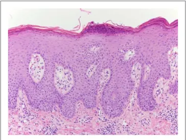
The patient then underwent 2 more doses of pembroli-zumab followed by a restaging PET/CT, which revealed a complete response with interval resolution of the hypermeta-bolic lymphadenopathy and the osseous lesions. Physical examination, however, revealed diffuse scaly plaques on the trunk and the upper and lower extremities (Figure 1). This was determined to be a grade 3 toxicity, and pembrolizumab therapy was held. Punch biopsy of a lesion on the right leg demonstrated psoriasiform epidermal hyperplasia with para-keratosis and intraepidermal pustules confirming the diagno-sis of psoriadiagno-sis (Figure 2). He had no signs or symptoms of worsening psoriatic arthritis. The patient began therapy with acitretin and narrow band ultraviolet B phototherapy with substantial improvement in the psoriatic lesions.
As the patient had a significant tumor response to pem-brolizumab therapy, it was restarted after 4 weeks, when the skin toxicity had improved to grade 1. The patient has since tolerated treatment well, without a psoriasis exacerbation.
Case 2
D.N. is a 75-year-old Caucasian man with a history of malig-nant melanoma of the left shoulder resected in 2011, who
presented with progressive dysphagia. An upper endoscopy revealed a gastric mass, and biopsy specimens demonstrated a poorly differentiated malignant neoplasm. Staging CT of the chest, abdomen, and pelvis demonstrated numerous pul-monary nodules bilaterally and scattered liver lesions consis-tent with metastatic disease. Biopsy of a liver lesion confirmed metastatic melanoma, negative for BRAF muta-tions. A brain MRI was negative for intracranial involve-ment. Pertinent medical history included acetylcholine receptor (AChR) autoantibody positive myasthenia gravis (MG) diagnosed at age 64. At that time, he had primarily ocular symptoms and AChR binding antibodies were posi-tive at a titer of 2.1 nmol/L (normal range = 0.0-0.4 nmol/L). Over the years, his MG progressed and he developed other symptoms such as dysphagia, chewing fatigue, dyspnea on exertion, and neck and limb weakness. He responded well to prednisone and was eventually transitioned to mycopheno-late mofetil. Just before the metastatic melanoma diagnosis, his MG was asymptomatic with mycophenolate mofetil 750 mg daily and pyridostigmine 60 mg 3 times daily.
For lack of a more robust treatment option, and after dis-cussing the potential side effects with the patient and the neu-rologist, pembrolizumab was started at 2 mg/kg every 3 weeks. The dose of mycophenolate mofetil was decreased to 500 mg daily and pyridostigmine was continued unchanged. After his second dose, the patient developed symptoms of diplopia and worsening bilateral ptosis concerning for an exacerbation of MG. Pembrolizumab was held, and pyr-idostigmine was titrated up to 180 mg 3 times daily, along with prednisone 30 mg daily. The patient’s condition contin-ued to deteriorate, and he was admitted to the intensive care unit with respiratory distress and worsening dysphagia, where he required noninvasive ventilation and gastrostomy tube placement. The patient’s AChR binding antibody titer
Figure 1. Sharply demarcated erythematous plaques with
overlying scale on the back.
Figure 2. Regular psoriasiform epidermal hyperplasia with
was measured during his hospitalization and was elevated at 0.77 nmol/L (normal range <0.02 nmol/L).
His MG symptoms improved with 7 treatments of plasma exchange, 2 doses of intravenous immunoglobulin, and 4 doses of rituximab. On discharge from the intensive care unit, the patient underwent a restaging PET/CT scan, which showed resolving lung nodules, consistent with a partial response. Due to the grade 4 toxicity, pembrolizumab therapy was not resumed. Temozolomide was started at 75 mg/m2 daily. The patient was readmitted 1 month later for pneumo-nia secondary to aspiration and Escherichia coli bacteremia. He quickly succumbed to the infection.
Discussion
Immune-related adverse events are well-recognized risks to checkpoint inhibitors such as pembrolizumab. However, patients with preexisting autoimmune disease were largely excluded from clinical trials evaluating checkpoint inhibitors due to concern that treatment would lead to unacceptable tox-icities from exacerbations of underlying disease. Therefore, less is known about the significance of these exacerbations.
Both the patients we describe in this report experienced a tumor response to pembrolizumab therapy while simultane-ously experiencing a flare of their autoimmune disease. For the patient in Case 1, we cannot entirely exclude the discon-tinuance of methotrexate as a contributing factor to his pso-riasis flare; however, in our experience, the timing (3 months instead of the typical 6-8 weeks after discontinuing metho-trexate) and severity of his flare (guttate in appearance and much different than his prior plaque type psoriasis) suggest that methotrexate withdrawal is unlikely to be the sole cause.
Checkpoint inhibitors induce irAE by nonspecific immu-nologic activation, decreasing the ability of effector T lym-phocytes to discriminate between self and nonself.6,7 Some studies suggest that the development of irAE correlates with tumor response.8,9 Accordingly, we speculate that exacerba-tions of preexisting autoimmune condiexacerba-tions may also corre-late with tumor response.
Isolated cases of MG were reported in studies evaluating PD-1 and PD-L1 inhibitors.10-12 Additionally, case reports of new diagnoses of MG secondary to checkpoint inhibitors have been described in the literature.13-16 There are also cases in the literature describing patients with preexisting MG who were treated with checkpoint inhibitors. One of these describes a patient with preexisting MG who developed an exacerbation while on the PD-1 inhibitor nivolumab for met-astatic melanoma. After temporary cessation of nivolumab therapy, the exacerbation resolved, and a CT scan revealed a response in the metastatic lymph nodes.17 Another discussed a patient with advanced melanoma and preexisting MG who developed a severe exacerbation after 3 doses of pembroli-zumab. He was reported to have stable disease at 4-month follow-up.18 Two published case reports describe patients with advanced melanoma experiencing exacerbations of
preexisting psoriasis while on nivolumab.19,20 In one of those reports, the patient was described as experiencing a clinical response to nivolumab therapy.19
While there was initially concern among oncologists regarding the safety of using checkpoint inhibitors in patients with preexisting autoimmune disease, a recent retrospective review examining the use of the CTLA-4 inhibitor ipilim-umab in such patients revealed that while ipilimipilim-umab ther-apy was associated with exacerbations of autoimmune disease, they were largely manageable with conventional immunosuppressive therapies.21 Additionally, one case report describes the use of immunotherapies in the treatment of metastatic melanoma in 2 patients with preexisting auto-immune diseases, and no exacerbations were observed.22 Of course, one must consider the potential morbidity from an exacerbation of underlying autoimmune disease prior to ini-tiating checkpoint inhibitor therapy.
Anecdotally, clinicians have noted a link between autoim-munity and tumor response, such as the development of vit-iligo after treatment with IL-2. In fact, Phan et al demonstrated an association between long-term immunologic side effects, especially vitiligo, and an antitumor response to IL-2 in patients with melanoma.23 These findings were validated in a meta-analysis that showed that patients who developed vitil-igo while receiving immunotherapy had 2 to 4 times less risk of disease progression and death compared to patients with-out vitiligo.24 A study by Hua et al suggested a similar link between vitiligo and response to pembrolizumab therapy with 71% of patients with vitiligo experiencing a response to ther-apy versus 28% without vitiligo.25 In a recent study published by Sanlorenzo et al, 42% of patients treated with pembroli-zumab developed cutaneous adverse events. These patients had a significantly longer progression-free survival than patients who did not develop cutaneous adverse events.26
Another study showed that administration of a CTLA-4 inhibitor in combination with an antimelanoma vaccine resulted in 14 patients developing grade 3 or 4 autoimmune toxicity with 36% of those patients experiencing a clinical response compared to 5% in those patients experiencing no autoimmunity.8 A single-institution study showed a signifi-cantly higher clinical benefit rate in patients on a CTLA-4 inhibitor who developed grade 3 or 4 irAE (60%) compared with those with grade ≤2 irAE (22%).9
and insulin-dependent diabetes mellitus. Interestingly, the authors were able to determine retrospectively that the patient had preexisting autoantibodies against striated muscle and insulin but without any clinical manifestations. He remained on chronic immunosuppression after discontinuation of IL-2. The authors note that while he did have regression of his met-astatic cancer, the continuing need for immunosuppression may have negated this effect.31
The mechanism by which checkpoint inhibitors trigger autoimmunity is incompletely understood, and may be related to dysregulation of a preexisting immune response to self-antigens, which had been formerly contained by immune checkpoints.32 Additionally, proliferation of effector T cells combined with impairment in regulatory T cells and dys-functional interactions with antigen presenting cells may play a role.33 Interestingly, while the 2 autoimmune disorders we presented have very different clinical manifestations, both involve CD4+ T cells. Psoriasis in particular is thought to be primarily a T-cell-mediated process, in which there is toxic injury to the epidermis resulting in the characteristic skin plaques and hyperproliferation.34 Accordingly, advances in therapy have been made through agents that block T cell functions.35 MG is driven by CD4+ T cells that are AChR specific, activating B cells, which, in turn, produce AChR autoantibodies.36 While the precise pathologic basis of auto-immunity is unknown, research is ongoing to identify bio-markers that predict response to checkpoint inhibitor therapy as well as predict for development of irAE.37
Conclusion
While intriguing, further investigation is needed into the relationship between irAE and response to immunotherapy. As the use of checkpoint inhibitors continues to broaden and biomarkers of response are discovered, perhaps the specula-tive link between autoimmunity and response to therapy will be elucidated, leading to more informative discussions with patients.
Declaration of Conflicting Interests
The author(s) declared no potential conflicts of interest with respect to the research, authorship, and/or publication of this article.
Funding
The author(s) received no financial support for the research, author-ship, and/or publication of this article.
References
1. Rosenberg SA, Yang JC, Topalian SL, et al. Treatment of 283 consecutive patients with metastatic melanoma or
renal cell cancer using high-dose bolus interleukin 2. JAMA.
1994;271:907-913.
2. Atkins MB, Lotze MT, Dutcher JP, et al. High-dose recombinant interleukin 2 therapy for patients with metastatic melanoma:
analysis of 270 patients treated between 1985 and 1993. J Clin
Oncol. 1999;17:2105-2116.
3. Atkins MB, Kunkel L, Sznol M, Rosenberg SA. High-dose recombinant interleukin-2 therapy in patients with metastatic
melanoma: long-term survival update. Cancer J Sci Am.
2000;6(suppl 1):S11-S14.
4. Creagan ET, Ahmann DL, Frytak S, Long HJ, Chang MN, Itri LM. Phase II trials of recombinant leukocyte A interferon in disseminated malignant melanoma: results in 96 patients.
Cancer Treat Rep. 1986;70:619-624.
5. Eggermont A. The role of interferon-alpha in malignant
melanoma remains to be defined. Eur J Cancer. 2001;37:
2147-2153.
6. Postow MA, Callahan MK, Wolchok JD. Immune checkpoint
blockade in cancer therapy. J Clin Oncol. 2015;33:1974-1982.
7. Khan H, Gucalp R, Shapira I. Evolving concepts:
immu-nity in oncology from targets to treatments. J Oncol.
2015;2015:847383.
8. Attia P, Phan GQ, Maker AV, et al. Autoimmunity correlates with tumor regression in patients with metastatic melanoma
treated with anti-cytotoxic T-lymphocyte antigen-4. J Clin
Oncol. 2005;23:6043-6053.
9. Ku GY, Yuan J, Page DB, et al. Single-institution experience with ipilimumab in advanced melanoma patients in the
com-passionate use setting. Cancer. 2010;116:1767-1775.
10. Patnaik A SM, Gubens MA, Gandhi L, et al. Phase 1 study of pembrolizumab plus ipilimumab as second-line therapy for advanced non-small cell lung cancer: KEYNOTE-021 Cohort D. J Clin Oncol. 2015;33(suppl):Abstract 8011.
11. Antonia SJ, Goldberg SB, Balmanoukian AS, et al. Phase Ib study of MEDI4736, a programmed cell death ligand-1 (PD-L1) antibody, in combination with tremelimumab, a cytotoxic T-lymphocyte-associated protein-4 (CTLA-4)
antibody, in patients with advanced NSCLC. J Clin Oncol.
2015;33(Suppl):Abstract 3014.
12. Brahmer JR, Tykodi SS, Chow LQM, et al. Safety and activity
of anti–PD-L1 antibody in patients with advanced cancer. N
Engl J Med. 2012;366:2455-2465.
13. Lopez D, Calvo A, Fershko A. Myasthenia gravis and rhabdo-myolysis in a patient with advanced renal cell cancer treated
with nivolumab: a case report and review of the literature. Br J
Med Health Res. 2015;2(12):11-16.
14. Johnson DB, Saranga-Perry V, Lavin PJ, et al. Myasthenia gra-vis induced by ipilimumab in patients with metastatic
mela-noma. J Clin Oncol. 2015;33(33):e122-e124.
15. Loochtan AI, Nickolich MS, Hobson-Webb LD. Myasthenia gravis associated with ipilimumab and nivolumab in the
treat-ment of small cell lung cancer. Muscle Nerve. 2015;52:307-308.
16. Shirai T, Sano T, Kamijo F, et al. Acetylcholine receptor bind-ing antibody-associated myasthenia gravis and rhabdomyolysis
induced by nivolumab in a patient with melanoma. Jpn J Clin
Oncol. 2016;46:86-88.
17. Maeda O, Yokota K, Atsuta N, Katsuno M, Akiyama M, Ando Y. Nivolumab for the treatment of malignant melanoma in a
patient with pre-existing myasthenia gravis. Nagoya J Med Sci.
2016;78:119-122.
18. Lau KH, Kumar A, Yang IH, Nowak RJ. Exacerbation of myasthenia gravis in a patient with melanoma treated with
19. Matsumura N, Ohtsuka M, Kikuchi N, Yamamoto T. Exacerbation of psoriasis during nivolumab therapy for
meta-static melanoma. Acta Derm Venereol. 2016;96:259-260.
20. Kato Y, Otsuka A, Miyachi Y, Kabashima K. Exacerbation of psoriasis vulgaris during nivolumab for oral mucosal
mel-anoma [published online September 21, 2015]. J Eur Acad
Dermatol Venereol. doi:10.1111/jdv.13336.
21. Johnson DB, Sosman JA. Therapeutic advances and
treat-ment options in metastatic melanoma. JAMA Oncol. 2015;1:
380-386.
22. Pedersen M, Andersen R, Norgaard P, et al. Successful treat-ment with Ipilimumab and interleukin-2 in two patients with metastatic melanoma and systemic autoimmune disease.
Cancer Immunol Immunother. 2014;63:1341-1346.
23. Phan GQ, Attia P, Steinberg SM, White DE, Rosenberg SA. Factors associated with response to high-dose
interleu-kin-2 in patients with metastatic melanoma. J Clin Oncol.
2001;19:3477-3482.
24. Teulings HE, Limpens J, Jansen SN, et al. Vitiligo-like depig-mentation in patients with stage III-IV melanoma receiving immunotherapy and its association with survival: a systematic
review and meta-analysis. J Clin Oncol. 2015;33:773-781.
doi:10.1200/JCO.2014.57.4756.
25. Hua C, Boussemart L, Mateus C, et al. Association of vitiligo with tumor response in patients with metastatic melanoma
treated with pembrolizumab. JAMA Dermatol. 2016;152:
45-51.
26. Sanlorenzo M, Vujic I, Daud A, et al. Pembrolizumab cutane-ous adverse events and their association with disease
progres-sion. JAMA Dermatol. 2015;151:1206-1212.
27. Whitehead RP, Hauschild A, Christophers E, Figlin R. Diabetes mellitus in cancer patients treated with combination interleukin
2 and alpha-interferon. Cancer Biother. 1995;10:45-51.
28. Chianese-Bullock KA, Woodson EM, Tao H, et al. Autoimmune toxicities associated with the administration of antitumor
vac-cines and low-dose interleukin-2. J Immunother. 2005;28:
412-419.
29. Sparano JA, Brandt LJ, Dutcher JP, DuBois JS, Atkins MB. Symptomatic exacerbation of Crohn disease after treatment
with high-dose interleukin-2. Ann Intern Med. 1993;118:
617-618.
30. Krouse RS, Royal RE, Heywood G, et al. Thyroid dysfunc-tion in 281 patients with metastatic melanoma or renal
carci-noma treated with interleukin-2 alone. J Immunother Emphasis
Tumor Immunol. 1995;18:272-278.
31. Fraenkel PG, Rutkove SB, Matheson JK, et al. Induction of myasthenia gravis, myositis, and insulin-dependent diabetes mellitus by high-dose interleukin-2 in a patient with renal cell
cancer. J Immunother. 2002;25:373-378.
32. Amos SM, Duong CPM, Westwood JA, et al. Autoimmunity
associated with immunotherapy of cancer. Blood.
2011;118:499-509.
33. Vudattu NK, Waldron-Lynch F, Truman LA, et al. Humanized mice as a model for aberrant responses in human T cell
immu-notherapy. J Immunol. 2014;193:587-596.
34. Nickoloff BJ, Wrone-Smith T. Injection of pre-psoriatic
skin with CD4+ T cells induces psoriasis. Am J Pathol.
1999;155:145-158.
35. Prinz JC. The role of T cells in psoriasis. J Eur Acad Dermatol
Venereol. 2003;17:257-270.
36. Hughes BW, Moro De Casillas ML, Kaminski HJ.
Pathophysiology of myasthenia gravis. Semin Neurol.
2004;24:21-30.
37. Topalian SL, Taube JM, Anders RA, Pardoll DM. Mechanism-driven biomarkers to guide immune checkpoint blockade in