ORIGINAL
ARTICLE
Virus C genotype predisposes to primary
hypothyroidism during interferon-
α
treatment
for chronic hepatitis C
Authors
Maria Helena Postal Pavan1 Elizabeth João Pavin2 Fernando Lopes Gonçales Jr3 Denise Engelbrecht Zantut Wittmann2
1MSc; MD, Infectology, Medical School, Universidade Estadual de Campinas
(FCM-UNICAMP), Campinas, SP, Brazil
2PhD; Professors of Endocrinology,
FCM-UNICAMP, Campinas, SP, Brazil
3PhD, Professor of Infectology, FCM-UNICAMP, Campinas, SP, Brazil
Submitted on: 02/02/2011 Approved on: 04/26/2011
Correspondence to: Maria Helena Postal Pavan Rua Tessalia Vieira de Camargo, 126 Barao Geraldo
13084-971, Campinas, SP, Brazil
Phone: (55) (19) 3521 7727 [email protected]
We declare no conflict of interest.
©2011 Elsevier Editora Ltda. All rights reserved.
ABSTRACT
Objective: The treatment of the chronic hepatitis C (HCV) with α-interferon is associated with thyroid dysfunction (TD). The aim of this study was to evaluate thyroid function out-come among patients with chronic HCV under treatment with conventional interferon (IFN)
or peguilated interferon (PEG-IFN) in association with ribavirin. Patients and Methods: We
studied 293 patients with chronic HCV, submitted to drug therapy for 24 or 48 weeks. Ini-tially, we evaluated FT4, TSH, TPOAb, TgAb, and continued to monitor FT4 and TSH every
three months during therapy and six months thereafter. Results: At baseline, TD prevalence
was 6.82% (n = 20); 6.14% hypothyroidism; 0.68% hyperthyroidism. TPOAb was present in 5.46% of euthyroid patients. Out of 273 euthyroid patients at baseline, 19% developed TD: 17.2% hypothyroidism; 1.8% hyperthyroidism; 5.1% destructive thyroiditis (DT). 90% of TPOAb-positive patients at baseline developed hypothyroidism vs 14.5% of TPOAb-negative patients (p < 0.001). On average, TD occurred after 25.8 ± 15.5 weeks of treatment. 87.2% of patients who developed hypothyroidism did so during the first therapeutic cycle (p = 0.004; OR = 3.52; 95% CI = 1.36-9.65). Patients infected with genotype 1 virus were 2.13 times more likely to develop hypothyroidism (p = 0.036; 95% CI = 1.04-4.38). Hypothyroid and DT patients presented higher TSH levels before-treatment than patients who had remained euthyroid (p < 0.001; p = 0.002, respectively). DT patients presented lower qALT (p = 0.012)
than euthyroid patients. Conclusion: Hypothyroidism was the most frequent TD, especially
during the first cycle of α-interferon. Genotype 1 virus was associated with a risk two times higher for developing the illness. There was no need to interrupt or to change HCV treatment. Therefore, approximately 34% of TD was transient.
Keywords: hypothyroidism; hepatitis C, chronic; interferon-α.
INTRODUCTION
Hepatitis C virus (HCV) affects from 1.5% to 2.5% of the Western population, and is the most common source of infections transmitted by blood transfusions. HCV should be diagnosed as early as possible as most patients without treatment develop hepatic cirrhosis and, eventually, hepatocel-lular carcinoma.1
Interferon-α (IFN-α) is a good option for treating HCV. The antiviral action and modulatory effect of the drug on the auto-immune response may elicit a production of antinuclear, antithyroid antibodies, as well as amplification of cellular cytotoxic re-sponse. Treatment using IFN-α and ribavi-rin (RBV), a synthetic guanosine-nucleotide
analog, increased remission rates from 20% to 40%. The therapeutic efficacy of this regi-men can be explained by the immunomod-ulatory and anti-inflammatory additional effects of RBV. Hence, this combined treat-ment favors the developtreat-ment of systemic and organ-specific autoimmune diseases, such as autoimmune thyroid disease (ATD) and thyroid dysfunction (TD).2-4
Patients infected with HCV present 40-42% of detectable antithyroid autoantibody levels; whereas in patients with hepatitis B virus, the index varies from 5 to 10%.4 The
IFN-α presents a wide spectrum of forms and intensi-ties, such as thyrotoxicosis in 2-3% and hypothyroidism in 2.4-19% of the cases.4-6 TD is frequently associated
with the female gender and HCV-related factors.7 There
seems to be no correlation between TD and IFN-α dos-age; however, the duration of treatment may interfere. Additionally, various authors have reported that 50% of patients with HCV and antithyroperoxidase antibodies detectable prior to IFN-α treatment developed ATDvs. 5.4% of patients with negative antibodies.5,6
Considering that ATD and TD occur frequently dur-ing HCV therapy with IFN-α and RBV, there is a recom-mendation of systematic evaluation for their presence during treatment and follow-up. Once TD is diagnosed, very often the treatment for HCV is unnecessarily inter-rupted, which drastically reduces therapeutic success.6
The aim of the present study was to assess thyroid function at baseline and during treatment for HCV with standard interferon-α (IFN-α) or peguilated interferon-α (PEG-IFN-α) combined with RBV.
MATERIAL AND METHODS
Patients
We prospectively studied 293 patients with HCV, treated with a combined regimen (IFN or PEG-IFN and RBV), and they were followed by the Infectious Disease Service between 2001 and 2007. Other etiologies of chronic hepa-titis were excluded and no patients presented hepahepa-titis B or AIDS.
Thyroid function was evaluated in patients before treatment, every three months during treatment and six months after treatment. Patients who presented TD at baseline underwent monthly reassessment; however, they were excluded from the follow-up group, com-prised by euthyroid individuals. The patients were from the city Campinas and region, state of São Paulo, an io-dine sufficient area.
Therapeutic plan
All patients were treated with IFN or PEG-IFN and RBV and received 1-3 therapeutic schedules. Treatment for vi-ral genotypes 2 and 3 lasted for 24 weeks, and 48 weeks for genotypes 1 and 4. IFN was indicated for patients infected by viral genotypes 2 and 3, and PEG-IFN was the first choice for those with genotype 1 or a second option for those with genotypes 2 and 3 who had failed therapy with IFN. Patients weighing less than 75 kg used PEG-INF-α 2b (Peg-Intron®), whereas patients over 75 kg were treated with PEG-INF-α 2a (Pegasys®). Virologic response was assessed at the end of treatment and 24 weeks thereafter.
A subcutaneous IFN dose of 3 MU was administrated three times a week. A subcutaneous PEG-IFN dose of
180 mg was administrated once a week for Pegasys® and 1.5 mg/kg/week for Peg-Intron®. The RBV dose varied from 1,000 to 1,250 mg/day. IFN/PEG-IFN/RBV dose was not modified by the presence of thyroid dysfunction. The patient was referred to the Endocrinology Service when thyroid dysfunction persisted for over 30 days.
Laboratory assessment
Chronic hepatitis C diagnosis was investigated through alteration of qALT (ALT of patient/ALT maximum ref-erence values, RV ≤ 1), through the presence of anti-HCV antibody, and was confirmed through the pres-ence of HCV-RNA (qualitative PCR-HCV, Amplicor 2.0, Roche). Viral genotype was determined by Line Probe assay, LIPA HCV, Innogenetics, Gent, Belgium. Hepatic damage was evaluated by biopsy and classified accord-ing to the recommendations of the Brazilian Society of Pathology.
Thyroid function was assessed by serum free T4
(FT4, RV = 0.9-1.8 ng/dL), thyrotrophin (TSH,
RV = 0.41-4.5 mIU/mL) (enzyme immunoassay kits, GenBio, San Diego, USA). ATD was verified by the detection of serum antithyroperoxidase (TPOAb, RV > 76 IU/mL) and antithyroglobulin antibodies (TgAb, RV > 120 IU/mL) (fluorimetric enzyme immu-noassay, Dade Behring Inc., Miami-FL, USA).
Thyroid disorders
The following thyroid disorders were considered:
• autoimmune thyroid disease: clinical and laboratory
euthyroid patients presenting elevated serum levels of TPOAb and/or TgAb;
• subclinical primary hypothyroidism: increased serum
TSH levels, less than 10 mUI/mL with normal FT4 levels;
• evident primary hypothyroidism: increased serum TSH
levels with reduced FT4 levels;
• hyperthyroidism: increased FT4 with reduced levels
of TSH;
• destructive thyroiditis: transient TD auto-limited.
Initial phase of thyrotoxicosis, intermediate phase of hypothyroidism and resolution phase of euthyroidism or permanent primary hypothyroidism.
Thyroid disorders were classified as transient or de-finitive, depending on whether or not they returned to normal levels after hepatitis treatment withdrawal. Au-toimmune etiology was based on the presence of elevat-ed serum levels of TPOAb and/or TgAb.
Statistical analysis
during treatment, sustained virological response, demon-strating the values of absolute (n) and percentage (%) fre-quency. Continuous descriptive variables are presented as mean, standard deviation, minimum, maximum and median values. The association between two categorical variables was analyzed by either Chi-square or Fisher’s test. The Mann-Whitney test was used to compare nu-merical variables between patients with and without thyroid dysfunction. The significance level was set at 5% (p < 0.05). Statistical analyses were performed using the SAS system for Windows (Statistical Analysis System) ver-sion 8.02. SAS Institute INC, 1999-2001, Cary, NC, USA.
RESULTS
Out of 293 patients included in the study, we verified TD prior to the use of IFN in 20 (6.82%); 18 of them present-ed primary hypothyroidism (prevalence = 6.14%) and two, hyperthyroidism (prevalence = 0.68%) (Table 1). Elevated serum TPOAb levels were detected in 13 (5.46%), and TgAb in five (1.68%) of 238 patients.
After excluding patients with previous TD, we stud-ied 273 euthyroid individuals who were treated with Interferon-α and ribavirin. Table 2 describes the base-line characteristics of the euthyroid study patients prior to treatment. Under treatment, 19% (n = 52) de-veloped TD, 18% of the men and 27% of the women
(p = 0.104). TD was diagnosed 25.8 ± 15.5 weeks
after treatment initiation. Hypothyroidism was verified in 17.2% (n = 47); hyperthyroidism in 1.8% (n = 5), and 5.1% (n = 14) presented destructive thyroiditis (DT).
Table 1. Baseline characteristics of patients with chronic hepatitis C and thyroid dysfunction before treatment with IFN-α
Patients
Hypothyroidism Hyperthyroidism (n = 20)
n 18 2
Gender
Female 6/92 (6.5%) 0
Male 12/236 (5.1%) 2
Age 47.9 ± 9.6 36.5
(years) (median = 46)
TPOAb 5/15 (33%) 0
TgAb 1/16 (6.25%) 0
Sustained virological response
Yes 8 (44.4%) 0
No 10 (55.6%) 1/1
Virus C genotype
1 12 (66.7%) 1
2 1 (5.5%) 0
3 5 (27.8%) 1
Table 2. Baseline characteristics of euthyroid patients with chronic hepatitis C before treatment with IFN-α
Patients (n total = 273)
Female 74 (27.1%)
Male 199 (72.9%)
Age (years) 43.9 ± 9.7 (18-71)
qALT 2.49 ± 1.7 (0.3-13.7)
Cirrhosis
Yes 46 (17.3%)
No 220 (82.7%)
Liver biopsy
Structural alterations
0/1 37 (14.6%)
2 120 (47.4%)
3 61 (24.1%)
4 35 (13.8%)
Activity
0/1 17 (13.2%)
2 67 (51.9%)
3 45 (34.9%)
Virus C genotype
1 151 (57.8%)
2 7 (2.7%)
3 103 (39.4%)
Number of therapeutic regimens
1 177 (64.8%)
≥ 2 96 (35.2%)
Treatment duration
< 48 weeks 123 (45.2%)
≥ 48 weeks 149 (54.8%)
Total treatment duration
37.7 ± 14.0 (8-120)
(weeks)
Interferon used
IFN 194
PEG 165
End of treatment response
Yes 184 (68.4%)
No 85 (31.6%)
Sustained virological response
Yes 144 (54.7%)
No 119 (45.2%)
Free T4 (ng/dL) 1.21 ± 0.19
TSH (mIU/L) 1.93 ± 0.89 TPOAb > 76 IU/ml (total n =200) 8 (4.0%) TgAb > 120 IU/mL (total n = 199) 3 (1.5%)
FT4, RV = 0.9-1.8 ng/dL TSH, RV = 0.41–4.5 mIU/L. TPOAb (RV ≤ 76 IU/mL) TgAb, RV ≤ 120 IU/mL.
patients became hypothyroid. Therefore, 90% of TPOAb-positive patients before treatment developed hypothyroidism vs. 14.5% of TPOAb-negative patients (p < 0.001). Moreover, 10 patients were infected with genotype 1 virus (76.9%); one patient with genotype 2 virus; and one with genotype 3 (one patient did not have the genotype identified).
Table 3 lists the characteristics of patients who developed hypothyroidism under treatment. 87.2% of the patients presented hypothyroidism during the first therapeutic regimen (p = 0.004; OR = 3.52;
95% CI = 1.36-9.65). The logistic regression analysis related to categorical variables established that patients infected with genotype 1 virus were 2.13 times more likely to develop primary hypothyroidism (p = 0.036; 95% CI = 1.04-4.38) during treatment. The variables age (p = 0.326), gender (p = 0.197), presence of cirrhosis (p = 0.984), liver biopsy characteristics (structural al-terations, p = 0.498 and activity, p = 0.892), use of IFN (p = 0.755) or PEG-IFN (p = 0.153), type of PEG-IFN used (p = 0.766) and sustained virological response (p = 0.946) were not significantly associated with TD.
Regarding patients under treatment, the euthyroid group had higher TSH serum levels before treatment (mean = 1.78vs.2.59; median = 1.6vs.2.72, p < 0.001) than the group that developed hypothyroidism. There was no significant difference between the two groups in relation to age (p = 0.567), weight (p = 0.148), FT4
lev-els before treatment (p = 0.126) and qALT (p = 0.219). Table 4 describes the types of TD encountered in this study.
Hyperthyroidism was confirmed in a small number of patients (n = 5), allowing only for descriptive analysis (Table 5).
Table 6 describes characteristics of the 14 patients who triggered DT during treatment. Comparing to those without such complication, DT patients were not significantly different in terms of age (p = 0.334), weight (p = 0.372), gender (p = 0.755), presence of cirrhosis (p = 1.000), liver biopsy characteristics (structur-al (structur-alterations, p = 0.701 and activity, p = 0.239), use of IFN (p = 0.765) or PEG-IFN (p = 0.147), type of PEG-IFN used (p = 0.510), sustained virological response (p = 0.823) and viral genotype (p = 0.082).
The comparative analysis of continuous variables between the euthyroid group and the DT group of pa-tients under treatment, demonstrated that the latter pre-sented TSH levels significantly higher before treatment (mean = 1.78vs.2.55; median = 1.6vs.2.72, p = 0.002), as well as lower qALT (mean = 1.68, median = 2.10vs.1.35, p = 0.012). The comparison between the two groups regarding age (p = 0.096), weight (p = 0.372), and FT4
levels prior to treatment (p = 0.727) was not significant.
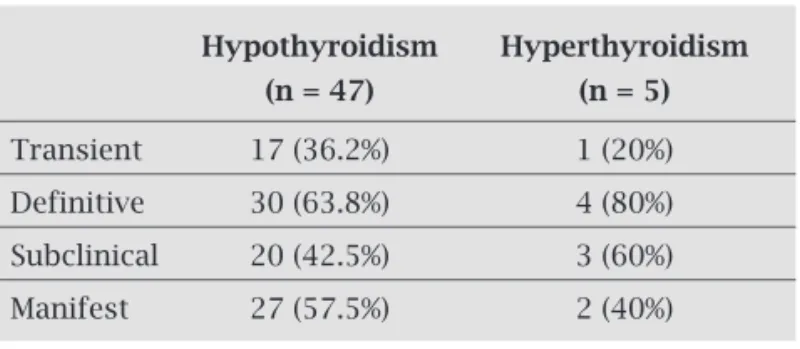
Table 4. Length and intensity of thyroid dysfunction
Hypothyroidism Hyperthyroidism
(n = 47) (n = 5)
Transient 17 (36.2%) 1 (20%) Definitive 30 (63.8%) 4 (80%)
Subclinical 20 (42.5%) 3 (60%)
Manifest 27 (57.5%) 2 (40%) Table 3. Characteristics of patients who developed
primary hypothyroidism under IFN-α treatment
Hypothyroid (n = 47)
Female 16 (34.0%)
Male 31 (66.0%)
Age (years) 44.6 ± 9.3 (25-68)
qALT 2.35 ± 1.61 (0.7-7.6)
Cirrhosis
Yes 8 (17.0%)
No 39 (83.0%)
Liver biopsy
Structural alterations
0/1 4 (8.5%)
2 20 (42.5%)
3 14 (29.8%)
4 7 (14.9%)
Virus C genotype (n = 253)
1 31 (72.1%)
2/3 12 (27.9%)
Event time under
26.52 ± 16.49 (8-96, median = 24) treatment (weeks)
Number of therapeutic regimens at the event
1 41 (87.2%)
2 6 (12.8%)
Total treatment
43.02 ± 15.64 (24-96; median = 48)
duration (weeks) Interferon used
IFN 14 (29.8%)
PEG 13 (27.6%)
IFN+PEG 20 (42.5%)
Final virological response
Yes 31 (68.9%)
No 14 (31.1%)
Sustained virological response (n = 254)
Yes 26 (57.8%)
No 19 (42.2%)
Free T4 (ng/dL) 0.79 ± 0.38 (0.04-1.6; median = 0.80) TSH (mIU/L) 27.5 ± 30.5 (4.7-100.0; median = 9.48)
DISCUSSION
Hepatitis C virus is a hepatotropic and lymphotropic RNA virus which may be associated with chronic infectious dis-ease.8 Although hepatocytes are the major site of HCV
rep-lication, extrahepatic complications of HCV infection may occur, such as autoimmune diseases and lymphoprolifera-tive disorders.9
HCV treatment with IFN-α and RBV can trigger adverse effects frequently leading to a reduction of the dose in over 40% of the patients or even withdrawal in approximately 14%.8
Standard or PEG-IFN-α may lead to influenza-like symptoms at the onset of treatment, as well as psychiatric, hematologic (neutropenia and thrombocytopenia)10,11 and
thyroid dysfunction.12 As referred by Sachithanandan et al.,13
thyropathies affect 26.3% of HCV treated patients. The as-sociation with RBV usually induces hemolytic anemia, with no additional risk of thyropathy.10
Generally, SVR is achieved in approximately 60% of the patients, 40% among patients of viral genotype 1 and 76% among those with viral genotypes 2 and 3. Other SVR predictive factors include: age less than 40 years, weight lower than 75 kg, female gender, Caucasian race, low vi-ral load before treatment, and absence of liver cirrhosis.10
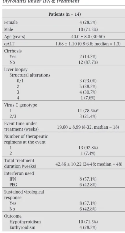
In our study, HCV patients obtained 53.9% as response rate of IFN-α therapy, in accordance to the literature. The study population showed a predominance of Caucasoid male patients infected by virus C of genotypes 1 and 3. Table 6. Patients who developed destructive thyroiditis under IFN-α treatment
Patients (n = 14)
Female 4 (28.5%)
Male 10 (71.5%)
Age (years) 40.0 ± 8.0 (30-60) qALT 1.68 ± 1.10 (0.8-6.6; median = 1.3)
Cirrhosis
Yes 2 (14.3%)
No 12 (87.7%)
Liver biopsy
Structural alterations
0/1 3 (23.0%)
2 5 (38.5%)
3 4 (30.7%)
4 1 (7.6%)
Virus C genotype
1 11 (78.5%)*
2/3 3 (21.4%)
Event time under
19.60 ± 8.99 (8-32, median = 18)
treatment (weeks) Number of therapeutic regimens at the event
1 13 (92.8%)
2 1 (7.4%)
Total treatment
42.86 ± 10.22 (24-48; median = 48)
duration (weeks) Interferon used
IFN 8 (57.1%)
PEG 6 (42.8%)
Sustained virological response
Yes 8 (57.1%)
No 6 (42.8%)
Outcome
Hypothyroidism 10 (71.5%)
Euthyroidism 4 (28.5%)
Table 5. Patients who developed hyperthyroidism under IFN-α treatment
Hyperthyroidism (n = 5)
Female 3 (60%)
Male 2 (40%)
Age (years) 46.4 ± 7.3 (39-56)
qALT 1.82 ± 0.94 (1.3-3.5; median = 1.4)
Cirrhosis
Yes 1 (20%)
No 4 (80%)
Liver biopsy
Structural alterations
0/1 0 (0%)
2 3 (60%)
3 1 (20%)
4 1 (20%)
Virus C genotype
1 2 (50%)
2/3 2 (50%)
Event time under
19.60 ± 8.99 (8-32, median = 18)
treatment (weeks)
Number of therapeutic regimens at the event
1 5 (100%)
Total treatment
43.2 ± 10.73 (24-48; median = 48)
duration (weeks) Interferon used
IFN 4 (80%)
PEG 1 (20%)
Sustained virological response
Yes 4 (80%)
No 1 (20%)
Free T4 (ng/dL) 2.57 ± 1.56 (1.36-4.46; median = 1.50) TSH (mIU/L) 0.13 ± 0.17 (0.01-0.33; median = 0.1)
Based on the literature,2 standard IFN was indicated
for patients infected with virus of genotypes 2 and 3, and PEG-IFN was the first choice for genotype 1. Non-responding patients with genotypes 2 and 3 could then be treated with PEG-IFN. More than one therapeutic regimen was prescribed to 35.2% of the patients, and 54.8% of them were treated for 48 weeks. PEG-IFN was administrated to 165 patients: those who weighed less than 75 kg used PEG–IFN-α 2b and those with more than 75 kg, PEG–IFN-α 2a.
Firstly, Pateron et al.14 reported a prevalence of 14% of
antithyroid autoantibodies in HCV patients. A review of published controlled studies15 observed that most reports
confirmed a higher prevalence of autoimmune thyroid dis-ease and hypothyroidism in chronic HCV-infected patients.
Several studies found that female gender and the pres-ence of TPOAb were considered major risk factors for the development of hypothyroidism.16-19 Our data demonstrated
that TPOAb-positive patients had a higher chance of de-veloping hypothyroidism; however, females were not more prone, similarly to the findings reported by Muratori et al.20
and Stefanova-Petrova et al.21
The prevalence of hypothyroidism in HCV patients is approximately 9% (0-13%) vs. 3% (0.5-4%) in healthy sub-jects.19,22-24 Thyroid autoimmunity was demonstrated in 15%
(5-28%) of the HCV patients and in 12% (0-11%) of healthy subjects, indicating a slight though significantly higher risk.15 In our study, we verified that the prevalence of
hypo-thyroidism (6.1%) and thyroid autoimmunity (5.5%) found in HCV patients before treatment was similar to the litera-ture reports, in spite of the differences existing in geographi-cal distribution, genetic variability, iodine intake or other infectious agents.25,26 Moreover, the prevalence of
hyperthy-roidism before IFN-α treatment (0.68%) found in our study population was not significantly different in HCV-infected patients, as described in the literature.15
After IFN-α therapy, 19% presented thyroid dysfunction: hypothyroidism in 17.2%, hyperthyroidism in 1.8% and destructive thyroiditis in 5.1% – very similar values were found by Sachithanandan et al.13 (26.3%), higher than those
described by Moncoucy et al.27 (7%) and Tran et al.16 (6.7%).
The rates of thyroid dysfunction triggered by the two types of IFN-α, standard and peguilated, were similar, as also found by Moncoucy et al.27 Moreover, thyroid
dysfunc-tion was not associated with sustained virological response of chronic hepatitis C to IFN-α therapy, as also demonstrat-ed by Hsieh et al.28 and in contrast to previous reports.29-31
In our patients, the presence of thyroid dysfunction did not lead to changes in the dose or therapy withdrawal.
After IFN-α plus RBV treatment 17.2% developed pri-mary hypothyroidism. However, this rate has varied in sev-eral studies.16,27 Among risk factors evaluated: age, gender,
presence of cirrhosis, qALT and FT4 levels before treatment,
type and commercial presentation of IFN-α used, and SVR, we found no association with primary hypothyroidism. In contrast, the likelihood of developing primary hypothyroid-ism increased in the presence of TPOAb, genotype 1, to be on the first therapeutic regimen, and having higher TSH lev-els before treatment. This last finding was also observed by Antonelli et al.19
Detection of TPOAb before therapy was associated with a risk 3.5 times greater of becoming hypothyroid on IFN-α use; 90% of such patients in our study developed the dysfunction vs. 14.5% of TPOAb-negative, corrobo-rating Prummel and Laurberg,7 who found a relative risk
of 3.9. Elevated endogenous interferon in response to viral diseases could possibly be associated with the de-velopment of IFN-α induced thyroiditis by in genetically predisposed individuals.17,27,32-34
Prummel and Laurberg7 have demonstrated that the
female gender has a relative risk of 4.4 for developing au-toimmune thyroiditis. This strong preponderance may perhaps be due to the effects of estrogen or secondary to X chromosome susceptibility genes.35,36 This fact was not
corroborated by our study, as well as by Muratori et al.20
and Stefanova-Petrova et al.21 It is important to highlight
that thyroid dysfunction could result from a direct effect of IFN-α on thyroid cell function.28,37,38
The higher prevalence of virus C genotype 1 in patients who developed hypothyroidism is not a consensus in the literature. Sachithanandan et al.13 have detected elevated
TPOAb levels before and during therapy only in geno-type 1 patients. Conversely, Huang et al.34 observed higher
prevalence of genotype 1b/2b in women with hepatitis C and detectable TPOAb, but with no correlation with hypo-thyroidism. Viral replication results in the production of a heterogeneous viral population within an infected indi-vidual.39 Some authors suggested that a portion of the HCV
genome could share a partial sequence homology in a few amino acid segments with thyroglobulin and microsome, rendering HCV patients susceptible to autoimmune thy-roid diseases.28 This hypothesis could explain the relation
between viral genotype and the predisposition to develop thyroid diseases in patients infected by virus C genotype 1, as verified in our patient population.
In the present study, patients had a relative risk of 3.5 for developing hypothyroidism during the first treatment course; however, Moncoucy et al.27 found no difference in
thyroid dysfunction rate between first and second courses of treatment. Previous studies have reported that thyroid dysfunction induced by IFN-α could develop in the first week or after a few months, during and after therapy, with a possible contribution of cumulative dose.17,29,32
syn-thesis and secretion in vitro.43 Our data did not suggest
a cumulative dose-effect, as only a few patients did not present thyroid dysfunction during the first treatment. Interestingly, after drug withdrawal, 64% of the patients remained hypothyroid, requiring L-thyroxin replacement therapy, and the other patients presented the subclinical form of the disease.
Hyperthyroidism seems a rare event during IFN-α ther-apy and was verified in 1.8% of the patients. Several studies found even lower prevalence of hyperthyroidism, ranging from 0.9% to 1.1%.16,27,29,44
Destructive thyroiditis occurred in 5.1% of our patients and approximately 70% developed permanent hypothyroid-ism. Almost 50% of non-autoimmune IFN-α-induced thy-roiditis manifests such a destructive thythy-roiditis, a self-lim-ited inflammatory disorder characterized by three phases of six to eight weeks each: thyrotoxicosis, hypothyroidism and resolution to euthyroidism or definitive hypothyroidism in less than 5% of the cases. The symptoms are usually mild, probably leading to a lower diagnostic rate; however, atrial fibrillation may occur.8,45 We observed that TSH levels before
treatment were higher and qALT was lower in patients who developed destructive thyroiditis under IFN-α. This finding has not been previously reported in the literature. Some au-thors found recurrent thyroiditis during retreatment;46
how-ever, in our study, 98% of the patients developed destructive thyroiditis only during the first treatment.
We concluded that in the screening for predictive factors of thyroid dysfunction in HCV patients before treatment with IFN-α it is especially important to evaluate TSH levels, as well as antithyroid antibodies and viral genotype. More-over, the first contact with IFN-α holds substantial risk to develop thyroid dysfunction. Special care is essential when there are laboratory alterations indicating thyrotoxicosis, which may be signaling an initial phase of destructive thy-roiditis, a benign and self-limited disturbance. We empha-size that a significant part of the dysfunction was transient or subclinical, not requiring treatment.
REFERENCES
1. Bellentani S, Tiribelli C. The spectrum of liver disease in the general population: lessons from the Dionysos study. J Hepatol 2001; 35:531-7.
2. Poynard T, Marcellin P, Lee SS et al. Randomized trial of inter-feron a2b plus ribavirin for 48 weeks or for 24 weeks versus in-terferon a2b plus placebo for 48 weeks for treatment of chronic infection with hepatitis C virus. Lancet 1998; 352:1426-32. 3. Tam RC, Pai B, Bard J et al. Ribavirin polarizes human T cell
responses toward a type 1 cytokine profile. J Hepatol 1999; 30:376-82.
4. Koh LKH, Greenspan FS, Yeo PPB. Interferon-a induced thy-roid dysfunction: three clinical presentations and review of the literature. Thyroid 1997; 7:891-6.
5. Dalgard O, Bjoro K, Hellum K et al. Thyroid dysfunction dur-ing treatment of chronic hepatitis C with interferon-a: no as-sociation with either interferon dosage or efficacy of therapy. J Int Med 2002; 251:400-6.
6. Carella C, Mazziotti G, Amato G et al. Interferon-α related thy-roid disease: pathophysiological, epidemiological, and clinical aspects. J Clin Endocrinol Metab 2004; 89:3656-61.
7. Prummel MF, Lauberg P. Interferon-α and autoimmune thy-roid disease. Thythy-roid 2003;13:547-51.
8. Tomer Y, Blackard JT, Akeno N. Interferon alpha treatment and thyroid dysfunction. Endocrinol Metab Clin N Am 2007; 36:1051-66.
9. Blackard JT, Kemmer N, Sherman KE. Extrahepatic replica-tion of HCV: insights into clinical manifestareplica-tions and biologi-cal consequences. Hepatology 2006;44:15-22.
10. Fried MW. Side effects of therapy of hepatitis C and their man-agement. Hepatology 2002; 36:237-44.
11. Evon DM, Verma A, Simpson K et al. Psychiatric symptoms during interferon treatment for hepatitis C: experiences from a tertiary care hepatology centre. Aliment Pharmacol Ther 2008; 27:1071-80.
12. Fentiman IS, Thomas BS, Balkwill FR et al. Primary hypothy-roidism associated with interferon therapy of breast cancer. Lancet 1985; 8438:1166.
13. Sachithanandan S, Clarke G, Crowe J et al. Interferon-associ-ated thyroid dysfunction in anti-D-relInterferon-associ-ated chronic hepatitis C. J Interf Cytok Res 1997; 17:409-11.
14. Pateron D, Hartmann DJ, Duclos-Vallee JC et al. Latent auto-immune thyroid disease in patients with chronic HCV hepati-tis. J Hepatol 1992; 16:244-5.
15. Antonelli A, Ferri C, Fallahi P et al. Thyroid disorders in chronic hepatitis C virus infection. Thyroid 2006;16:563-72. 16. Tran A, Quaranta JF, Benzaken S et al. High prevalence of
thyroid autoantibodies in a prospective series of patients with chronic hepatitis C before interferon therapy. Hepatol 1993; 18:253-7.
17. Preziati D, La Rosa L, Covini G et al. Autoimmunity and thy-roid function in patients with chronic active hepatitis treated with recombinant interferon alpha-2a. Eur J Endocrinol 1995; 132:587-93.
18. Fernandez-Soto L, Gonzalez A, Escobar-Jimenez F et al. In-creased risk of autoimmune thyroid disease in hepatitis C vs
hepatitis B before, during, and after discontinuing interferon therapy. Arch Int Med 1998; 158:1445-8.
19. Antonelli A, Ferri C, Pampana A et al. Thyroid disorders in chronic hepatitis C. Am J Med 2004; 117:10-13.
20. Muratori L, Bogdanos DP, Muratori P et al. Susceptibility to thyroid disorders in hepatitis C. Clin Gastroenterol Hepatol 2005; 3:595-603.
21. Stefanova-Petrova DV, Tzvetanska AH, Naumova EJ et al. Chronic hepatitis C virus infection: prevalence of extrahepatic manifestations and association with cryoglobulinemia in Bul-garian patients. World J Gastroenterol 2007; 13:6518-28. 22. Matsuda J, Saitoh N, Gotoh M et al. High prevalence of
anti-phospholipid antibodies and anti-thyroglobulin antibody in patients with hepatitis C virus infection treated with interfer-on-alpha. Am J Gastroenterol 1995; 90:1138-41.
23. Custro N, Montalto G, Scafidi V et al. Prospective study on thyroid autoimmunity and dysfunction related to chronic hepatitis C and interferon therapy. J Endocrinol Invest 1997; 20:374-80.
25. Prentice LM, Phillips DI, Sarsero D et al. Geographical distri-bution of subclinical autoimmune thyroid disease in Britain: a study using highly sensitive direct assays for autoantibod-ies to thyroglobulin and thyroid peroxidase. Acta Endocrinol 1990;123:493-8.
26. Minelli R, Braverman LE, Giuberti T et al. Effects of excess io-dine administration on thyroid function in euthyroid patients with a previous episode of thyroid dysfunction induced by interferon-alpha treatment. Clil Endocrinol 1997; 47:357-61. 27. Moncoucy X, Leymarie F, Delemer B et al. Risk factors and
long-term course of thyroid dysfunction during antiviral treat-ments in 221 patients with chronic hepatitis C. Gastroenterol Clin Biolog 2005; 29:339-45.
28. Hsieh MC, Yu ML, Chuang WL et al. Virologic factors related to interferon-alpha-induced thyroid dysfunction in patients with chronic hepatitis C. Eur J Endocrinol 2000; 142:431-7. 29. Lisker-Melman M, Di Bisceglie AM, Usala SJ et al.
Develop-ment of thyroid disease during therapy of chronic viral hepati-tis with interferon alfa. Gastroenterol 1992; 102:2155-60. 30. Primo J, Hinojosu J, Molés JR et al. Development of thyroid
dysfunction after alpha-interferon treatment of chronic hepa-titis C. Am J Gastroenterol 1993; 88:1976-7.
31. Reid I, Sharpe I, McDevitt J et al. Thyroid dysfunction can pre-dict response to immunotherapy with interleukin-2 and inter-feron-2 alpha. Brit J Cancer 1991; 64:915-8.
32. Watanabe U, Hashimoto E, Hisamitsu T et al. The risk factor for development of thyroid disease during interferon-alpha therapy for chronic hepatitis C. Am J Gastroenterol 1994; 89:399-403.
33. Vanderpump MP, Tunbridge WM, French JM et al. The inci-dence of thyroid disorders in the community: a twenty-year follow-up of the Whickham Survey. Clin Endocrinol 1995; 43:55-68.
34. Huang JF, Chuang WL, Dai CY et al. The role of thyroid au-toantibodies in the development of thyroid dysfunction in Tai-wanese chronic hepatitis C patients with interferon-alpha and ribavirin combination therapy. J Vir Hepat 2006; 13:396-401. 35. Grossman CJ, Roselle GA, Mendenhall CL. Sex steroid
regula-tion of autoimmunity. J Ster Biochem Mol Biol 1991; 40:649-59.
36. Tomer Y, Davies TF. Searching for the autoimmune thyroid disease susceptibility genes: from gene mapping to gene func-tion. Endocr Rev 2003; 24:694-717.
37. Carella C, Amato G, Biondi B et al. Longitudinal study of anti-bodies against thyroid in patients undergoing interferon-alpha therapy for HCV chronic hepatitis. Horm Res 1995; 44:110-4. 38. Katabami S, Kamijo K, Kodama T et al. An episode of silent
thyroiditis in a patient with chronic thyroiditis and papillary adenocarcinoma following alpha interferon treatment for hep-atitis C. Endocr J 1993; 40:311-6.
39. Simmonds P. Genetic diversity and evolution of hepatitis C vi-rus - 15 years on. J Gen Virol 2004; 85:3173-88.
40. Gisslinger H, Gilly B, Woloszczuk W et al. Thyroid autoim-munity and hypothyroidism during long-term treatment with recombinant interferon-alpha. Clil Exp Immunol 1992; 90:363-7.
41. Nagayama Y, Ohta K, Tsuruta M et al. Exacerbation of thyroid autoimmunity by interferon alpha treatment in patients with chronic viral hepatitis: our studies and review of the literature. Endocr J 1994; 41:565-72.
42. Chung YH, Shong YK. Development of thyroid autoimmun-ity after administration of recombinant human interferon-alpha 2b for chronic viral hepatitis. Am J Gastroenterol 1993; 88:244-7.
43. Yamazaki K, Kanaji Y, Shizume K et al. Reversible inhibition by interferons alpha and beta of 125I incorporation and thy-roid hormone release by human thythy-roid follicles in vitro. J Clin Endocrinol Metab 1993; 77:1439-41.
44. Hollowell JG, Staehling NW, Flanders WD et al. Serum TSH, T(4), and thyroid antibodies in the United States population (1988 to 1994): National Health and Nutrition Examina-tion Survey (NHANES III). J Clin Endocrinol Metab 2002; 87:489-99.
45. Weetman AP, Smallridge RC, Nutman TB et al. Persistent thy-roid autoimmunity after subacute thythy-roiditis. J Clin Lab Im-munol 1987; 23:1-6.