C
a s eR
e p o Rt3 3 9 Arq Bras Oftalmol. 2016;79(5):339-41 http://dx.doi.org/10.5935/0004-2749.20160097
INTRODUCTION
Scleritis is currently recognized as a heterogeneous group of diseases characterized by inflammation of the sclera, which may be caused by a local or systemic infection, an immune-mediated di sease, or a primary manifestation of an acquired connective tissue disorder or vasculitic disease, often signaling a life-threatening situa-tion(1,2). Surgically induced necrotizing scleritis (SINS) occurs after
ocular surgeries for cataract extraction, trabeculectomy, strabismus,
and pterygium retinal detachment(3).
The diagnosis and treatment of these special cases using immu-nosuppressive patch grafts or amniotic membrane grafts should be
prompt(4). Immunosuppressive agents used to treat these conditions
include azathioprine, cyclophosphamide, tacrolimus, and high-dose
pulse methylprednisolone(4). Although other biological agents can be
used, particularly for the treatment of necrotizing scleritis, which po-tentially represents vasculitis, rituximab is generally recommended for the treatment of systemic vasculitis(5).
CASE REPORT
Our patient was a 51-year-old woman who presented with a chief complaint of redness and pain affecting both eyes. She pre-viously underwent (2-3 months before) pterygium excision using the bare sclera technique, without the use of adjunctive irradiation or mitomycin C. An ophthalmological examination revealed bilateral conjunctival hyperemia and scleral thinning with peripheral corneal ulcers measuring 5 × 2 mm in the right eye and nasal scleral thinning with peripheral corneal swelling in the left eye that further promoted scleral thinning, staphyloma formation, and subsequent ulcerative
necrosis. Complete hematology and immunology profiles for rheu-matoid arthritis, antinuclear antibody (ANA), and anti-neutrophil cy-toplasmic antibodies (ANCA) were determined, which showed that only ANA was positive and extractable nuclear antigen antibodies (ENA) were negative. Serology tests, such as venereal disease re search
laboratory test, Treponema pallidum hemagglutination assay, and
tuberculin test, were all negative.
She was further evaluated in the Rheumatology Department because of generalized pain and ANA positivity. Combined with la-boratorial analysis showing normal renal, hematological, and hepatic functions and negativity for anti-double stranded DNA (dsDNA), ANCA, ENA, rheumatoid factor, and anti-cyclic citrullinated peptide antibody (anti-CCP), a diagnosis of fibromyalgia and SINS was made, and the patient was promptly treated with high-dose corticoste-roids. However, 1 month later, she presented with aggravated scleral thinning; thus, immunosuppressive agents (2 mg/kg of azathioprine
and 1 g/m2/month of intravenous cyclophosphamide) were added
to corticosteroid therapy for 12 months. However, remission was not achieved and necrotizing scleritis of the right eye continued. There-fore, rituximab treatment was initiated, which resulted in improve-ment after the first cycle of two infusions of 1 g each administered 2 weeks apart. Visual acuity was preserved in the left eye but decreased from 20/20 to 20/160 in the right eye. The prednisone dose was tapered off, and after 3 months, the patient was free from all drugs (Figures 1-4). She has remained symptom-free for 6 months after the second rituximab cycle. The laboratory workup was repeated twice, and ANA was the only altered parameter with a 1/320 speckled pat-tern, and ENA remained negative. There was no symptom that could be attributed to a collagen disease.
Management of necrotizing scleritis after pterygium surgery with rituximab
Tratamento com rituximabe de esclerite necrosante após cirurgia de pterigeo
Tania SaleSde alencar Fidelix1, luiS anTonio Vieira1, Virginia FernandeS Moca TreViSani1
Submitted for publication: August 5, 2015 Accepted for publication: December 9, 2016
1 Departamento de Oftalmologia e Ciências Visuais, Universidade Federal de São Paulo (UNIFESP),
São Paulo, SP, Brazil.
Funding: No specific financial support was received for this study.
Disclosure of potential conflicts of interest: The authors declare no potential conflict of interest.
Corresponding author: Tania Fidelix. TSA Reumatologia. Rua Barão do Triunfo, 156 - São Paulo, SP 04602-000 - Brazil - E-mail: [email protected]
ABSTRACT
The authors present a case of necrotizing scleritis after pterygium excision successfully treated with rituximab after attempts with high doses of corticos-teroids and immunosuppressive drugs. A literature review revealed case reports and a phase I/II dose-ranging randomized clinical trial using rituximab for necrotizing scleritis with or without association with autoimmune disease. This is the only case report on rituximab treatment for necrotizing scleritis after pterygium surgery. In cases with refractoriness to immunosuppressive drugs, a CD20 antibody can be used.
Keywords: Scleritis; Pterygium/surgery; Rituximab/therapeutic use; Antibodies, monoclonal/administration and dosage
RESUMO
Os autores apresentam um caso de sucesso no tratamento com rituximabe de esclerite necrosante após cirurgia de pterígio refratário a altas doses de corticoste-roides e drogas imunossupressoras. Uma revisão da literatura direcionada ao uso de rituximabe para tratamento de esclerites necrosantes revelou relatos de casos e um estudo clínico randomizando fase I/II. Este é o único caso descrito de rituximabe para o tratamento de esclerite necrosante pós cirúrgica. O uso de anticorpo anti-CD20 pode ser uma opção em casos refratários aos imunossupressores no tratamento da esclerite necrosante pós-cirúrgica.
Ma n a g e M e n to fn e c r o t i z i n gs c l e r i t i sa f t e rp t e ry g i u Ms u r g e ryw i t hr i t u x i M a b
3 4 0 Arq Bras Oftalmol. 2016;79(5):339-41 DISCUSSION
The local and systemic treatment of scleritis has recently under-gone significant changes. These changes include more aggressive and early treatment of patients to maintain vision and achieve rapid remission.
Increased use of combination immunosuppressive therapy and biological agents in patients with severe and refractory ocular inflam-matory disease has contributed to modifications in the prognosis of
these conditions1.
Scleral necrosis and melting may occur after pterygium surgery
due to the use of adjunctive irradiation(6) and mitomycin C(7), although
the use of these gents reportedly contributes to prolonged inhibition of wound healing. In addition, excessive cauterization during the
bare sclera technique may cause scleral necrosis(8). According to a
review by Doshi et al.(9), the technique used for pterygium surgery can
alter the risk of necrotizing or even infectious scleritis. In 203 cases, SINS occurred in 17.2% and scleritis in 68.8% of cases that underwent the bare sclera technique, as in our case.
Medical management of SINS after pterygium surgery includes immunosuppression with the oral steroids methylprednisolone + cyclophosphamide or tacrolimus for patients who do not respond
to cyclophosphamide and azathioprine(4). Surgical intervention in the
form of patch grafts of scleral, corneal, or amniotic membranes(10) has
also been reported.
Autoimmunity or hypersensitivity is now well accepted as an etiological factor in the development of SINS. Immune complexes have been found in and around episcleral vessel walls by immuno-fluorescence techniques, and systemic immunosuppressive regimes
have been successful in the treatment of SINS(3,10). Clinical or
serolo-gical markers of connective tissue disorders are present in as many as 62% of cases. Our patient was ANA-positive without evidence of any associated connective tissue disorder. The search for an associated autoimmune disease was exhaustive and included tests for anti-CCP, ENA panel, complement system proteins, and anti-dsDNA. These tests were performed on more than two occasions, particularly for ANA and the ENA panel, because it was important to exclude the
Figure 1. Right eye before rituximab. Conjunctival and scleral nasal hyperemia with local thinning and a peripheral corneal ulcer.
Figure 2. Left eye before rituximab. Conjunctival hyperemia, scleral thinning, and asso ciated scleral staphyloma with necrotic ulceration.
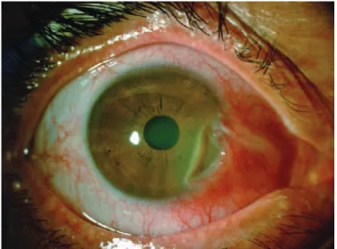
Figure 3. Right eye after rituximab. Sectoral scleral thinning with no signs of active inlammation.
Fi d e l i x TSA, e TA l.
3 4 1 Arq Bras Oftalmol. 2016;79(5):339-41
possibility of an autoimmune disease associated with ANA positivity, such as systemic lupus erythematosus or systemic vasculitis.
In this case, the scleritis was refractory to classical immunosup-pressive therapy, although all recommended steps, including high prednisone doses, azathioprine and cyclophosphamide (12 pulses), were attempted.
A review of the literature for studies of biologic therapy for scleri-tis revealed case reports and a randomized clinical trial of necrotizing scleritis treated with rituximab after immunosuppressive therapy,
which achieved good responses(11,12).
Rituximab is a chimeric anti-CD20 monoclonal antibody that continues to increase in popularity for the treatment of ocular
in-flammatory diseases and intraocular lymphoma(13). SINS should be
considered as a differential diagnosis in patients with scleritis or scle-ral melting following pterygium surgery, particularly after radiation or mitomycin C therapy. Evidence of a connective tissue disease may or may not be found on clinical examination or laboratory inves-tigations, although it remains uncertain whether an autoimmune disease is present before or is unleashed by the section of sclera. Therefore, it is important to be alert to the risks of scleritis after such surgical procedures, particularly those that can lead to the damage of avascular tissue, such as sclera, because the risks of necrosis and infections are considerably increased. Early diagnosis and prompt immunosup pression is required for the successful management of
this complication(4). The CD20 antibody rituximab is now an option
that can be used for the treatment of these special cases.
REFERENCES
1. Wakefield D, Di Girolamo N, Thurau S, Wildner G, McCluskey P. Scleritis: immunopa-thogenesis and molecular basis for therapy. Prog Retin Eye Res. 2013;35:44-62.
2. Foster CS. Ocular manifestations of the potentially lethal rheumatologic and vascu-litic disorders. J Fr Ophtalmol. 2013;36(6):526-32.
3. Galanopoulous A, Snibson G, O’Day J. Necrotising anterior scleritis after pterygium surgery. Aust NZ J Ophthalmol. 1994;22(3):167-73.
4. Beardsley RM, Suhler EB, Rosenbaum JT, Lin P. Pharmacotherapy of scleritis: current paradigms and future directions. Expert Opin Pharmacother. 2013;14(4):411-24. 5. Stone JH, Merkel PA, Spiera R, Seo P, Langford CA, Hoffman GS, Kallenberg CG, St Clair
EW, Turkiewicz A, Tchao NK, Webber L, Ding L, Sejismundo LP, Mieras K, Weitzenkamp D, Ikle D, Seyfert-Margolis V, Mueller M, Brunetta P, Allen NB, Fervenza FC, Geetha D, Keogh KA, Kissin EY, Monach PA, Peikert T, Stegeman C, Ytterberg SR, Specks U; RAVE-ITN Research Group. Rituximab versus cyclophosphamide for ANCA-associated vasculitis. N Engl J Med. 2010;363(3):221-32. Comment in: Nat Rev Rheumatol. 2010; 6(10):556; N Engl J Med. 2010;363(21):2072-3; author reply 2073-4; Curr Rheumatol Rep. 2010;12(6):395-8; N Engl J Med. 2010;363(21):2072-3; author reply 2073-4. 6. Mackenzie FD, Hirst LW, Kynaston B, Bain C. Recurrence rate and complications after
beta-irradiation for pterygium. Ophthalmology. 1991;98(12):1776-81. Comment in: Oph thalmology. 1992;99(6):841. Ophthalmology. 1992;99(6):841-2.
7. Rubenfeld RS, Pfister RS, Stein RM, Foster CS, Martin NF, Stoleru S, et al. Serious compli-cations of topical mitomycin-C after pterygium surgery. Ophthalmology. 1992;99(11): 1647-54.
8. Alzagoff Z, Tan DT, Chee SP. Necrotising scleritis after bare sclera excision of ptery-gium. Br J Ophthalmol. 2000;84(9):1050-2.
9. Doshi RR, Harocopos JH, Schwab IR, Cunningham Jr ET. The spectrum of postopera-tive scleral necrosis. Surv Ophthalmol. 2013;58(6):620-33.
10. O’Donoughue E, Lightman S, Tuft S, Watson P. Surgically induced necrotizing scle-rokeratitis (SINS)-precipitating factors and response to treatment. Br J Ophthalmol. 1992;76(1):17-21
11. Suhler EB, Lim LL, Beardsley RM, Giles TR, Pasadhika S, Lee ST, et al. Rituximab therapy for refractory orbital inflammation: results of a phase 1/2, dose-ranging, randomized clinical trial. JAMA Ophthalmol. 2014;132(5):572-578.
12. Joshi L, Tanna A, McAdoo SP, Medjeral-Thomas N, Taylor SR, Sandhu G, et al. Long- term outcomes of rituximab therapy in ocular granulomatosis with polyangiitis: impact on localized and nonlocalized disease. Ophthalmology. 2015;122(6):1262-8. 13. Shetty RK, Adams BH, Tun HW, Runyan BR, Menke DM, Broderick DF. Use of rituximab
for periocular and intraocular mucosa-associated lymphoid tissue lymphoma. Ocul Immunol Inflamm. 2010;18(2):110-2.