Using Group II Introns for Attenuating the
Vitro
and
In Vivo
Expression of a Homing
Endonuclease
Tuhin Kumar Guha, Georg Hausner*
Department of Microbiology, University of Manitoba, Winnipeg, Canada
Abstract
InChaetomium thermophilum(DSM 1495) within the mitochondrial DNA (mtDNA) small ribosomal subunit (rns) gene a group IIA1 intron interrupts an open reading frame (ORF) encoded within a group I intron (mS1247). This arrangement offers the opportunity to exam-ine if the nested group II intron could be utilized as a regulatory element for the expression of the homing endonuclease (HEase). Constructs were generated where the codon-opti-mized ORF was interrupted with either the native group IIA1 intron or a group IIB type intron. This study showed that the expression of the HEase (in vivo) inEscherichia colican be reg-ulated by manipulating the splicing efficiency of the HEase ORF-embedded group II introns. Exogenous magnesium chloride (MgCl2) stimulated the expression of a functional HEase but the addition of cobalt chloride (CoCl2) to growth media antagonized the expression of HEase activity. Ultimately the ability to attenuate HEase activity might be useful in precision genome engineering, minimizing off target activities, or where pathways have to be altered during a specific growth phase.
Introduction
Homing endonucleases (HEases) are site-specific DNA cleaving enzymes that are encoded by homing endonuclease genes (HEGs) which are frequently found embedded within archaeal introns, composite mobile genetic elements such as group I introns, group II introns, inteins [1–5] and sometimes HEGs are freestanding [5–9]. The LAGLIDADG family (LAG HEase) are frequently encoded within fungal mitochondrial group I introns [10,11] and HEases in general recognize long asymmetrical 12–40 bp of DNA sequences as their target sites and cleave in a manner that generates four nucleotide 30-overhangs [6–8]. As LAG HEases require long DNA target sequences they cut infrequently within a genome [8] and this feature has been utilized for various applications in genome editing as it relates to agriculture [12], population control of disease vectors [13–15], and human health [16–19]. In this study we are testing the possibil-ity of utilizing mitochondrial group II intron sequences as an on/off“switch”system that pro-vides the opportunity for temporal control of HEase activity inEscherichia coli.
OPEN ACCESS
Citation:Guha TK, Hausner G (2016) Using Group II Introns for Attenuating theIn VitroandIn Vivo Expression of a Homing Endonuclease. PLoS ONE 11(2): e0150097. doi:10.1371/journal.pone.0150097
Editor:Cecile Fairhead, Institut de Genetique et Microbiologie, FRANCE
Received:October 27, 2015
Accepted:February 9, 2016
Published:February 24, 2016
Copyright:© 2016 Guha, Hausner. This is an open access article distributed under the terms of the
Creative Commons Attribution License, which permits unrestricted use, distribution, and reproduction in any medium, provided the original author and source are credited.
Data Availability Statement:All relevant data are within the paper and its Supporting Information files.
Funding:This research is supported by a Discovery Grant from the Natural Sciences and Engineering Research Council of Canada to GH. The authors also gratefully acknowledge support for TKG from a University of Manitoba Faculty of Science Graduate Award and the University of Manitoba Faculty of Graduate Studies GETS program.
Previously a molecular switch was developed that controlled the endonuclease activity of PI-SceIin vitro; here two cysteine amino acid residue pairs were separately inserted into the HEase DNA binding loops to allow for disulfide bond formation that lock the endonuclease into a nonproductive conformation [19]. This essentially is a redox switch and the activity of the protein could be controlled by adding or removing a reducing agent [19]. Other strategies suggested for regulating endonuclease activity included manipulating metal ion cofactors (such as Mg+2) or developing temperature sensitive versions of HEases [20]. However, in order for a true on/off“switch”it was suggested that manipulating cellular concentrations of ion cofactors might be difficult and using different temperatures might pose a problem with some cell lines or some temperature sensitive HEases once misfolded could not be reactivated (i.e. refolded) [19]. Redox switches as developed for PI-SceI have potential forin vitroapplications but are not practical forin vivoapplications as the targeted cell would probably suffer damage if oxidiz-ing conditions were applied.
Recently we characterized a twintron-like arrangement (or‘nested’intron) at position S1247 in the mitochondrial DNA (mtDNA) small ribosomal subunit (rns) gene of Chaeto-mium thermophilumvar.thermophilumLa Touche (strain DSM 1495). The external intron encodes a double-motif LAG HEase ORF (I-CthI) which is interrupted by an internal ORF-less group IIA1 intron [21]. The group IIA1 intron was shown byin vitroself-splicing experiments to be excised and thus generating a transcript where the LAG HEase ORF is contiguous and could allow for the expression of a functional endonuclease [22]. Splicing of the internal group II intron could be a regulatory step that allows for the maturation of the HEase transcript and translation of the open reading frame. Splicing of group II introns requires the intron RNA to assume a splicing competent tertiary fold that includes interactions between intron and flank-ing exon sequences. Foldflank-ing of the intron RNA is in part facilitated by base pair complementar-ities [23]. In addition various intron and/or host genome encoded factors tend to assistin vivo RNA folding and splicing of group II intron from transcripts [23,24]. Group II intron RNAs are ribozymes composed of six helical regions, referred to as domains I through VI, emerging from a central wheel and these domains interact to form a conserved tertiary structure that brings together distant sequences to form an active site. The active site binds the splice sites and the branch-point nucleotide residue and uses bound Mg+2ions to activate the appropriate bonds for catalysis [25,26]. Terbium cleavage assay determined domain V (DV) to be an important region of the active site as it contains the catalytic triad AGC and an AY bulge, both of which bind Mg+2[27,28]. Experimental results from phosphorothioate substitutions at the splice sites suggested that group II introns either use separate active sites with different Mg+2 ions to catalyze the two splicing (transesterification) steps or a single active site which is rear-ranged between the steps [29,30]. Moreover, X-ray crystallographic structure of the catalytic core of anOceanobacillus iheyensisIIC intron revealed that two helices of DV are bent to bring the AC bulge near the CGC triad, thus juxtaposing the phosphate backbones of the most con-served DV sequences, rather than stacking coaxially [31]. Nine potential Mg+2binding sites were assigned in or near DV for this intron [32]. Hence, a potential trigger for splicing might involve the availability of Mg+2insideE.colicells and this property has been applied in this research to develop a mechanism for thein vivoregulation of HEase (I-CthI) activity.
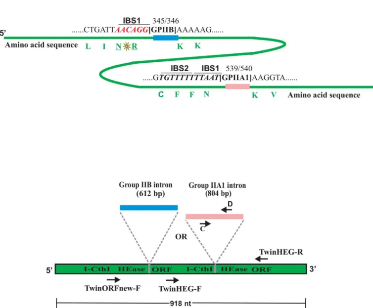
part to establish a splicing competent fold [32]. A second construct was designed where the native IIA1 intron was replaced by a mitochondrial group IIB intron (no ORF) [rI1 of Scene-desmus obliquus] along with its corresponding IBS1 sequence [33].
It has been suggested that thein vivocontrol of HEase activity would allow for more precise temporal inactivation or modification of genes [34,35]; for example, to shift metabolic cesses during a particular growth phase of the bacteria that are being manipulated for the pro-duction of certain metabolites or proteins [36].
Materials and Methods
Design of the
Escherichia coli
expression vectors and substrate
An expression plasmid with the codon-optimized version of the I-CthI ORF along with its native internal group IIA1 intron (GenBank accession number: JX139037.1) was synthesized by Genscript (New Jersey, USA). However, the IBS1 and IBS2 sequences which are upstream of the intron insertion site (IBS1: 50TGTTTT 30, IBS2: 50TTTAAT 30) and thus located within the HEase ORF were not modified in order to maintain the splicing potential of the group IIA1 intron. The synthesized ORF sequence (1722 bp) was inserted into the pET28b (+) plasmid as a NheI/BamHI fragment. The vector provides the ORF with an N-terminal 6X histidine-tag; this construct was named I-CthI-[IIA1]-pET28b (+).
In order to expand the concept of the“intron based on switch”as a potential regulatory ele-ment for HEase expression, a non-native group IIB intron (rI1) fromScenedesmus obliquus (GenBank accession number X17375.2) [37] was inserted in a suitable position within the I-CthI ORF. The intron was inserted at a location that allowed for maintaining the IBS/EBS interactions with minimal modification of the HEase coding sequence; fortuitously for the rI1 intron only the IBS1 sequence is essential for splicing [33]. Prior to the insertion of this intron, a suitable region in the HEase ORF sequence was located, that matched the required IBS1 (aac coding for arginine andaggcoding for asparagine) element for this group IIB intron. However, the presence of this sequence at the carboxyl terminal of the HEase ORF made it less suitable. An alternative approach was undertaken. A sequence located further upstream,cgcaac encod-ing asparagine and arginine, was rearranged toaaccgcessentially introducing two conservative amino acid replacements. Thecgcnucleotides were further modified toaggwhich did not change the amino acid composition of the protein (Fig 1A). The codon-optimized ORF (1530 bp) including the intron was synthesized and inserted as a NdeI/BamHI fragment into the pET28b (+) plasmid (same as for the native intron). The construct was named I-CthI-[IIB]-pET28b (+). Both of the constructs were transformed intoE.coliBL21 (λDE3) (chemi-cally competent cells, New England Biolab, MA, USA) for testing thein vivogroup II intron splicing competency and for additional biochemical studies.
A substrate plasmid was previously constructed in order to evaluate the activity of I-CthI [22]. Briefly, a segment of thernssequence (GenBank accession number: JN007486.1) flanking the mS1247 nested intron was synthesized by Genscript (New Jersey, USA) and cloned in pUC57 vector as an EcoRV fragment. This substrate plasmid was named Cth-rns.pUC57 and the overall size of the construct is 3.1 kb. Most of the chemicals and the reagents used in this study were purchased from Thermo Fisher Scientific unless otherwise mentioned in the text.
In vivo
RNA splicing assay
transformed with either I-CthI-[IIA1]-pET28b (+) or the empty pET28b (+) vector and inoculated in 10 mL of Luria Bertani (LB; peptone 10 g/L, yeast extract 5 g/L and NaCl 5 g/L; pH 7.0) media supplemented with 100μg/mL kanamycin and 0.25% w/v glucose and
incu-bated overnight (O/N) with agitation at 37°C. Five hundredμL from the O/N culture was
Fig 1. (A) Homing endonuclease ORF and location of introns.Schematic representation (not drawn to scale) for the location of the native group IIA1
intron (GPIIA1, shown in pink) and the non-native group IIB intron (GPIIB, shown in blue) within the I-CthI HEase ORF sequence (shown in green). Number 539 and 540 represent the exact location (insertion site) of the GPIIA1 in the ORF sequence. The IBS1 and the IBS2 elements are in italics (underlined), both are located upstream from the intron insertion site. The corresponding amino acids (marked in green) have been indicated to their respective codons. In another construct, GPIIB was inserted in the I-CthI HEase ORF sequence at position 345/346 that allowed for maintaining the IBS/EBS interactions with minimal modification of the HEase coding sequence (see text for details). The conservative amino acid substitution (indicated with a yellow star) between Arginine (R) and Asparagine (N) residues has been introduced to form the correct IBS1 (marked in red, italics and underlined) for supporting GPIIB splicing. The corresponding amino acids (marked in green) have been indicated to their respective codons.(B) Overview of primer location for RT-PCR.Diagram (not drawn to scale) showing the relative location of the RT-PCR primers within I-CthI HEase ORF sequence utilized for detecting splicing products during the
in vivoRNA splicing assay.
used to inoculate 50 mL of LB medium supplemented with kanamycin (kan) and glucose (as described above). Additionally, each of the culture flasks was supplemented with either 1 mM, 5 mM, 10 mM or 20 mM magnesium chloride (MgCl2). Initially, exogenous MgCl2
concentrations up to 100 mM were tested. Even though such high concentrations of MgCl2
was not detrimental to the bacterial cells (as evident from checking O.D.600), there was no
appreciable difference in the splicing activity as compared to the culture containing 20 mM MgCl2. Therefore, MgCl2concentrations up to 20 mM were considered for further studies. A
culture flask with no exogenously added MgCl2was used as the negative control. A second
set of negative controls consisted of cultures either: a) 10μM of cobalt chloride (CoCl2) was
added to the LB media or b) the LB media was supplemented with both 10μM CoCl2and 5
mM MgCl2. The cultures were grown at 37°C with agitation till the O.D.600reached 0.65.
Ten mL of the bacterial cells from each of the above cultures were centrifuged for 3 minutes at 7000 rpm. The cells were lysed and RNA was extracted using the GENEzol TriRNA Pure kit (FroggaBio, North York, Ontario) following the manufacturer’s protocol. To ensure com-plete removal of any contaminating DNA, the RNA was treated with 2 units of DNaseI (Fer-mentas, Ottawa, Canada) and incubated at 37°C for 15 minutes; the reaction was stopped by adding 1μL EDTA (50 mM) followed by 10 minute incubation at 65°C. Furthermore, in
order to confirm the elimination of the DNA, 2μL of the reaction mixture was applied to
perform a standard PCR reaction using the forward primer TwinHEG-F (50-ATGTGGT TATCCCGCATTTCG-30) and the reverse primer TwinHEG-R (50-TTGAAGTTTT CGTTCTTGATGCC-30;Fig 1B).
The ThermoScript RT-PCR system (Life Technologies) was used to make cDNA from the RNA extracted from bacterial cells grown in the presence of various concentrations of MgCl2.
Briefly, for the first strand synthesis a 20μL reaction mix was prepared containing 1μg RNA,
0.5 mM of the reverse primer TwinHEG-R, 0.1 M DTT, 4μL of 5X cDNA synthesis buffer, 1
mM of each dNTP, 40 units RNase-OUT (Life Technologies) and 15 units of Thermoscript reverse transcriptase. Reverse transcription was performed at 55°C for 1 hour and stopped by heating the reaction mixture to 85°C for 10 minutes. Finally, 1μL of RNase H (2 units) was
added to the reaction mixture followed by incubation at 37°C for 20 minutes.
In vitro
and
in vivo
protein expression and purification
For both types of constructs I-CthI-[IIA1]-pET28b (+) and I-CthI-[IIB]-pET28b (+), protein expression was first evaluated with anin vitrotranslation assay. RNA extracted fromE.coli BL21 cells grown under different Mg+2concentrations were further subjected toin vitro trans-lation. The PURExpressIn VitroProtein Synthesis Kit (New England Biolab, MA, USA) which is a cell-free transcription/translation system was utilized to assess if the proteins can be expressed in an“E.coli”environment. Although the PURExpress kit is designed for coupled transcription and translation from an expression construct, direct translation from an mRNA template is also possible provided purified RNA (1–5μg) is added to the reaction mixture,
albeit a proper ribosome binding site must be present for efficient translation. After three hours of incubation at 37°C, 2.5μL of the reaction sample was mixed with 2.5μL of the 2x
pro-tein loading dye [65.8 mM Tris-HCl, pH 6.8, 26.3% (w/v) glycerol, 2.1% SDS, 0.01% Bromo-phenol blue] and subjected to 12.5% SDS PAGE (BioRad, Mississauga, Ontario). The gel was stained in Coomasie Brilliant Blue (Roche, Mississauga, Ontario) and analyzed for the presence of the desired protein at 29 kDa.
Forin vivoprotein overexpression inE.coliand protein purification, the same protocols were applied for both types of constructs I-CthI-[IIA1]-pET28b (+) and I-CthI-[IIB]-pET28b (+). In order to check for the expression of the HEase proteinin vivo, the remaining 40 mL of culture (first 10 mL from each 50 mL culture were removed for RNA extraction to performin vivoRNA splicing assay; see above) was induced with Isopropylβ-D-1-thiogalactopyranoside (IPTG; Life Technologies) to a final concentration of 0.5 mM when the O.D.600of the cells
reached 0.65. The cultures were then shifted to 28°C and incubated with agitation for 4 hours. Cells were resuspended in the lysis buffer [50 mM Tris-HCl (pH 8.0), 100 mM NaCl, 10% (w/ v) glycerol, 6 mMβ-mercaptoethanol] at a ratio of 5 mL of buffer to 1 g of cells (wet weight) and sonicated in short pulses of 15 seconds using the Sonic Dismembrator model 300 (Thermo Fisher Scientific). Eightμg of crude lysate from each of the induced samples were subjected to
12.5% SDS PAGE. Gels were stained in Coomasie Brilliant Blue (Roche, Mississauga, Ontario) and analyzed for the presence of the desired protein band. Ni-NTA super flow column (Qia-gen, Toronto) was used to purify the protein following the methods described previously [22].
In vitro
endonuclease assay
The I-CthI HEases as expressed from constructs containing either the native group IIA1 intron or the rI1 group IIB intron were evaluated for activity by performingin vitroendonuclease assays as previously described [22]. Briefly 1μg of substrate plasmid Cth-rns.pUC57 was
treated with 8μL of purified HEase protein (3 mg/mL) in the endonuclease reaction buffer [50
mM Tris-HCl (pH 8.0), 10 mM MgCl2,100 mM NaCl] and the reactions were stopped at
spe-cific time intervals 0, 30, 60, 90 and 120 minutes. The digested products were resolved via aga-rose gel electrophoresis. Two non-substrate sequences were also challenged with the HEase preparations to evaluate the specificity and purity of the HEases extracts. AC.thermophilum rnssegment containing the mS1247 nested intron plus flanking exon sequences (GenBank accession number: JN007486.1) cloned in the pUC57 vector, and a fragment of the cytochrome oxidase (cox) genefrom Annulohypoxylon stygium(GenBank accession number:
NC_023117.1) cloned into the pUC57 vector served as negative controls. These non-substrates (1μg) were challenged with the same concentration of the HEase protein and incubated for
two hours at 37°C. In one set of reactions, in order to rule out the possibility that addition of CoCl2has any inhibitory effect on the I-CthI HEase’s activity, 10μM of CoCl2was added to
combination with MgCl2), the protein was incubated with the substrate while 10μM of CoCl2
was added to the endonuclease reaction buffer minus 10 mM MgCl2.
Cleavage site mapping assay
The Cth-rns.pUC57 substrate construct was incubated with the I-CthI protein expressed from both types of intron containing constructs. The linearized substrate plasmids were recovered and treated with T4 DNA polymerase (Life Technologies) in order to remove the expected four nucleotide 30-overhangs. Treated DNA fragments were religated with Quick ligase (New England Biolab, MA, USA) following the manufacturer’s protocol. After religation these plas-mids were transformed intoE.coliand eventually reisolated and sequenced. The cleavage map-ping assay was performed as previously described [38,39] and the cleavage site can be
designated by noting which nucleotides have been removed during the T4 DNA polymerase treatment from the substrate plasmid.
Evaluating the role of MgCl
2in stimulating HEase expression
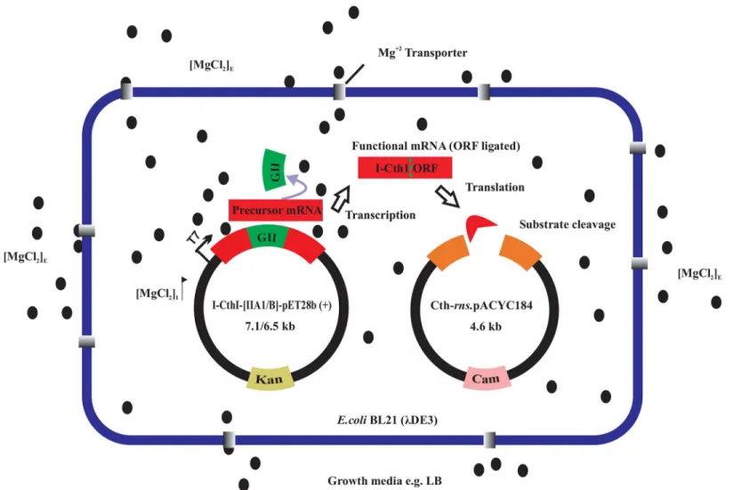
In order to evaluate if intron splicing could be manipulated as a potential“on switch”, the addi-tion of exogenous Mg+2was investigated with regards to expression of the HEases. Anin vivo endonuclease assay was established to evaluate the expression of functional HEases at various Mg+2concentrations. Here two compatible plasmids were maintained inE.coliBL21 (λDE3) based on antibiotic selection [kanamycin (kan) and chloramphenicol (cam)]. The I-CthI-[IIA1]-pET28b (+)—kan (ColE1 origin of replication) construct (7.1 kb) allowed for the expression of the HEase ORF and a second plasmid with cam and the appropriate HEase target site sequence served as the substrate plasmid. For constructing the substrate plasmid, the Cth-rns.pUC57 was digested with BamHI (Life Technologies) and XbaI (Life Technologies) and a 469 bp containing thernssegment with the HEase target site was cloned in the pACYC184 plasmid [ATCC 37033 (American Type Culture Collection, Manassas, VA, USA; p15A origin of replication].The substrate plasmid was named Cth-rns.pACYC184—cam (4.6 kb). Excision of the intron would permit the expression of an active HEase from the I-CthI-[IIA1]-pET28b (+)—kan plasmid and this could be detected by plating cells on various media with different antibiotics, ultimately the presence of the HEase should lead to the loss of the Cth-rns. pACYC184 containing the cam resistance marker (Fig 2).
For thein vivoendonuclease assay, co-transformedE.coliBL21 (λDE3) cells were grown overnight in duplicates in culture tubes containing 5 mL LB media plus the appropriate antibi-otics. One percent glucose was added to the media containing the HEase-co-transformed con-struct to prevent leaky expression from the T7 promoter. A 0.5 mL aliquot from the 5 mL O/N cultures was used to inoculate 50 mL LB broth cultures supplemented with 100μg/mL kan,
60μg/mL cam, 1% glucose and 5 mM MgCl2. For convention, we refer to this culture flask as
‘LB+Mg+2’. Another culture flask designated‘LB’which contained no added MgCl2was
inocu-lated with the same amount of O/N culture and this served as the negative control for this experiment. The cells were grown at 37°C with vigorous shaking (210 rpm) and the cultures were either induced with 0.5 mM IPTG when the O.D.600reached ~ 0.56 or not induced. The
cultures were then shifted to 28°C for the production of the HEase. After 4 hours, both the induced and uninduced cultures from‘LB+Mg+2’and‘LB’were diluted to 10−6and 100
μL of
the diluted cultures were plated on LB agar plates containing 60μg/mL cam (done in triplicate).
endonuclease activity, 50 ng of the vector was also co-transformed along with Cth-rns.
pACYC184 into 100μL of chemically competentE.coliBL21 (λDE3) cells and the above
proto-col was followed. To evaluate the effect of 5 mM exogenous MgCl2on thein vivosplicing
potential of the rI1 group IIB intron, the I-CthI-[IIB]-pET28b (+)—kan construct was cotrans-formed with the substrate plasmid and thein vivoendonuclease protocol was performed as described above.
In order to antagonize the stimulatory effect of MgCl2on splicing of group II introns,
10μM of cobaltous chloride (CoCl2) was added to the culture media along with 5 mM MgCl2.
It has been previously shown that CoCl2perturbs the import of Mg+2inE.colicells [40–42].
Moreover, in order to negate the possibility that CoCl2can promote splicing of the group IIA1
or group IIB,E.coliBL21 (λDE3) cells containing each of the constructs I-CthI-[IIA1]-pET28b (+) and I-CthI-[IIB]-pET28b (+) were exposed to 10μM of CoCl2in the culture media. The
cultures containing both the salts (MgCl2and CoCl2) as well as CoCl2alone were either
Fig 2. Impact of MgCl2on splicing and the expression of a homing endonuclease.Anin vivoendonuclease assay was established where two
compatible plasmids were maintained inE.coliBL21 (λDE3) based on antibiotic selection [kanamycin (kan) and chloramphenicol (cam)]. Cells were grown in absence and presence of either 5 mM or 10 mM added MgCl2(E = external) and induced with 0.5 mM IPTG (O.D.600= 0.56). The internal concentration (I) of
MgCl2increases probably due to the activities of magnesium transporters. Free Mg+2ions are available to bind to the catalytic center of the group II intron
[domain V (not shown)] and initiate efficient splicing and religation of the ORF. The expressed HEase cleaves the target site in the substrate plasmid resulting in the loss of cam resistance marker. Cells grown in the absence of MgCl2fail to splice out the internal group II thus yielding non-functional HEase thereby
resulting in the maintenance of the substrate plasmid and hence the survival of colonies on cam plates. TheE.coligenome is not shown for simplicity.
uninduced or induced with 0.5 mM IPTG when the O.D.600reached ~ 0.5. The cultures were
further incubated at 28°C for the production of the HEase. After 4 hours, the cultures were diluted to 10−6and 100
μL of the diluted cultures were plated on each of the LB agar cam
selec-tion plates (done in triplicate). The plate assays were performed as described above in order to evaluate the splicing of the internal group II introns in the presence of CoCl2.For statistical
analysis, unpaired student’s t test was performed to determine the significance of the results obtained. Graphpad Prism 6.01 statistical analysis software was used to calculate the Student’s t test and the respective bar graphs were drawn using the same software.
Results
Exogenous Mg
+2induces
in vivo
splicing of group IIA1 and group IIB
introns
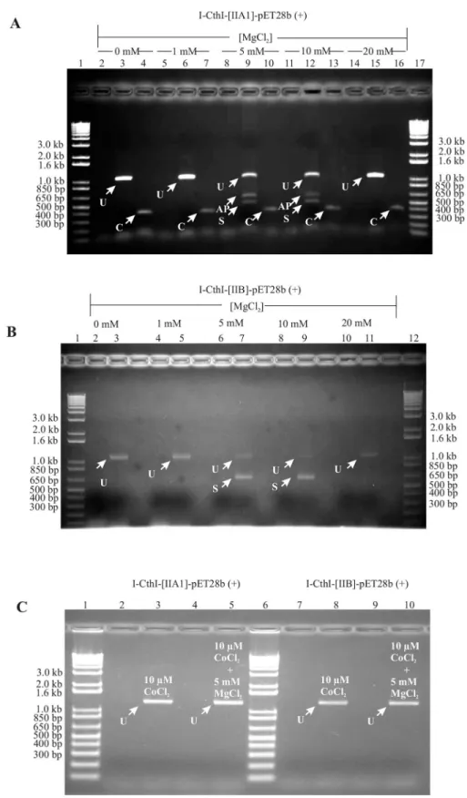
To demonstrate the splicing competency of group IIA1 and IIB intron in the presence of vari-ous added concentrations of exogenvari-ous Mg+2, RNA was extracted fromE.coliBL21 cells con-taining the constructs I-CthI-[IIA1]-pET28b (+) or I-CthI-[IIB]-pET28b (+). The splicing reaction products for the native intron were recovered with RT-PCR utilizing primers Twin-HEG-F and TwinHEG-R. Among the observed cDNAs obtained from RNA extracted from bacterial cells grown in the presence of various concentrations, only cells grown at 5 mM and 10 mM MgCl2showed evidence of alternate products. A PCR product near the 500 bp marker,
the expected size (538 bp) for cDNAs from transcripts where the group IIA1 intron spliced out, was further investigated (Fig 3A). Bacterial cells grown in either lower (0 mM, 1 mM) or higher (20 mM) MgCl2did not show any evidence for splicing and only the full length
unspliced PCR product (1296 bp) was recovered. A 400 bp PCR product was observed when internal group IIA1 intron specific primers (‘C’and‘D’) were used and this served as the posi-tive control showing the presence of the intron in all the samples examined. PCR amplicons derived from the unspliced and spliced cDNAs were gel excised and submitted for DNA sequence analysis and the resulting data were compared with the control non-spliced DNA template. Comparative sequence analysis showed that the 556 bp RT-PCR product was the result of the group IIA1 intron being spliced out and the joining of the flanking exon seg-ments. Based on a previous study [22] it was expected that the RT-PCR product obtained from a spliced transcript should be 538 bp in length. The additional 18 bp noted was due to a shift of the 50splice junction which was 18 nucleotides downstream from the original IBS1 and IBS2 elements. This is probably due to a cryptic/alternate splice site that was utilized inE. coli. However, the 30splice site was found to be consistent with previous studies on this intron in its“native”mitochondrial environment [22].
Similar results were obtained for demonstrating the splicing competency of the group IIB intron RNA extracted from bacterial cells grown in the presence of increasing concentrations of MgCl2(seeMaterial and Methodsection—in vivoRNA splicing assay) yielded a RT-PCR
product of 658 bp representing the spliced version of the HEase transcript. However, no splic-ing was observed when the cells were grown in the absence or in the presence of 1 mM and 20 mM concentrations of external MgCl2(Fig 3B). Sequence analysis of the RT-PCR product
revealed that the splicing of this group IIB intron occurred as predicted based on its IBS1 sequence [37] and the splicing followed the conventional intron/splice sites yielding a continu-ous I-CthI ORF. To demonstrate the splicing competency of group IIA1 and IIB intron in the presence of CoCl2or both MgCl2and CoCl2in the growth media, RNA was extracted fromE.
Fig 3. (A) A mtDNA group IIA1 intron can splice inE.coli.A 1% agarose gel showing the RT-PCR results (Primers: TwinHEG-F/R) forin vivogroup IIA1 intron splicing under various concentrations of external MgCl2
addition of both 10μM of CoCl2and 5 mM MgCl2in the growth media did not show any
evi-dence for splicing and only the full length unspliced PCR product was recovered (Fig 3C).
Alternate splice site for the group IIA1 does not affect I-CthI functionality
Sequence analysis of the cDNA (556 bp) from the group IIA1 intron construct derived tran-scripts had revealed a cryptic splice site which is 18 nucleotides downstream of the original (native) IBS sequences. The alternate IBS sequences can potentially H-bond with sequences that are near or overlap with the native (original) EBS sequences (S1A Fig). In order to evaluate if the addition of the extra six amino acids could affect the active site or the protein’s tertiary structure, the online program Protein Homology/analogY Recognition Engine V2.0 (PHYRE2) (http://www.sbg.bio.ic.ac.uk/phyre2/html/page.cgi?id+index) [43] was used to map the posi-tion of these newly added amino acids onto the predicted tertiary structure of I-CthI. The pro-gram showed that the protein is composed of ten alpha helices (39%) and nine beta stands (26%), the confidence key of these regions were found to be 100% when compared to the crys-tal structure of the HEase I-SmaMI (PDB accession number: c4loxA). The additional six amino acids V, R, R, C, G and Y are located in a linker region of the HEase protein and do not appear to disrupt the active sites (LAGLIDADG motifs) or beta sheets required for making contact with the DNA target site (S1B Fig). The HEase protein derived from the group IIA1 intron con-taining construct was purified and challenged with its substrate. The enzyme completely linear-ized the circular substrate plasmid (3.1 kb) within 90 minutes. However, when this protein was challenged with two different non-substrates to test its specificity, even after two hours of incu-bation at 37°C, the protein failed to cleave the non substrates (S1C Fig). It was also noted that the addition of 10μM CoCl2in thein vitroendonuclease reaction buffer did not inhibit the
functionality of the I-CthI HEase and this protein could cleave its substrate in 90 minutes at 37°C. However, when the substrate was incubated just in the presence of 10μM CoCl2without
MgCl2in the endonuclease reaction buffer, the protein did not initiate any cleavage activity (S2 Fig).
presence of low (1 mM) or high (20 mM) concentration of MgCl2in the culture media respectively. Lanes 9
and 12 (see white arrows) show the PCR product (556 bp) that indicates splicing occurred (S) and possible alternative products (AP) are also indicated in lanes. Lanes 4, 7, 10, 13 and 16 (see white arrows) are positive controls (Primers C and D) that show the presence of the internal group IIA1 (indicated by C; 400 bp) in all the samples examined. Lane 1 and 17 contain the 1kb plus DNA ladder (Life Technologies). For more
information on primers see text andFig 1B.(B) A mtDNA group IIB intron can splice inE.coli.A 1% agarose gel showing the RT-PCR results (Primers: TwinORFnew-F/TwinHEG-R) forin vivogroup IIB intron splicing under various concentrations of external MgCl2in the culture media. Lanes 2, 4, 6, 8 and 10 (see
white arrows) show the results for standard PCR (Primers: TwinORFnew-F/TwinHEG-R) on RNA samples testing for the absence of genomic DNA prior to cDNA synthesis. Lanes 3, 5 and 11 (see white arrows) show the PCR product (~1.2 kb) that represent unspliced transcripts (U) in the absence (0 mM), presence of low (1 mM) or high (20 mM) concentration of MgCl2in the culture media respectively. Lanes 7 and 9 (see white
arrows) show a PCR product that represents spliced (S) transcripts (658 bp) in the presence of 5 mM and 10 mM MgCl2in the culture media. Lane 1 and 12 contain 1kb plus DNA ladder (Life Technologies).(C) Effect of
CoCl2and/or MgCl2on intron splicing. A 1% agarose gel showing the RT-PCR results forin vivosplicing of
group IIA1 intron and group IIB intron when 10μM CoCl2alone or 10μM CoCl2in combination with 5 mM
MgCl2were added to the LB growth media. Lanes 2 and 4 show the results for standard PCR on RNA
samples testing for the absence of genomic DNA prior to cDNA synthesis. Lane 3 and 5 show that splicing cannot be detected (see band marked with U) when I-CthI-[IIA1]-pET28b (+) [BL21] was grown with the addition of 10μM CoCl2in one LB culture media and combination of 10μM CoCl2and 5 mM MgCl2in the
other LB media respectively. For the second construct I-CthI-[IIB]-pET28b (+) [BL21], lanes 7 and 9 represent the results for standard PCR on RNA samples testing for the absence of genomic DNA prior to cDNA synthesis. Lanes 8 and 10 show that splicing cannot be detected (see band marked with U) when I-CthI-[IIB]-pET28b (+) [BL21] was grown with the addition of 10μM CoCl2in one LB culture media and combination of
10μM CoCl2and 5 mM MgCl2in the other LB media respectively. Lanes 1 and 6 represent 1kb plus DNA
ladder (Life Technologies).
In vitro
and
in vivo
translation show evidence of HEase protein
production under specific [Mg
+2]
SDS PAGE analysis showed the presence of the protein I-CthI at the desired location (~ 29 kDa) only fromin vitrotranslation assays that used RNA extracted from cells grown in the cul-ture media supplemented with either 5 mM or 10 mM MgCl2. RNA extracted from cells grown
in the absence or at 1 mM and 20 mM MgCl2failed to yield the desired protein in thein vitro
translation assays. This was observed for both of the tested constructs I-CthI-[IIA1]-pET28b (+) and I-CthI-[IIB]-pET28b (+) when the cells were grown at the same concentrations of MgCl2(Fig 4A).
Fig 4. (A) The effect of MgCl2onin vitroprotein expression.A 12.5% SDS-PAGE showingin vitroprotein expression for constructs I-CthI-[IIA1]-pET28b
(+) [left] and I-CthI-[IIB]-pET28b (+) [right] in the presence of various concentrations of external MgCl2in the culture media. Lane 1 represents theE.coli
dihydrofolate reductase (marked with arrow) when 125 ng/μL was used as the template (positive control) for the PURExpressIn VitroProtein Synthesis kit. Lanes 2 and 10 show thein vitroprotein expression profiles when empty pET28b (+) vectors (without the above constructs) were used as the negative control. Lanes 3 and 11 represent thein vitroprotein expression profile when RNA (extracted from the culture in the absence of MgCl2) was used as the
template. Lanes 4 through 7 represent the protein expression profiles when RNA (extracted from the cultures in the presence of 1 mM, 5 mM, 10 mM and 20 mM respectively) was used as the template for thein vitroprotein synthesis. The expression of the protein (I-CthI) has been marked with arrows. Forin vitro
expression from the I-CthI-[IIB]-pET28b (+) construct, lanes 12 through 15 follow the same order as depicted for the I-CthI-[IIA1]-pET28b (+) construct (i.e. lanes 4–7). Lanes 8 and 9 represent the Blueye prestained protein ladder (FroggaBio, North York, Ontario).(B) The effect of MgCl2onin vivoprotein
expression.A 12.5% SDS-PAGE showingin vivoprotein expression for constructs I-CthI-[IIA1]-pET28b (+) [left] and I-CthI-[IIB]-pET28b (+) [right] in the
presence of various concentrations of external MgCl2in the culture media. Lanes 1 and 9 represent thein vivoprotein expression profiles from the empty
pET28b (+) vector (without the constructs). Lanes 2 through 6 represent the protein expression profiles when I-CthI-[IIA1]-pET28b (+) [BL21] was grown under increasing concentrations of external MgCl2starting from 0 mM, 1 mM, 5 mM, 10 mM and 20 mM. Lane 10 through 14 represent the protein expression
profiles when I-CthI-[IIB]-pET28b (+) (BL21) was grown under increasing concentrations of external MgCl2. Lanes 10 through 14 follow the same order as for
the protein expression profiles when I-CthI-[IIA1]-pET28b (+) [BL21] was grown under increasing concentrations of external MgCl2(i.e. lanes 2–6). The
overexpressed I-CthI (migrate at ~29 kDa) has been marked with arrows. Lanes 7 and 8 represent the Blueye prestained protein ladder (FroggaBio, North York, Ontario).
The codon-optimized HEase ORF (from both constructs) expressed inE.coliBL21 cells only when the cells were grown in the culture media supplemented with 5 mM or 10 mM MgCl2
(Fig 4B). The recovery and purification of the HEase protein was achieved by affinity column chromatography involving Superflow1nickel resin (Qiagen, Toronto). A step up gradient of 25 mM, 50 mM and 100 mM imidazole in the wash buffer was used to remove the background proteins while the pure HEase protein was obtained in the elution buffer supplemented with 250 mM imidazole which was later dialyzed to remove any salts. This purified protein was pooled and concentrated (3 mg/mL) in order to perform thein vitroendonuclease assay.
I-CthI ORF interrupted with either a group IIA1 or IIB introns results in the
expression of an active HEase
The primary objective of this work was to evaluate if group II introns can be utilized as regula-tory elements for expressing a mtDNA fungal HEase inE.coli. Two types of group II introns were utilized, group IIA1 and IIB introns. As previously mentionedin vitroendonuclease assays showed that functional HEase were expressed inE.coliunder conditions that favour group II intron splicing. It appears that both types of mitochondrial group II introns can splice inE.coliunder suitable Mg+2concentrations ultimately yielding HEase that can linearize their substrates in one hour at 37°C; and non-substrates were not cleaved even after two hours of incubation indicating that the enzyme is still highly specific for its cleavage site (Fig 5).
Fig 5.In vitroendonuclease assay.A 1% agarose gel showing thein vitroendonuclease assay with
construct I-CthI-[IIB]-pET28b (+) encoded HEase. Lanes C and L represent uncut substrate plasmid and linearized substrate plasmid (cleaved with BamHI), respectively. Numbers on the top of each lane represent incubation times in minutes at 37°C. For each of the above endonuclease assay, 1μg of substrate DNA was treated with 8μL of the purified HEase (2.5 mg/mL). The arrow shows the linearized band at 3.1 kb when the substrate was incubated for 60 minutes. Two negative controls were applied in this assay. Cnsand Coxswere
used to examine the specificity of this nested intron encoded I-CthI. Coxsis the substrate for another HEase
encoded acoxgene intron fromAnnulohypoxylon stygiumwhile Coxs+ E represents the assay with the
same plasmid incubated with purified I-CthI (encoded from the above stated construct) for 120 minutes at 37°C. Cnsrepresents the negative control plasmid previously used [22] in characterizing the I-CthI HEase;
this pUC57 based construct contains thernsgene plus the mS1247 nested intron. Cns+ E represents the
negative control plasmid incubated with the same concentration of the HEase for 120 minutes at 37°C. For both the negative controls, no endonuclease activities were observed. Lanes marked with M represent the 1 kb plus DNA ladder (Life Technologies).
Endonuclease cleavage mapping of HEases derived from ORFs
interrupted by group II introns shows cleavage sites have not changed
LAGLIDADG HEases tend to generate cohesive termini by generating staggered cuts with four nucleotide 30-single stranded overhangs. The T4 DNA polymerase treated and religated I-CthI cleaved substrate plasmid sequences when compared with the sequence of the uncut substrates showed that a 50-AAGA-30segment was removed from the sense strand. The endonuclease cleavage mapping data is shown inS3 Fig. So the cleavage mapping site experiments showed that the intron contained HEases ORFs (containing either the group IIA1 or IIB intron) ulti-mately allowed for the expression of a functional I-CthI protein that cleaves 8 bp downstream of the mS1247 nested intron insertion site (sense strand) or 4 bp downstream of position S1247 at the antisense strand. These results are in agreement with previous experiments utilizing I-CthI constructs that did not contain introns within the HEase ORF [22]; so the presence of the introns and subsequent RNA processing events inE.colidid not alter the target site speci-ficity of the HEase.
In vivo
endonuclease assays for HEase activity in the presence of MgCl
2and/or CoCl
2In vivoendonuclease assays were performed to evaluate the effect of the addition of either MgCl2and/or CoCl2on the expression and functionality of the I-CthI HEase. The results of
thein vivoendonuclease assays for HEase activity are depicted inS1,S2andS3Tables. Since three technical and two biological replicates (i.e. six independent values) were performed for each of the assay plates, the mean value for the bacterial colony forming units (cfu/mL) are pro-vided along with their respective standard deviations (σ).
First, in order to check whether the protein expressed from I-CthI-[IIA1]-pET28 b (+) con-struct is functional (i.e. can it cleave) or is toxic to theE.coliBL21 genome, I-CthI-[IIA1]-pET28b (+) [BL21] was either uninduced or induced with 0.5 mM IPTG in the presence of 5 mM exogenously added MgCl2(S1 Table, left panel). When the uninduced culture was plated
on kan plates, the bacterial colony count was calculated 3.2 x 10¹⁰cfu/mL,σ= 1.8 x 10⁹and for
the induced culture, the count was 3.0 x 10¹⁰cfu/mL,σ= 2.0 x 10⁹. Moreover, a bacterial lawn
was observed in the absence of the antibiotic. These data showed that I-CthI is not toxic toE. coliand it does not appear to have any target specificity within theE.coligenome. The plate assay results from the negative control pET28b (+) vector cotransformed with Cth-rns. pACYC184 substrate are also listed (S1 Table, right panel). The results showed that even in the absence or in the presence of 0.5 mM IPTG, when the cells were plated on cam plates, the bac-terial colony count was 4.4 x 10¹⁰cfu/mLσ= 2.2 x 10⁹and 4.2 x 10¹⁰cfu/mLσ= 1.8 x 10⁹
respectively. Bacterial lawn was also observed when no antibiotic was applied. These data sug-gested that the proteins encoded from the empty pET28b (+) vector were not detrimental to the substrate plasmid carrying the cam resistance marker as the cells were viable in the pres-ence of the antibiotic.
The plate assay results from theE.colicells cotransformed with I-CthI-[IIA1]-pET28b (+) and Cth-rns.pACYC184 and grown in the absence (LB, left panel) or presence of 5 mM exoge-nously added MgCl2(LB+Mg+2, right panel) are depicted inS2 Table. The results in the left
panel show that when the growth media had no exogenously added MgCl2, even with or
with-out induction with 0.5 mM IPTG, viable colonies were observed when plated on LB agar-cam plates. The colony count for the uninduced and the induced cultures were 3.0 x 10¹⁰cfu/mLσ
internal group II intron thereby not yielding functional HEase. In contrast, in the presence of 0.5 mM IPTG and in the presence of 5 mM MgCl2in the growth media (right panel) allowed
for the expression of a functional HEase which probably happened due to the splicing out of the internal intron. This functional protein could have cleaved the target site, thereby degrad-ing the cam resistance substrate plasmid. The bacterial colony count was 2.3 x 10⁹cfu/mLσ=
1.3 x 10⁹(marked with asterisk inS2 Table, also seeS4 Fig, plate 2). It is worthwhile to mention that there is an approximately 12.6 fold decrease in the bacterial viability when compared to cfu/mL of the cells grown under inducible conditions but in the absence of exogenous MgCl2.
Student’s t test performed on the cfu/mL indicated significant difference (P value<0.0001) in
the number of viable colonies (decrease) when compared to the cells grown under inducible conditions but in the absence of exogenous MgCl2(Fig 6A).
The results for CoCl2acting as a possible antagonist for the uptake of MgCl2are provided in S3 Table. The left panel of the table shows the results of thein vivoendonuclease assay in the presence of only 10μM of CoCl2in the culture media. This set of assays was performed to rule
out the possibility that CoCl2was involved in splicing of the internal intron. Even with or
with-out induction with 0.5 mM IPTG, viable colonies were observed when plated on LB agar-cam plates. The colony count for the uninduced and the induced cultures were 2.2 x 10¹⁰cfu/mLσ
= 0.9 x 10⁹and 2.5 x 10¹⁰cfu/mLσ= 1.2 x 10⁹respectively indicating that CoCl2was not
involved in facilitating the excision of the group II introns thus yielding a non-functional pro-tein. The right panel shows the results of thein vivoendonuclease assay in the presence of both 10μM of CoCl2and 5 mM MgCl2in the culture media. Indeed, viable cells with colony count
of 2.8 x10¹⁰cfu/mL,σ= 1.3 x 10⁹(marked with asterisk inS3 Table, also seeS4 Fig, plate 3)
were observed on the LB agar cam plates. Student’s t test performed on the cfu/mL indicated significant difference (P value<0.0001) in the number of viable colonies (increase) when the
Fig 6. Bar graphs showing the results of thein vivoendonuclease assay.Bar graph shown in panel (A)represents the Student’s t test performed for
assessing the significance of the decrease (p<0.0001) in the number of viable colonies (cfu/mL) for I-CthI-[IIA1]-pET28b (+) and Cth-rns.pACYC184 co-transformed cell lines when grown with the addition of 5 mM MgCl2in the LB media compared to the cells grown in only LB media. Moreover, the graph also
shows significant difference (p<0.0001) with regards to an increase in the number of viable colonies (cfu/mL) when the respective co-transformed cells were
grown with the addition of 5 mM MgCl2and 10μM CoCl2in the LB media compared to the cells grown with 5 mM MgCl2in the LB media. Bar graph shown in
panel (B) represents the similar results when Student’s t test performed for assessing the significance of the decrease / increase (p<0.0001) in the number of viable colonies (cfu/mL) for I-CthI-[IIB]-pET28b (+) and Cth-rns.pACYC184 co-transformed cell lines grown under the same conditions as described in panel A. Graphpad Prism 6.01 statistical analysis software was used to calculate the Student’s t test and the respective bar graphs were drawn using the same software.
cells were grown with the addition of both 5 mM MgCl2and 10μM CoCl2compared to the
cells grown with the addition of only 5 mM MgCl2under inducible conditions (Fig 6A).
Assuming that CoCl2antagonizes somehow MgCl2uptake one would predict that under both
inductive and non-inductive conditions cells should be viable when plated on LB cam plates as the substrate plasmid carrying the antibiotic resistance marker (cam) was not targeted hence maintained.
The plate assay results from the second cotransformed construct I-CthI-[IIB]-pET28b (+) and Cth-rns.pACYC184 are provided in theS4andS5Tables, and the images of the LB agar cam plates are presented inS5 Fig, plates 1–3. For this construct 19 fold decrease in the bacte-rial viability was observed when compared to cfu/mL of the cells grown under inducible condi-tions but in the absence of exogenous MgCl2. Student’s t test performed on the cfu/mL
indicated significant difference (p value<0.0001) in the number of viable colonies (decrease)
when the cells were grown under inducible conditions but in the absence of exogenous MgCl2
(Fig 6B). Student’s t test performed on the cfu/mL indicated significant difference (p value
<0.0001) in the number of viable colonies (increase) when the cells were grown with the
addi-tion of both 5 mM MgCl2and 10μM CoCl2compared to the cells grown with the addition of
only 5 mM MgCl2under inducible conditions (Fig 6B).
Discussion
Group II introns by manipulating their EBS elements are currently utilized as genome editing tools in the form of targetrons which can be applied for targeted insertional mutagenesis [44,
45]. Previously we described a mtDNA encoded HEase that is encoded within a group I intron (mS1247) where the intron encoded ORF is disrupted by an ORFless group IIA1 intron [22]. This arrangement hinted at the possibility that the group II intron could be regulatory in nature with regards to the expression of the HEase. Herein we are applying group II introns as regula-tory element that allowed for the expression of a fungal mtDNA HEase withinE.coli.
Sequences representing either a group IIA1 or a group IIB type intron where inserted into the ORF for the HEase I-CthI at positions that allows for proper intron/exon (i.e. EBS/IBS) interac-tions so that splicing competent folds could be achieved.
It has been previously shown that an organellar group IIB intron can splice inE.coli[37], in this study we show that group IIA1 introns also have the potential to splice inE.coli. It is assumed that group II introns for efficient splicing require intron and/or host encoded factors [25,26,46,47] so this would suggest that withinE.colifactors are available that can be recruited for the removal of the organellar introns that were investigated in this study. In both cases HEase expressed from constructs where the HE ORFs were disrupted by group II introns were active and cut their respective substrates at the expected cleavage sites.
For the group IIB intron the intron/exon junctions based on RT-PCR on RNA extracted fromE.coliwere as expected based on previous reports [37]. However, for the group IIA1 intron the intron/exon junction shifted by 18 nucleotides adding 6 amino acid residues to the HEases. These alternate IBS/EBS interactions might be fortuitous but might suggest that this group IIA1 intron splices differently inE.colicompared to its native environment. Therefore, with regards to designing HEases with an“intron based”regulatory element it is important to evaluate the intron/exon junction in the alternate host environment to ensure HEase function-ality/specificity has not been compromised. With regards to I-CthI the altered splicing of the group IIA1 intron added six amino acids to a segment of the protein that apparently did not alter the HEases cleavage specificity or the stability of the protein.
replacements), (c) as a gene targeting tool by promoting mutation inducing non homologous end-joining repair, or (d) as rare cutting enzymes that are part of cloning vectors and cloning strategies [34,35]. In some instances, such asin vivogene targeting temporal regulation of HEase activity might be desirable in order to minimize nonspecific activity of the enzyme. This study showed that modulating the activity of I-CthI inE.colican be accomplished by inserting group II intron sequences into the HEase ORF as splicing of the intron can be stimulated by the addition of Mg+2or antagonized by the addition of Co+2. This strategy would have applica-tions in bacterial systems which are more emendable to group II intron splicing unlike eukary-otic cells [48–50]. Group II intron sequences in general are readily available [51] and unlike previous attempts to control HEase activity via redox switches (see PI-SceI [19])in vivo appli-cations are possible.
The Mg+2transport systems inE.colihave not yet been fully elucidated [52]. In one study, magnesium has been shown to modulate the function of riboswitches by facilitating the ligand-riboswitch interactions e.g. btuB ligand-riboswitch fromE.coli[53]. In our study, exogenous Mg+2 concentration was evaluated for manipulating intron splicing which allowed for attenuating the expression of a HEase. In the presence of certain concentrations of Mg+2(5 mM or 10 mM) in the growth media the group II introns appeared to splice and thus functional HEases were produced. Magnesium appears to act as a cationic trigger which might enter theE.colicells through the magnesium transport systems, raising the intracellular magnesium concentration hence facilitating intron splicing. In order to assess if intron splicing is occurring due to the import of Mg+2in the bacterial cells, CoCl2was used to antagonize the Mg+2effect. Earlier
studies have shown that cobaltous ion, at concentrations as low as 10μM, inhibits the
energy-dependent transport of Mg+2into cells ofE.coli[40,41].In vivoendonuclease assays in the presence of MgCl2and 10μM of CoCl2showed a reduction in the expression of HEase. This is
probably due to Co+2interfering with the entry of magnesium into the cells, leading to Mg+2 levels that are not amenable to intron splicing. The failure of the addition of 20 mM MgCl2to
stimulate splicing might be an indication that excess Mg+2can interfere with the proper folding of the group II introns [54–56].
Recently the RNA-guided CRISPR-associated (Cas) endonuclease Cas9 has been developed into a genome editing tool [56–60] and it appears well suited for mammalian systems although off-target activity is a concern [61,62]; Cas9 also appears to be less effective inE.coli[62–64]. With regards to addressing the off-target activity several methods have been developed to con-trol the nuclease activity of Cas9, such as generating versions of Cas9 that are split into two components and these have been engineered to combine by the addition of a chemical signal such as rapamycin or by blue light irradiation (i.e. a photoactivatable form of Cas9) [65,66]. Another strategy has been to place an“intein”sequence within Cas9 and the intein has been engineered to splice from the host protein when a ligand (4-hydroxytamoxifen) is added to the media [67]. This ligand-dependent intein is somewhat analogous to our“self-splicing”group II introns that can be promoted to splice at the RNA level when suitable levels of Mg+2are present in the media. One can foresee the application of group II intron sequences as agents that allow for inducible genome editing in cell types that are amendable to support the splicing of these elements and can uptake suitable amounts of Mg+2. The ability to antagonize splicing with Co+2provides a“switch like”mechanism where the production of HEase can be stopped or at least attenuated to limit the amount of endonuclease that is produced in a cell and thus poten-tially avoid nonspecific activities.
LtrB group II intron (including a version where the ORF was deleted) from the Gram-positive bacteriumLactococcus lactiscan splice intranswhen fragmented at various locations through-out its structure [70]. Therefore, a HEase ORF could be split and encoded by two compatible plasmids, carrying different selectable markers and different promoters; with one construct bearing the amino terminal part of the HEase ORF plus the 5' segment of a group II intron sequence and the other construct carrying the 3' segment of group II intron sequence plus the carboxyl terminal part of the HEase ORF. Upon expression, these two RNAs can assemble via the intron segments into a tertiary structure that promotes trans-splicing of the intron sequences and thus the exons get ligated together to produce a functional HEase transcript.
The current study is“a proof of principle”that shows that the expression of a gene can be controlled or at least attenuated by the activity of autocatalytic group II intron sequences. The exact nature of Mg+2or Co+2transport from the media intoE.coliis not clear but based on our data we can conclude that manipulation of the concentration of positive cations such as Mg+2 and Co+2can influence splicing of heterologous introns withinE.coli. Group II introns could be applied to other heterologous or native proteins that are components of biochemical path-ways to allow for temporal control of their expression and possibly promote a shift in metabolic processes. Therefore, in the future group II introns could be a potential tool that can be applied not only to genome editing but also to metabolic engineering [71–73].
Supporting Information
S1 Fig. (A) Intron and exon binding sites for the mS1247 nested group IIA1 intron. Wat-son-Crick base pairing (shown by solid black dots) between the newly discovered cryptic (marked by asterisk sign) splice site sequence (IBS1and IBS2) and corresponding exon bind-ing sequences (EBS1and EBS2) of the mS1247 internal group IIA1 intron. The original IBS1, IBS2 and EBS1, EBS2 for mS1247 nested intron fromC.thermophilumare indicated.(B) Anin silicomodel for the expressed I-CthI protein.Anin silicomodel for the I-CthI protein
derived from the I-CthI-[IIA1]-pET28b (+) construct generated by the PHYRE2 program. The program identified the double motif LAGLIDADG I-SmaMI (PDB: c4loxA) HEase protein as a template for folding I-CthI. Alpha helices and beta sheets along with amino terminal (N) and carboxyl terminal(C)have been marked. The LAGLIDADG motifs contribute towards the active site of the enzyme while the beta sheets arrange in a configuration that forms the DNA binding surface. The extra six amino acids (V, R, R, C, G and Y) were not present in any of the active sites of the HEase instead they are located in a linker region between the two beta sheets near the carboxyl terminal of the protein. The linker region showing the extra six amino acids has been magnified for better illustration. The amino acid positions are also mentioned.(C)In vitroendonuclease assay for I-CthI.A 1% agarose gel showing thein vitroendonuclease assay
withC.thermophilumHEase ORF intron containing construct I-CthI-[IIA1]-pET28b (+). Lane C and L represent uncut substrate plasmid and linearized (L) substrate plasmid (cleaved with BamHI), respectively. Numbers on the top of each lane represent incubation time in min-utes at 37°C. For each of the above endonuclease assays, 1μg of substrate DNA was treated
with 8μL of the purified HEase (3 mg/mL). The arrow shows the linearized band at 3.1 kb
when the substrate was incubated for 90 minutes. Cnsrepresents the negative control plasmid
while Cns+ E represents the negative control plasmid incubated with the same concentration
of the HEase for 120 minutes at 37°C. Another negative control plasmid (Coxs) was used to
examine the specificity of this nested intron encoded I-CthI. Coxsis the substrate for another
HEase encoded from the intron of thecoxgene fromAnnulohypoxylon stygiumwhile Coxs+ E
observed. Lane denoted with M represents the 1 kb plus DNA ladder (Life Technologies). (TIF)
S2 Fig. CoCl2does not affect I-CthI endonuclease activity.A 1% agarose gel showing the
effect of addition of 10μM CoCl2during thein vitroendonuclease assay with construct
I-CthI-[IIA1]-pET28b (+) and I-CthI-[IIB]-pET28b (+) encoded I-CthI HEase. Lane 2 represents the uncut substrate (C) plasmid. Lane 3 shows the endonuclease activity of I-CthI on the substrate plasmid in the presence of 10μM CoCl2alone in the endonuclease reaction buffer without 10
mM MgCl2. Lane 4 represents the linearized substrate (L) when treated with BamHI. Numbers
on the top of the lanes (30, 60, 90) represent incubation time in minutes at 37°C. The white arrow shows the linearized band at 3.1 kb. The same order (i.e. lanes 9 through 14) was main-tained for the endonuclease activity of the I-CthI HEase derived from I-CthI-[IIB]-pET28b (+) [BL21] construct. Lanes 1, 8 and 15 contain the 1 kb DNA ladder (Life Technologies).
(EPS)
S3 Fig. Endonuclease cleavage mapping for HEases derived from ORFs interrupted by group II introns (IIA or IIB). (A)The cleavage sites were mapped by comparing uncut sub-strate with I-CthI treated subsub-strate DNAs. Cleavage by I-CthI generates a staggered cut with 4 nucleotide 3’overhang in the substrate plasmid at the enzyme’s target site. T4 DNA polymerase was used to blunt the cleaved ends. The religated plasmid was sequenced and compared to the sequence of the untreated substrate plasmid in order to map the cleavage site by scanning for a 4 bp deletion in the T4 DNA polymerase treated cleaved substrate plasmid.(B)Schematic representation of the I-CthI cleavage site near the mS1247 intron insertion site. Proposed cleavage sites are indicated by open triangles; and a vertical line represents the intron insertion site. The HEase cleavage site is 8 nt downstream of the intron insertion site with regards to the sense strand or 4 nt downstream with regards to the antisense strand.
(TIF)
S4 Fig. Effect of MgCl2on cell viability due to I-CthI activity.Images of LB agar cam plates
depictingin vivoendonuclease assays performed to evaluate the effect of the addition of either MgCl2and/or CoCl2on the expression and functionality of the I-CthI HEase within cells
cotransformed with I-CthI-[IIA1]-pET28b (+) and Cth-rns.pACYC184. Two biological and three technical replicates were performed however, one representative from each has been shown. Plate 1, 2 and 3 represent the viable number of colonies when the 100μL of 10−6
cotransformed cells (induced with 0.5 mM IPTG) from 0 mM MgCl2, 5 mM MgCl2and 10μM
CoCl2+ 5 mM MgCl2in the LB growth media were plated on LB agar plates supplemented
with 60μg/mL cam respectively.
(TIF)
S5 Fig. Effect of CoCl2on cell viability due to I-CthI activity.Images of LB agar cam plates
depictingin vivoendonuclease assays performed to evaluate the effect of the addition of either MgCl2and/or CoCl2on the expression and functionality of the I-CthI HEase in cells
cotrans-formed with I-CthI-[IIB]-pET28b (+) and Cth-rns.pACYC184. Two biological and three technical replicates were performed however, one representative from each has been shown. Plate 1, 2 and 3 represent the viable number of colonies when the 100μL of 10−6cotransformed cells (induced
with 0.5 mM IPTG) from 0 mM MgCl2, 5 mM MgCl2and 10μM CoCl2+ 5 mM MgCl2in the LB
growth media were plated on LB agar plates supplemented with 60μg/mL cam respectively.
(TIF)
S1 Table.In vivoactivity of I-CthI expressed from I-CthI-[IIA1]-pET28b (+).In vivo
and pET28b (+) and challenged with the substrate plasmid Cth-rns.pACYC184 [BL21]; results reported cfu/mL. Three technical and two biological replicates were performed for each of the constructs.The numbers represent the mean of six independent cfu/mL. Standard deviations are also indicated for each of the above observations.
(DOCX)
S2 Table. Effect of 5 mM MgCl2on thein vivoactivity of I-CthI-[IIA1].In vivoendonuclease
activity of I-CthI-[IIA1]-pET28b (+) + Cth-rns.pACYC184 [BL21] cotransformed constructs presented in cfu/mL. This table presents the plate assay results of the above construct under different conditions, one is without added MgCl2and the other is with the addition of 5 mM
MgCl2. Three technical and two biological replicates were performed for each of the constructs
and the numbers represent the mean of six independent cfu/mL. Standard deviations are also indicated for each of the above observations.mark on specific boxes indicates that the images of the plates (Plate D) are provided in theS4 Fig.
(DOCX)
S3 Table. Effect of CoCl2on thein vivoactivity of I-CthI-[IIA1].Cobalt chloride antagonism
on the possible uptake of magnesium inE.colicells as shown by thein vivoendonuclease activ-ity of the HEase expressed from I-CthI-[IIA1]-pET28b (+) and challenged witht the substrate plasmid Cth-rns.pACYC184 [BL21]; results are presented in cfu/mL.This table shows the plate assay results of the above constructs under different conditions, one is with the addition of 10μM CoCl2and the other is with the addition of both 10μM CoCl2and 5 mM MgCl2in the
LB media.Three technical and two biological replicates were performed for each of the con-structs and the numbers represent the mean of six independent cfu/mL. Standard deviations are also indicated for each of the above observations.mark on specific boxes indicates that the images of the plates (Plate D) are provided in theS4 Fig.
(DOCX)
S4 Table.In vivoactivity of I-CthI expressed from I-CthI-[IIB]-pET28b (+).In vivo
endonu-clease assay showing the HEase activity as demonstated in cells that were cotransformed with I-CthI-[IIB]-pET28b (+) and Cth-rns.pACYC184 [BL21]; results are reported in cfu/mL. The plate assay results of the above construct under different conditions, one is without added MgCl2and the other is with addition of 5 mM MgCl2. Standard deviations are also indicated
for each of the above observations.mark on specific boxes (Plate D) indicates that the images of the plates (Plate D) are provided in theS5 Fig.
(DOCX)
S5 Table.In vivoactivity of I-CthI-[IIB] in the presence of CoCl2.Cobalt chloride antagonism
on the possible uptake of magnesium inE.colicells during thein vivoHEase endonuclease assay in cells cotransformed with I-CthI-[IIB]-pET28b (+) and Cth-rns.pACYC184 [BL21]; results reported in cfu/mL. This table presents the plate assay results of the above construct under differ-ent conditions with the addition of either exogeneous CoCl2(10μM) or 10μM CoCl2and 5 mM
MgCl2in the LB media. Standard deviations are also indicated for each of the above results.
mark on specific boxes indicates that the images of the plates (Plate D) are provided in theS5 Fig. (DOCX)
Acknowledgments
Author Contributions
Conceived and designed the experiments: TKG GH. Performed the experiments: TKG. Ana-lyzed the data: TKG GH. Contributed reagents/materials/analysis tools: GH. Wrote the paper: TKG GH.
References
1. Gimble FS. Invasion of a multitude of genetic niches by mobile endonuclease genes. FEMS Microbiol Lett. 2000; 185: 99–107. PMID:10754232
2. Hausner G. Introns, Mobile Elements, and Plasmids. In: Bullerwell CE, editor. Organelle Genetics. Springer Berlin Heidelberg; 2012. p. 329–357.
3. Edgell DR, Belfort M, Shub DA. Barriers to intron promiscuity in bacteria. J Bacteriol. 2000; 182: 5281–
5289. PMID:10986228
4. Toor N, Zimmerly S. Identification of a family of group II introns encoding LAGLIDADG ORFs typical of group I introns. RNA. 2002; 8: 1373–1377. PMID:12458791
5. Dujon B. Group I introns as mobile genetic elements: facts and mechanistic speculations—a review. Gene. 1989; 82: 91–114. PMID:2555264
6. Belfort M, Perlman PS. Mechanisms of intron mobility. J Biol Chem. 1995; 270: 30237–30240. PMID: 8530436
7. Mueller JE, Smith D, Belfort M. Exon coconversion biases accompanying intron homing: battle of the nucleases. Genes Dev. 1996; 10: 2158–2166. PMID:8804310
8. Stoddard BL. Homing endonuclease structure and function. Q Rev Biophys. 2006; 38: 49–95. 9. Dalgard JZ, Klar AJ, Moser MJ, Holley WR, Chatterjee A, Mian IS. Statistical modeling and analysis of
the LAGLIDADG family of site specific endonucleases and identification of an intein that encodes a site-specific endonucleases of the H-N-H family. Nucleic Acids Res. 1997; 25: 4626–4638. PMID: 9358175
10. Haugen P, Bhattacharya D. The spread of LAGLIDADG homing endonuclease genes in rDNA. Nucleic Acids Res. 2004; 32: 2049–2057. PMID:15069127
11. Silva G, Poirot L, Galetto R, Smith J, Montoya G, Duchateau P, et al. Meganucleases and other tools for targeted genome engineering: perspectives and challenges for gene therapy. Curr Gene Ther. 2011; 11: 11–27. PMID:21182466
12. Gao H, Smith J, Yang M, Jones S, Djukanovic V, Nicholson MG, et al. Heritable targeted mutagenesis in maize using a designed endonuclease. Plant J. 2010; 61: 176–187. doi:10.1111/j.1365-313X.2009. 04041.xPMID:19811621
13. Deredec A, Burt A, Godfray HC. The population genetics of using homing endonuclease genes in vec-tor and pest management. Genetics. 2008; 179: 2013–2026. doi:10.1534/genetics.108.089037PMID: 18660532
14. Windbichler DA, Papathanos PA, Catteruccia F, Ranson H, Burt A, Crisanti A. Homing endonuclease mediated gene targeting inAnopheles gambiaecells and embros. Nucleic Acids Res. 2007; 35: 5922–
5933. PMID:17726053
15. Chan YS, Naujoks DA, Huen DS, Russell S. Insect population control by homing endonuclease–base gene drive: an evaluation inDrosophila melanogaster. Genetics, 2011; 188: 33–44. doi:10.1534/ genetics.111.127506PMID:21368273
16. Davé UP, Akagi K. Tripathi R, Cleveland SM, Thompson MA, Yi M, et al. Murine leukemias with retrovi-ral insertions at Lmo2 are predictive of the leukemias induced in SCID-X1 patients following retroviretrovi-ral gene therapy. PLoS Genet. 2009; 5: e1000491. doi:10.1371/journal.pgen.1000491PMID:19461887 17. Grizot S, Smith J, Daboussi F, Prieto J, Rendondo P, Merino N, et al. Efficient targeting of a SCID gene
by an engineered single chain homing endonuclease. Nucleic Acids Res. 2009; 38: 2006–2018. 18. Takeuchi R, Lambert AR, Mak AN, Jacoby K. Tapping natural reservoirs of homing endonucleases for
targeted gene modification. Proc Natl Acad Sci USA. 2011; 108: 13077–13082. doi:10.1073/pnas. 1107719108PMID:21784983
19. Posey KL, Gimble FS. Insertion of reversible redox switch into a rare cutting DNA endonuclease. Bio-chemistry. 2002; 41; 2184–2190. PMID:11841209
21. Hafez M, Majer A, Sethuraman J, Rudski SM, Michel F, Hausner G. The mtDNA rns gene landscape in Ophiostomatales and other fungal taxa: Twintrons, introns and intron encoded proteins. Fungal Genet Biol. 2013; 53: 71–83. doi:10.1016/j.fgb.2013.01.005PMID:23403360
22. Guha TK, Hausner G. A homing endonuclease with a switch: Characterization of a twintron encoded homing endonuclease. Fungal Genet Biol. 2014; 65: 57–68. doi:10.1016/j.fgb.2014.01.004PMID: 24508098
23. Lambowitz AM, Zimmerly S. Mobile group II introns. Annu Rev Genet. 2004; 38: 1–35. PMID: 15568970
24. Olga F, Nora Z. Group II introns: structure, folding and splicing mechanism (Review). Biol Chem. 2007; 388: 665–678. PMID:17570818
25. Michel F, Costa M, Westhof E. The ribozyme core of group II introns: a structure in want of partners (Review). Trends Biochem Sci. 2009; 34: 189–199. doi:10.1016/j.tibs.2008.12.007PMID:19299141 26. Lambowitz AM, Zimmerly S. Group II introns: Mobile ribozymes that invade DNA. Cold Spring Harb
Per-spect Biol. 2011; 3: a003616. doi:10.1101/cshperspect.a003616PMID:20463000
27. Gordon PM, Piccirilli JA. Metal ion coordination by the AGC triad in domain 5 contributes to group II intron catalysis. Nature Struct Biol. 2002; 8: 893–898.
28. Gordon PM, Fong R, Piccirilli JA. A second divalent metal ion in the group II intron reaction center. Chem Biol. 2007; 14: 607–612. PMID:17584608
29. Pyle AM, Lambowitz AM. Group II introns: ribozymes that splice RNA and invade DNA. In: The RNA world ( 3rd ed). 2006. pp. 469–505.
30. Zhang L, Doudna JA. Structural insights into group II intron catalysis and branch-site selection. Sci-ence. 2002; 295: 2084–2088. PMID:11859154
31. Toor N, Keating KS, Taylor SD, Pyle AM. Crystal structure of a self-spliced group II intron. Science. 2008; 320:77–82. doi:10.1126/science.1153803PMID:18388288
32. Zimmerly S, Semper C. Evolution of group II introns. Mob DNA. 2015; 6:7. doi: 10.1186/s13100-015-0037-5PMID:25960782
33. Hollander V, Kück U. Group II intron splicing inEscherichia coli: phenotypes of cis-acting mutations resemble splicing defects observed in organelle RNA processing. Nucleic Acids Res. 1999; 27: 2339–
2344. PMID:10325423
34. Hafez M, Hausner G. Homing endonucleases: DNA scissors on a mission. Genome. 2012; 55: 553–
569. doi:10.1139/g2012-049PMID:22891613
35. Stoddard BL. Homing endonucleases from mobile group I introns: discovery to genome engineering. Mob DNA. 2014; 5: 7. doi:10.1186/1759-8753-5-7PMID:24589358
36. Hoefel T, Faust G, Reinecke L, Rudinger N, Weuster-Botz D. Comparative reaction engineering studies for succinic acid production from sucrose by metabolically engineered Escherichia coli in fed-batch-operated stirred tank bioreactors. Biotechnol J. 2012; 7: 1277–1287. doi:10.1002/biot.201200046 PMID:22588847
37. Kück U, Godehardt I, Schmidt U. A self-splicing group II intron in the mitochondrial large subunit rRNA (LSUrRNA) gene of the eukaryotic alga Scenedesmus obliquus. Nucleic Acids Res. 1990; 18: 2691–
2697. PMID:1692614
38. Nishioka M, Fujiwara S, Takagi M, Imanaka T. Characterization of two intein homing endonucleases encoded in the DNA polymerase gene ofPyrococcus kodakaraensisstrain KOD1. Nucleic Acids Res. 1998; 26: 4409–4412. PMID:9742242
39. Bae H, Kim KP, Song JM, Yang JS, Kwon ST. Characterization of intein homing endonuclease encoded in the DNA polymerase gene ofThermococcus marinus. FEMS Microbiol Lett. 2009; 297: 180–188. PMID:19634205
40. Nelson DL, Kennedy EP. Magnesium transport inEscherichia coli. Inhibition by cobaltous ion. J Biol Chem. 1971; 246: 3042–3049. PMID:4928897
41. Nelson DL, Kennedy EP. Transport of magnesium by a repressible and a nonrepressible system in
Escherichia coli. Proc Natl Acad Sci USA. 1972; 69: 1091–1093. PMID:4556454
42. Truong DM, Sidote DJ, Russell R, Lambowitz AM. Enhanced group II intron retrohoming in magne-sium-deficientE.colivia selection of mutations in the ribozyme core. Proc Natl Acad Sci USA, 2013; 110: 3800–3809.
43. Kelley LA, Mezulis S, Yates CM, Wass MN, Sternberg MJ. The Phyre2 web portal for protein modeling, prediction and analysis. Nat Protoc. 2015; 10: 845–858. doi:10.1038/nprot.2015.053PMID:25950237 44. Enyeart PJ, Mohr G, Ellington AD, Lambowitz AM. Biotechnological applications of mobile group II
![Fig 4. (A) The effect of MgCl 2 on in vitro protein expression. A 12.5% SDS-PAGE showing in vitro protein expression for constructs I-CthI-[IIA1]-pET28b (+) [left] and I-CthI-[IIB]-pET28b (+) [right] in the presence of various concentrations of external Mg](https://thumb-eu.123doks.com/thumbv2/123dok_br/16280618.184592/12.918.63.861.353.791/expression-protein-expression-constructs-presence-various-concentrations-external.webp)
![Fig 5. In vitro endonuclease assay. A 1% agarose gel showing the in vitro endonuclease assay with construct I-CthI-[IIB]-pET28b (+) encoded HEase](https://thumb-eu.123doks.com/thumbv2/123dok_br/16280618.184592/13.918.305.830.515.834/endonuclease-agarose-showing-endonuclease-construct-cthi-encoded-hease.webp)

