Structural and functional studies with mytoxin II from
Bothrops moojeni
reveal remarkable similarities and
differences compared to other catalytically inactive
phospholipases A
2
-like
Guilherme H.M. Salvador
a,1, Walter L.G. Cavalcante
a,b,1, Juliana I. dos Santos
a,1,
Márcia Gallacci
b, Andreimar M. Soares
c, Marcos R.M. Fontes
a,*aDepto. de Física e Biofísica, Instituto de Biociências, UNESP
–Univ Estadual Paulista, Botucatu, SP, Brazil
bDepto. de Farmacologia, Instituto de Biociências, UNESP
–Univ Estadual Paulista, Botucatu, SP, Brazil
cCentro de Estudos de Biomoléculas Aplicadas à Saúde, CEBio, Fundação Oswaldo Cruz, FIOCRUZ Rondônia e Departamento de Medicina, Universidade Federal de Rondônia, UNIR, Porto Velho, RO, Brazil
a r t i c l e
i n f o
Article history:
Received 15 February 2013
Received in revised form 16 May 2013 Accepted 11 June 2013
Available online 25 June 2013
Keywords:
Lys49-phospholipase A2 Bothrops moojenivenom Oligomeric assembly X-ray crystallography Myographic studies
a b s t r a c t
Lys49-phospholipases A2(Lys49-PLA2s) are proteins found in bothropic snake venoms (Viperidae family) and belong to a class of proteins which presents a phospholipase A2 scaffold but are catalytically inactive. These proteins (also known as PLA2s-like toxins) exert a pronounced local myotoxic effect and are not neutralized by antivenom, being their study relevant in terms of medical and scientific interest. Despite of the several studies reported in the literature for this class of proteins only a partial consensus has been achieved con-cerning their functional–structural relationships. In this work, we present a comprehensive structural and functional study with the MjTX-II, a dimeric Lys49-PLA2fromBothrops moojenivenom which includes: (i) high-resolution crystal structure; (ii) dynamic light scattering and bioinformatics studies in order to confirm its biological assembly; (iii) myographic and electrophysiological studies and, (iv) comparative studies with other Lys49-PLA2s. These comparative analyses let us to get important insights into the role of Lys122 amino acid, previously indicated as responsible for Lys49-PLA2s catalytic inactivity and added important elements to establish the correct biological assembly for this class of proteins. Furthermore, we show two unique sequential features of MjTX-II (an amino acid insertion and a mutation) in comparison to all bothropic Lys49-PLA2s that lead to a distinct way of ligand binding at the toxin’s hydrophobic channel and also, allowed the presence of an additional ligand molecule in this region. These facts suggest a possible particular mode of binding for long-chain ligands that interacts with MjTX-II hydrophobic channel, a feature that may directly affect the design of structure-based ligands for Lys49-PLA2s.
Ó2013 Elsevier Ltd. All rights reserved.
1. Introduction
Envenoming by snakebites represents a relevant and neglected global health problem, particularly in tropical
regions (Gutierrez et al., 2006; Harrison et al., 2009;
Russell, 1991). Recent estimates indicate that at least 421,000 envenomations and 20,000 deaths related to ophidian accidents occur each year, mainly in Latin
Amer-ica, Asia and Africa (Kasturiratne et al., 2008); however, this
same study suggests that these numbers can be as high as
1,841,000 envenomations and 94,000 deaths (Kasturiratne
et al., 2008). Even so, the mortality caused by snakebite is
*Corresponding author. Tel.:þ55 14 38800271; fax:þ55 14 38153744. E-mail address:[email protected](M.R.M. Fontes).
1 These authors contributed equally to this work.
Contents lists available atSciVerse ScienceDirect
Toxicon
j o u r n a l h o m e p a g e : w w w . e l s e v i e r . c o m / l o c a t e / t o x i c o n
0041-0101/$–see front matterÓ2013 Elsevier Ltd. All rights reserved.
http://dx.doi.org/10.1016/j.toxicon.2013.06.013
much higher than the given by several neglected tropical diseases, such as dengue haemorrhagic fever, leishmani-asis, cholera, schistosomiasis and Chagas disease, which leads the World Health Organization to include the ophidian accidents in the list of neglected tropical diseases (Williams et al., 2010).
Snakes from Viperidae family are found in many parts of
the world causing several accidents every year (Gutierrez
and Lomonte, 1995; Kasturiratne et al., 2008). Particularly in Brazil, the majority of ophidian accidents occur with the
Bothrops genus (Viperidae family) (Rosenfeld and Kelen, 1971; Saúde, 2001) that are characterized by pronounced local effects, including hemorrhage, edema, pain and
myonecrosis (Gutierrez and Chaves, 1980; Gutierrez and
Ownby, 2003; Homsi-Brandeburgo et al., 1988; Mebs et al., 1983; Queiroz and Petta, 1984; Rosenfeld and Kelen,
1971). These local effects are very relevant in terms of
medical and scientific interest since the proteins
respon-sible for the toxic process which may lead to permanent tissue loss, disability and, in some cases may require the amputation of the victim’s affected limb are not efficiently
neutralized by antivenom administration (Gutierrez and
Lomonte, 1995).
Phospholipases A2 (PLA2s) are enzymes that catalyze
the hydrolysis of glycerophospholipids, in a calcium-dependent manner, and represent the most abundant
myotoxic components in Viperidae snake venoms
(Gutierrez and Ownby, 2003). These proteins can be clas-sified into two groups according to their evolutionary pathway: i) the catalytically active enzymes, such as
Asp49-, Asn49- and Gln49-PLA2s and ii) the catalytically inactive
PLA2variants (Lys49-, Arg49-, and some Asp49-PLA2s) (dos
Santos et al., 2011b). In this latter group, the most studied
toxins are the basic and homodimeric Lys49-PLA2s that
induce noticeable local myonecrosis by means of a
calcium-independent mechanism (Lomonte and Rangel, 2012). In
addition, Lys49-PLA2s exhibit some effects found
exclu-sively in vitro, as the blockade of neuromuscular
trans-mission in isolated preparations, which has been directly associated to their ability in destabilizing cell membranes (Gallacci and Cavalcante, 2010; Correia-de-Sa et al., 2013). Although these toxins are not able to block neuromuscular
transmission in vivo, it may be a useful experimental
approach to investigate both the mechanism of action and the structural–activity relationship of the myotoxic
Lys49-PLA2s.
In this work we report structural and functional studies
with a basic Lys49-PLA2fromBothrops moojeni, known as
Myotoxin II or MjTX-II. B. moojeni snakes are found in
central and southeastern part of the Brazil and also in some parts of Argentina, Paraguay and Bolivia, living mainly in “cerrado” and“araucaria forests” ecosystems (Borges and Araujo, 1998). Their study have clinical and scientific importance because of the number of accidents caused by these snakes due to their aggressive behavior, their large size compared to other snakes from the same genus and because their adaptive capacity against environmental
changes (Melgarejo, 2003). MjTX-II has 122 amino acids,
molecular weight of approximately 13.5 kDa (Lomonte
et al., 1990; Watanabe et al., 2005), and presents myo-toxic activity that is characterized by increase of serum
creatine kinase and morphologic changes in mice muscles
when studied in vivo and in vitro (Stabeli et al., 2006;
Cavalcante et al., 2007). In addition, it was demonstrated that this protein presents antimicrobial, antitumoral and antiparasitic effects, having therefore potential to
ther-apeutical applications (Stabeli et al., 2006).
Although the crystal structure of MjTX-II had been
re-ported in the literature in 1997 (de Azevedo et al., 1997), the
article just presents the comparison of this structure with
BaspTX-II (myotoxin II fromBothrops asper) that was the
only Lys49-PLA2 structure known at that data.
Further-more, the authors did not deposit the coordinates of MjTX-II structure in PDB data bank making any comparison with other structures impossible. In 2005, the structure of the complex formed between MjTX-II and stearic acid was
solved (Watanabe et al., 2005), revealing the ligand binding
sites and comparing it to PrTX-II/fatty acid structure that
was solved in 2001 (Lee et al., 2001). Since then, several
structures of native and complexed Lys49-PLA2s have been
solved revealing some consensual features of these pro-teins (e.g. homodimeric conformation) but bringing many controversial and intriguing issues (e.g. biological
assem-bly, myotoxic site, the role of Lys122 residue) (Murakami
et al., 2005; dos Santos et al., 2009; Fernandes et al., 2010; Marchi-Salvador et al., 2009; dos Santos et al., 2011b). Then, in this article we try to definitively address
these issues for Lys49-PLA2s in general and to highlight
some specific characteristics of MjTX-II which may be very
important considering the medical and scientific
impor-tance of Lys49-PLA2s proteins for the establishment of
myonecrosis.
2. Materials and methods
2.1. Crystallization, X-ray data collection and data processing
A lyophilized sample of MjTX-II was dissolved in
ultra-pure water at a concentration of 11 mg mL 1. MjTX-II
crystals were obtained by the hanging-drop
vapor-diffu-sion method (Ducruix and Giegé, 1992) in which 1
ml
pro-tein solution and 1
ml reservoir solution were mixed and
equilibrated against 500
ml of the precipitant solution.
Single crystals were obtained using a solution containing 20% (v/v) 2-propanol, 20% (w/v) polyethylene Glycol 4000 and 1.0 M Sodium Citrate pH 5.6. The crystals measured
0.300.250.15 mm after growing approximately one
month at 291 K.
X-ray diffraction data were collected using wavelength
of 1.423A at a synchrotron-radiation source (MX2
beam-line–Laboratório Nacional de Luz Síncrotron, LNLS,
Cam-pinas, Brazil) using a MAR CCD imaging-plate detector
(MAR ResearchÔ). The crystals submitted to X-ray
diffrac-tion experiments were held in appropriate nylon loops and
flash-cooled in a stream of nitrogen at 100 K without
cryoprotectant. The best data set was collected with a crystal-to-detector distance of 75 mm and an oscillation
range of 1resulting in 104 images collected. The data were
processed at 1.92 A resolution using the HKL program
package (Otwinowski and Minor, 1997) showing the
crys-tals belong to P212121 space group and that they are
isomorphous to the crystals of MjTX-II complexed to stearic
acid (Watanabe et al., 2005). X-ray diffraction data
pro-cessing and refinement statistics are shown inTable 1.
2.2. Structure determination and refinement
The crystal structure was solved by the Molecular
Replacement Method using the program MOLREP (Vagin
and Teplyakov, 1997) from CCP4 package v.6.1.13 (Potterton et al., 2004) and atomic coordinates of MjTX-II/ stearic acid complex (monomer A with the stearic acid ligand omitted was used - PDB access code 1XXS) (Watanabe et al., 2005). Rounds of crystallographic
refine-ment with CNS v.1.3 (Brunger et al., 1998) and manual
modeling using the program Coot v.0.7 (Emsley and Cowtan,
2004) were used to improve the model, considering Rcryst
and free R-factors. Polyethylene glycol (PEG) 4000, iso-propanol and solvent molecules were added by CNS v.1.3 and Coot v.0.7 programs. Due to the lack of electron density in some regions of the model, the following amino acid side
chains were not modeled: monomer A–Lys16, Lys 36, Lys70,
Glu86, Asn88 and Lys128; monomer B–Lys16, Lys57, Lys69,
Lys70 and Lys128. Thefinal model was checked in
MolPro-bity program (http://molprobity.biochem.duke.edu/) (Chen
et al., 2010). The coordinates were deposited in the Protein Data Bank with identification code 4KF3.
2.3. Comparative analysis
Molecular comparisons of the structures were
per-formed using the Coot v.0.7 program (Emsley and Cowtan,
2004) with onlyCacoordinates. The structures of MjTX-II/
stearic acid (PDB ID 1XXS) (Watanabe et al., 2005),
BaspTX-II (PDB ID 1CLP) in its native form (Arni et al., 1995)
and complexed to suramin (PDB ID 1Y4L) (Murakami et al.,
2005), BthTX-I (PDB ID 3HZD), BthTX-I/PEG4000 (PDB ID
3IQ3), BthTX-I/BPB (PDB ID 3HZW), PrTX-I/BPB (PDB ID
2OK9) (Fernandes et al., 2010), BthTX-I/a–tocopherol (PDB
ID 3CXI), PrTX-I (PDB ID 2Q2J), PrTX-I/a–tocopherol (PDB
ID 3CYL) (dos Santos et al., 2009), PrTX-I/Rosmarinic acid
(PDB ID 3QNL) (dos Santos et al., 2011a), PrTX-II/fatty acid
(PDB ID 1QLL) (Lee et al., 2001), BnSP-6 and BnSP-7 (PDB ID
1PC9 and 1PA0 respectively) (Magro et al., 2003) and BnIV/
Myristic acid (PDB ID 3MLM) (Delatorre et al., 2011) were
used in the comparative analysis.
All the structuralfigures were generated using the Pymol
program (DeLano, 2002). Analysis of the quaternary
as-semblies and interfacial contacts of the crystallographic models were performed using the online interactive tool
PISA (Krissinel and Henrick, 2007) available at the European
Bioinformatics Institute server (http://www.ebi.ac.uk).
2.4. Dynamic light scattering
Dynamic light scattering (DLS) experiments were
executed at 283 K using a DynaPro TITANÔ(Wyatt
Tech-nologyÔ) device. One hundred measurements were acquired
with the protein dissolved in ultra-pure water at 3.5 mg mL 1
concentration. Analyses of these data were performed
with Dynamics v.6.10 program (Wyatt TechnologyÔ).
2.5. Myographic study
Adult male Swiss mice (20–25 g) were killed by exsan-guination after cervical dislocation. The mouse phrenic nerve-diaphragm muscle was removed and mounted vertically under a tension of 5 g in a conventional isolated organ bath chamber containing 15 ml of Ringer solution, with the following composition (mol/l): NaCl, 135; KCl, 5;
MgCl2, 1; CaCl2, 2; NaHCO3, 15; Na2HPO4, 1; glucose, 11. This
solution was gassed with O2(95%)þCO2(5%) and kept at
352C. The preparation was attached to an isometric
force transducer (Grass, FT03) coupled to a signal amplifier (Gould Systems, 13-6615-50). The recordings were made on a computer through data acquisition system (Gould Sytems, Summit ACQuire and Summit DataViewer). The preparation was stabilized for at least 45 min before the toxin addition. Indirect contractions were evoked by supramaximal strength pulses (0.2 Hz; 0.5 ms; 3 V), delivered by an electronic stimulator (Grass S88K) and applied on the phrenic nerve by suction electrode. Direct contractions were evoked by supramaximal pulses (0.2 Hz; 5 ms; 13 V) through a bipolar electrode positioned on opposite sides of the muscle. Experiments of direct
con-tractions were performed in the presence ofD-tubocurarine
(5
mg/ml) previously to toxin addition. The amplitudes of
indirect and direct twitches were evaluated during 90 and 120 min respectively and the time required to reach 50%paralysis (t1/2) was determined in each situation.
2.6. Electrophysiological study
The mouse phrenic diaphragm muscle was removed and fixed in an isolated organ bath chamber containing 5 ml of Ringer solution. The resting membrane potentials Table 1
X-ray data collection and refinement statistics for the MjTX-II crystallo-graphic structure.
Unit cell (A) a¼50.0;b¼62.2;c¼86.0
Space group P212121
Resolution (A) 50.00–1.92 (1.99–1.92)a
Unique reflections 19571 (2036)a
Completeness (%) 92.4 (97.6)a
I/s(I) 10.4 (2.0)a
Redundancy 3.9 (3.7)a
Molecules in ASU 2
Matthews coefficient VM(A3Da 1) 2.5
Rmergeb(%) 11.0 (41.9)a
Rcryst 22.8
Rfree 25.7
Number of non-hydrogen atoms
Protein 1916
Waters 186
PEG molecules 4
Isopropanol molecules 6 Mean B-factor (A2)c
Overall 56.12
Ramachandran plot (%)d
Residues were in favored 96.7 Residues were in allowed 3.3
aNumbers in parenthesis are for the highest resolution shell. b R
merge¼Shkl[Si(Ihkl,i <Ihkl>j)]/Shkl,<Ihkl>, whereIhkl,iis the intensity of an individual measurement of the reflection with Miller indicesh,kand l, and<Ihkl>sthe mean intensity of that reflection. Calculated forI> 3s (I).
c Calculated with CNS program. d Calculated with MolProbity program.
(MP) and miniature endplate potentials (MEPP) were
measured by standard microelectrode techniques (Fatt and
Katz, 1951). The glass microelectrodes werefilled with 3 M
KCl and introduced intracellularly in the musclefibers with
a micromanipulator (Leitz). Microelectrodes were attached
to a preamplifier (World Precision Instruments, Electro 70s)
coupled to an amplification system (Biopac Systems, MP450) and monitored on an oscilloscope (Tektronix, 2232) and on a computer with a data acquisition and
analysis system (AcqKnowledgeÒ
, version 3.8.2). The resting MP was recorded at times 5, 15, 30, 60 and 90 min and MEPPs at 5, 30, 60 and 90 min after MjTX-II adminis-tration. Recording sites were rejected if the membrane
potential was less than–65 mV on the initial impalement.
2.7. Ethics
Institutional Animal Care and Use Committee (Institute of
Biosciences–Sao Paulo State University–UNESP) approved
this study under the number 033/05. Animal procedures were in accordance with the guidelines for animal care prepared by the Committee on Care and Use of Laboratory Animal Resources, National Research Council, USA.
2.8. Statistical analysis
Results are expressed as meanS.E. Data were analyzed
by ANOVA complemented by the Tukey–Kramer test.
Values ofP<0.05 were considered significant.
3. Results and discussion
3.1. Overall crystallographic structure
The crystal structure of MjTX-II was solved at 1.92A
resolution reveling an asymmetric unit containing two
monomers. As shown in Table 1, the refinement of the
model converged to afinal Rcrystof 22.8% and an Rfree of
25.7%. Thefinal model is constituted by 1916 non-hydrogen
protein atoms, 186 water, four polyethylene glycol 4000 (PEG4K) and six isopropanol molecules. The overall
ste-reochemical quality of the final MjTX-II structure was
judged as satisfactory since 96.7% and 100% of the total number of amino acid residues are located in the favored and allowed regions of the Ramachandran plot
respec-tively, according to their4/jangle combinations. MjTX-II
structure is stabilized by seven disulfide bridges and pre-serves the classical secondary structure elements found in
this group of proteins, i.e., an N-terminal
a-helix, a
“short”helix, a non-functional Ca2þ-binding loop, two anti-parallel
a-helices (2 and 3), two short strands of anti-parallel
b-sheet (known as
b-wing), and a C-terminal loop (
Fig. 1A).MjTX-II structure presents four PEG4K molecules
interact-ing with it (Fig. 2): (i) two PEG4K (PEG 1 and 2) molecules
are found inside of the hydrophobic channels (one mole-cule in each protein protomer), displaying hydrogen bond
with Gly30 and also other interactions with“active site”
residues; (ii) one PEG4K (PEG 3) molecule interacts at the same time with the residues Lys49 and Tyr52 from both
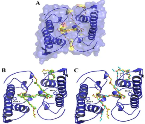
Fig. 1.Crystal structure of MjTX-II (A) and its superposition with MjTX-II/stearic acid (B) and with BthTX-I/PEG4K (C). The protein is represented in cartoon and ligand molecules are shown in sticks: in yellow, PEG4K from MjTX-II structure, in green, stearic acid molecules from MjTX-II/stearic acid structure and in cyan, PEG4K from BthTX-I/PEG4K structure. Drawn using PyMol program (DeLano, 2002). (For interpretation of the references to colour in thisfigure legend, the reader is referred to the web version of this article.)
monomers and (iii) one PEG4K (PEG 4) molecule interacts with Lys7, Trp77 and several other residues of monomer A (Fig. 3).
Dynamic light scattering experiments indicates a mean
hydrodynamic radius (RH) of 2.3 nm with a polydispersity
of 12.0%. ThisRHvalue corresponds to a molecular weight of
approximately 23 kDa and is, thus, equivalent to a dimer. These results are in agreement with other literature data
for Lys49-PLA2s since electrophoresis, spectroscopic (Arni
et al., 1999; da Silva Giotto et al., 1998), crystallographic (Arni and Ward, 1996; dos Santos et al., 2009; Magro et al., 2003; Murakami et al., 2005), small angle X-ray scattering (Murakami et al., 2007) and dynamic light scattering (Fernandes et al., 2010) experiments demonstrates that
bothropic Lys49-PLA2s are dimeric in solution.
In addition, the inspection of unit-cell packing showed that there are two possible dimeric configurations for the
MjTX-II structure. Thefirst possibility is similar to the
so-called“conventional dimer”and the second one is similar
to the“alternative dimer”(Murakami et al., 2005). In the
conventional dimer, the monomers are stabilized by
in-teractions between the tips of
b-wings and the residues of
the N-terminal helices (Arni and Ward, 1996) while in the
alternative dimer they are stabilized by contacts between the putative calcium-binding loops and C-termini forming
a connection route between the “active sites” of both
monomers (dos Santos et al., 2009). Examination of the
unit-cell packing using PISA software (Krissinel and
Henrick, 2007) points the alternative dimeric confi gura-tion is the most probable to occur in solugura-tion. According to this analysis, MjTX-II/PEG4K crystallographic structure
presents an interfacial area of 552.6A2,Gint¼ 9.8 kcal/mol
andGdiss¼0.145 kcal/mol. Furthermore, this choice is also
supported by previous small angle X-ray scattering
exper-iments (Murakami et al., 2007) and functional aspects of
Lys-PLA2s myotoxins (dos Santos et al., 2009; Murakami
et al., 2005).
3.2. Comparison between MjTX-II and MjTX-II complexed to stearic acid structures
The crystal structure of MjTX-II co-crystallized with stearic acid (a fatty acid) has been previously solved (Watanabe et al., 2005) and evidenced six stearic acid molecules interacting with the protein: two of them in each hydrophobic channel (two molecules in each protomer) and other two in the dimeric interface interacting with Lys7 residue. Contrasting with the co-crystallized structure (MjTX-II/stearic acid), the native MjTX-II (this study) only presents four PEG4K molecules: three of them are inside the hydrophobic channels and the fourth one interacts with
Lys7 residue (Fig. 1A). However, the comparison of both
structures reveals that all ligands occupy similar positions: (i) PEG 1 and PEG 2 occupy the same sites that two stearic acids from the MjTX-II/stearic acid complex (inside of the
hydrophobic channels) (Fig. 1B); (ii) PEG 3 is at the
hy-drophobic channels entrance (N-terminal face of the dimeric structure), connecting both protomers of the dimeric structure and is located approximately at the same position that two stearic acids molecules in the MjTX-II
complexed structure (Fig. 1B); (iii) PEG 4 occupies
approximately the same position of two stearic acids in the dimeric interface of MjTX-II/stearic acid structure which presents 50% occupancy values and are sited in a tail-to-tail
conformation (Fig. 1B) (Watanabe et al., 2005). Due to the
alternative dimeric configuration adopted for the native Fig. 2.Electron density difference maps for PEG4K molecules from the MjTX-II structure: (A) PEG4K (1) in the hydrophobic channel region of monomer A; (B) PEG4K (2) in the hydrophobic channel region of monomer B; (C) PEG4K (3) in the hydrophobic channel region connecting the monomers and (D) PEG4K (4) around Lys7 residue from monomer A. The maps were calculated with coefficientsjFobsj jFcalcjand contoured at 1.0 standard deviations. The ligands molecules were not considered for electron density maps calculation but are drawn for clarity. Drawn using PyMOL program (DeLano, 2002).
Fig. 3.Interaction of PEG4K molecules in MjTX-II structure. (A) and (B) represents the interactions of PEG4K (1 and 2) molecules that occupies the hydrophobic channel of the structure. (C) Hydrophobic interactions of PEG4K (3) molecule that connects both monomers. (D) Hydrophobic interactions of PEG4K (4) molecule in the protein surface (around Lys7 from monomer A). Drawn using Ligplot program (Wallace et al., 1995).
MjTX-II only one PEG ligand with 100% occupancy was modeled at this site. Therefore, despite the differences between both ligands (PEG4K and stearic acid), both structures are essentially identical as evidenced by the
root-mean-square deviation (r.m.s.d.) of 0.52A for theirCa
atoms superposition. Important regions for this toxin
biological functions (e.g. the active site, Ca2þ-binding
loop, C-termini (dos Santos et al., 2009; Fernandes et al.,
2010; Magro et al., 2003)) are conserved indicating that PEG4K may structurally simulate a fatty acid molecule bound to toxin’s hydrophobic channels since its backbone
is structurally similar to the protein substrate (Watanabe
et al., 2005). For this reason, we can state that the MjTX-II structure may represent the protein in its active state
(attached to the membrane) (dos Santos et al., 2009).
3.3. Comparison of MjTX-II and other Lys49-PLA2structures
Several myotoxic Lys49-PLA2s in the apo and complexed
forms have been solved (Arni et al., 1999; dos Santos et al.,
2011a; dos Santos et al., 2009; Fernandes et al., 2010; Lee et al., 2001; Magro et al., 2003; Marchi-Salvador et al., 2009; Murakami et al., 2005, 2007; Watanabe et al., 2005). Table 2 shows a structural comparison between the monomers of MjTX-II and the same analysis for several
other apo and complexed Lys49-PLA2s. As previously
observed (dos Santos et al., 2009), all complexed structures
present lower r.m.s.d. values compared to their respective apo structures. In other words, there is a clear structural
pattern for Lys49-PLA2s whose apo and complexed states
can also be distinguished by the“two angle”model
previ-ously suggested (dos Santos et al., 2009). Applying this
model to MjTX-II structure, the aperture and torsional
an-gles between its monomers are 55and 25, respectively.
These values are in agreement to those calculated for
MjTX-II/stearic acid structure (52and 20) and are also similar to
values found for other complexed Lys49-PLA2s (Table 3)
(dos Santos et al., 2009).
In 2001, Lee and colleagues solved the PrTX-II/fatty acid structure and suggested an important role played by Lys122. According to the authors, Lys122 interacts with the
main chain carbonyl of Cys29 causing hyperpolarization of the Cys29/Gly30 peptide bond and, consequently, increases
the affinity of the toxin for fatty acids (Lee et al., 2001). This
hypothesis suggested that Lys49-PLA2s are enzymes that
are able to hydrolyze phospholipids but fail to release the products of its action. The fatty acid would stay retained in the hydrophobic channel of the toxin consequently
inhib-iting it, therefore explaining why Lys49-PLA2s toxins do not
display significant catalytic activity. In contrast with this
hypothesis, Fernandes and colleagues (Fernandes et al.,
2010) performed a very comprehensive study using 16
different dimeric Lys49-PLA2s and showed that Lys 122 is a
veryflexible residue that may adopt random configurations even though it usually interacts with different negative charged sites. Despite the highlighted absence of pattern for Lys122 interaction, PrTX-II complexed to fatty acid and MjTX-II complexed to stearic acid structures are two
observed exceptions (Lee et al., 2001; Watanabe et al.,
2005). The Lys122 side chain interacts with main chain
carbonyl of Cys29 in both monomers of these dimeric complexes indicating that the phenomena just occur when fatty acids are present in the hydrophobic channels (Fernandes et al., 2010). Interesting, the results presented here for the MjTX-II can clarify this issue. As discussed in the item 3.2, the structure of MjTX-II presents PEG4K molecules in its hydrophobic channels at the same regions where fatty acids are found in the MjTX-II/stearic acid
structure. Therefore, thisfinding suggests that the
Cys29-Lys122 interaction is not exclusive for Lys49-PLA2s-fatty
acid bound structures.
Additionally, comparison between BthTX-I/PEG4K
(Fernandes et al., 2010) and MjTX-II/PEG4K (this work) structures reveals important features of MjTX-II in
com-parison to BthTX-I and other bothropic Lys49-PLA2s
myo-toxins. Despite the high sequential and structural similarity between MjTX-II and BthTX-I (they share 95% of identity), and the presence of two PEG4K molecules in the hydro-phobic channels of both structures, their superposition clearly demonstrates the ligand way of binding at these
proteins are somewhat different (Fig. 1C). The reason seems
to be the insertion of the residue Asn120 and the Leu32Gly mutation since these characteristics apparently shifts the orientation of PEG molecules inside the hydrophobic
channel by modification of the channel ends. Interestingly,
Asn120 insertion and Leu32Gly mutation are only found in MjTX-II when comparing this protein to all the other
bothropic Lys49-PLA2s whose structures are solved to date
(Fig. 4). Therefore, the Asn120 insertion is probably the responsible for the binding of the interchain PEG 3 mole-cule since this insertion leads to a distortion of the protein
protomers (Table 3). This evidence is enhanced by the fact
that other Lys49-PLA2s toxins that presents 121 amino
acids do not bind this interchain PEG4K or PEG3350 (Fernandes et al., 2010) whereas MjTX-II (122 residues) allow a PEG4K interaction at this region. It is important to
highlight that bothropic Lys49-PLA2s that do not have
structures solved yet but present mutations and/or
in-sertions at the positions 32 and 120 are known (Fig. 4);
however their structural characteristics upon ligand bind-ing will just be evidenced when these structures are elucidated.
Table 2
Superposition between Lys49-PLA2s monomers (root mean square devi-ation ofCaatoms).
Protein (PDB id) r.m.s.d. (A)
MjTX-II/PEG4000 0.23
MjTX-II/Stearic acid (1XXS) 0.26 BaspTX-II/Suramin (1Y4L) 0.68 BthTX-I/a-tocopherol (3CXI) 0.76 PrTX-I/a-tocopherol (3CYL) 0.82
BthTX-I/BPB (3HZW) 0.67
PrTX-I/BPB (2OK9) 0.47
BthTX-I/PEG4000 (3IQ3) 0.79
PrTX-I/Rosmarinic acid (3QNL) 0.32 BnIV/Myristic acid (3MLM) 0.55 PrTX-II/Fatty acid (1QLL) 0.25
BaspTX-II (1CLP) 0.60
BnSP-6 (1PC9) 0.96
BnSP-7 (1PA0) 0.96
PrTX-I (2Q2J) 1.04
BthTX-I (3HZD) 1.05
The C-termini of Lys49-PLA2s toxins has been pointed as
containing their myotoxic site (Arni and Ward, 1996; Magro
et al., 2003; Ward et al., 2002). Afterward, dos Santos and colleagues reviewed several native and complexed
Lys49-PLA2s and proposed that Arg118, Lys20 and Lys115 residues
constitute these toxins myotoxic site (dos Santos et al.,
2009). In the same study, the authors propose a two-step
mechanism for Lys49-PLA2s which allow the
destabiliza-tion of biological membranes. Thefirst step of the
mecha-nism would be the interaction of the Lys20, Lys115 and Arg118 of these proteins with the phospholipid head group
region (dos Santos et al., 2009); subsequently, a quaternary
rearrangement takes place in Lys49-PLA2s monomers
allowing long-chain hydrophobic portions of membrane phospholipids to be inserted into the hydrophobic channel
of the toxin (dos Santos et al., 2009; dos Santos et al., 2011a).
Therefore, the hydrophobic channel was demonstrated to be
involved in one of the steps required for Lys49-PLA2s action
mechanism (dos Santos et al., 2009; dos Santos et al., 2011a).
It is also interesting to highlight that if the alternative dimer is considered as biological dimer, the myotoxic sites from both monomers are aligned at the same plane (side by side) for the complexed structures (active state) and an interchain
Tyr119-Tyr119 hydrogen bond is formed (Table 3) which
increasing the toxin potency (dos Santos et al., 2009). The
sequence alignment of bothropic Lys49-PLA2s (Fig. 4) shows
that the residues of the myotoxic site (Lys20, Lys115 and Arg118) and Tyr119 are conserved in MjTX-II, however, the interchain Tyr119–Tyr119 hydrogen bond is not present in its
dimeric interface (these residues are at a distance of 4.7A).
Analyzing MjTX-II sequence (Fig. 4) it is possible to observe
that the C-terminal region of this toxin presents some par-ticularities as an insertion of a residue at position 120 and a mutation at position 121 (His/Tyr) if compared to other
bothropic Lys49-PLA2s whose structures are known.
There-fore, Asn120 insertion may be the responsible for a diversion of this region as evidenced by the lack of Tyr119-Tyr119 hydrogen bond which is probably compensated by the cre-ation of two new hydrogen bonds with the participcre-ation of
Tyr121 residue (Table 3).
Then, taking into account these facts (Asn120 insertion and mutations of residues 32 and 121) and their conse-quences to PEG4Ks mode of binding, it is reasonable to suggest that MjTX-II may require specific or modified in-hibitors when compared to molecules that are able to
inhibit bothropic Lys49-PLA2s by interaction with their
hydrophobic channels. This is due to the different profile of
ligand binding presented at this region (Fig. 1C.) and may
have implications when considering structure-based ligand
design for Lys49-PLA2s.
Table 3
Torsional angles (qT), aperture angles (qA) and hydrogen bonds at the interface of Lys49-PLA2s fromBothropsgenus.
Protein (PDB id) qT(torsional angle)a q
A(aperture angle)a Hydrogen bonds at the interfacesb
Monomer A Monomer B DistanceA
MjTX-II 55 25 Ala19 [N] Tyr121 [OH] 2.88
Asn17 [ND2] Tyr121 [OH] 2.29
MjTX-II/Stearic acid (1XXS) 52 20 Tyr121 [OH] Asn17 [ND2] 2.36
Tyr121 [OH] Ala19 [N] 2.58 Ala19 [N] Tyr 121 [OH] 3.64
BaspTX-II/Suramin (1Y4L) 51 15 – – –
BthTX-I/a-tocopherol (3CXI) 40 11 Tyr119 [O] Asn17 [ND2] 2.84
Tyr 119 [OH] Tyr119 [OH] 2.57
PrTX-I/a-tocopherol (3CYL) 41 12 Tyr119 [O] Asn17 [ND2] 2.84
Tyr119 [OH] Tyr119 [OH] 2.48
BthTX-I/BPB (3HZW) 53 22 Tyr119 [OH] Asn17 [ND2] 3.25
Tyr119 [OH] Tyr119 [OH] 3.09
PrTX-I/BPB (2OK9) 41 29 Tyr119 [O] Asn17 [ND2] 2.82
Tyr119 [OH] Tyr119 [OH] 2.16
BthTX-I/PEG4000 (3IQ3) 41 29 Tyr119 [O] Asn17 [ND2] 2.82
Tyr119 [OH] Tyr119 [OH] 2.56
PrTX-I/Rosmarinic acid (3QNL) 43 23 Tyr119 [O] Asn17 [ND2] 3.16
Tyr119 [OH] Tyr119 [OH] 2.18 Asn17 [ND2] Tyr119 [O] 3.22
BnIV/Myristic acid (3MLM) 41 27 Tyr119 [OH] Tyr119 [OH] 2.42
Tyr119 [O] Asn17 [ND2] 2.49
PrTX-II/fatty acid (1QLL)c 81 23 Tyr119 [OH] Tyr119 [OH] 3.09
BaspTX-II (1CLP)d 44 22 Tyr119 [OH] Tyr119 [OH] 2.35
Tyr119 [OH] Lys20 [NZ] 3.86
BnSP-6 (1PC9) 60 6 Tyr119 [O] Asn17 [ND2] 2.51
BnSP-7 (1PA0) 60 6 Lys69 [NZ] Val31 [O] 2.74
Tyr119 [O] Asn17 [ND2] 2.68
PrTX-I (2Q2J) 60 7 Val31 [O] Lys69 [NZ] 2.57
Asn17 [ND2] Tyr 119 [O] 2.91 Asn17 [ND2] His120 [O] 3.36
BthTX-I (3HZD) 60 14 Val31 [O] Lys69 [NZ] 2.72
aTorsional and aperture angles were calculated according to the proposal ofdos Santos et al., 2009.
bHydrogen bonds were inferred using the online interactive tool PISA (http://www.ebi.ac.uk/msd-srv/prot_int/cgi-bin/piserver).
cPrTX-II/fatty acid (PDB id 1QLL) crystallographic structure do not follow the torsional/aperture angle pattern for complexed structures as pointed bydos
Santos et al., 2009.
dBaspTX-II (PDB id1CLP) crystallographic structure was deposited as an apo structure but according todos Santos et al., 2009it can be a complex.
3.4. The crystal structure of MjTX-II and the biological dimer of Lys49-PLA2structures
As discussed in the last two sections, MjTX-II structure
was solved in the oligomeric assembly known as
“alterna-tive dimer”given that it has higher probability of
occur-rence in solution due to bioinformatic analyses and also
due to several experimental and functional reasons (dos
Santos et al., 2011a; dos Santos et al., 2009; Fernandes et al., 2010; Marchi-Salvador et al., 2009; Murakami et al., 2005, 2007). However, as discussed in a recent review in thisfield (Lomonte and Rangel, 2012), this subject is still controversial for some authors.
Although no experiment was able to definitively prove
the correct assembly adopted by Lys49-PLA2s toxins,
MjTX-II structure added an important experimental evidence for the choice of the alternative dimer as the probable qua-ternary assembly found in solution for these proteins. As
shown in theFig. 2, the PEG 3 binds simultaneously to both
monomers of the protein. This mode of binding is only possible for the alternative dimeric conformation because if the conventional dimer had been used to solve this struc-ture a large portion of the ligand would be exposed to the solvent which is energetically unfavorable. Similar results were also obtained for PrTX-I/a-tocopherol and BaspTX-II/
suramin structures (dos Santos et al., 2009; Murakami
et al., 2005), leading these authors to choose the alterna-tive assembly when solving both complexed structures.
3.5. Functional studies
Myographic studies show that MjTX-II (1.0
mM)
pro-duced an irreversible and time-dependent blockade of both
directly (t1/2¼40.02.3 min,n¼7) and indirectly (t1/
2¼32.14.8 min,n¼5) evoked twitches (Fig. 5A and B).
The present findings were consistent with those already
described for other Lys49-PLA2s (Cavalcante et al., 2007;
Gallacci et al., 2006; Randazzo-Moura et al., 2008; Rodrigues-Simioni et al., 1995; Soares et al., 2000, 2001; Stabeli et al., 2006). Statistical comparison of the indirectly
evoked contractionst1/2of the MjTX-II with those of other
Lys49 PLA2s, such as PrTX-I from Bothrops pirajai (t1/
2¼49.06.9 min,n¼8) and BthTX-I fromBothrops
jar-aracussu(t1/2¼40.33.5 min,n¼6), obtained at same experimental conditions, indicates that these toxins have
similar potency (Cavalcante et al., 2007). In contrast,
MjTX-II presents a more potent neuromuscular blockade when
compared to MjTX-I fromB. moojeni,since at 1.0
mM this
toxin depressed in only about 20% the twitches amplitude
after 90 min of contact with the preparation (Salvador et al.,
2013). Then, these results indicate MjTX-II has similar or
superior neuromuscular blockade action compared to other
Lys49-PLA2s.
It has been suggested that in vitro neuromuscular
blockade effect observed for Lys49-PLA2s may be a
conse-quence of their membrane depolarizing activity (Gallacci
and Cavalcante, 2010). In order to clarify this issue, we Fig. 4.Amino acid sequence alignment of Lys49-PLA2s fromBothropsgenus. The natural substitution Gly32 and the Asn120 insertion are highlighted. NCBI GI entry codes for the proteins represented are BnIV (Bothrops pauloensis): 333361256, BnSP-6 (Bothrops pauloensis): 49258448, BnSP-7 (Bothrops pauloensis): 239938675, BthTX-I (Bothrops jararacussu): 51890398, MjTX-II (Bothrops moojeni): 62738542; BaspTX-II (Bothrops asper): 166215047, PrTX-I (Bothrops pirajai): 17433154, PrTX-II (Bothrops pirajai): 17368328, Basp-M1-3-3 (Bothrops asper): 17433168, BaTX (Bothrops alternatus): 292630846, Batr-1(Bothrops atrox): 82201805, BlK-PLA2 (Bothrops leucurus): 353678055, Myo-IVa (Bothrops asper): 166216293.
performed an electrophysiological study to evaluate membrane depolarizing activity induced by MjTX-II. This technique measures the resting membrane potential of the sarcolemma and has been used as a more direct and sen-sitive method to assess the initial events in myotoxicity (Aragao et al., 2009). The results show a progressive in-crease in the resting membrane potential of skeletal muscle
fibers from 15 min onwards (Fig. 6) and the increase of the
frequency of miniature endplate potentials after 5 min (data not shown), probably as a consequence of the cell depolarization. Taken together, these results show a direct effect of MjTX-II increasing the permeability of the skeletal
musclefibers plasma membrane.
Although the Lys49-PLA2s mechanism of action is not
yet fully elucidated, several studies point the sarcolemma
as the initial target of these myotoxins (Gutierrez and
Lomonte, 1995; Lomonte et al., 2003; Lomonte and Rangel, 2012). A recent study suggests Arg118, together with Lys20 and Lys115, are the residues responsible for
protein-membrane anchorage initial steps (dos Santos
et al., 2009; dos Santos et al., 2011a). After the establish-ment of these electrostatic interactions, the protein
un-dergoes a quaternary rearrangement that allows
hydrophobic portions of membrane phospholipids to be inserted in the protein hydrophobic channels, therefore
culminating with membrane destabilization (dos Santos
et al., 2011a). Thefirst consequence of this destabilization is the loss of ionic permeability regulation, leading to a reduction of the resting membrane potential, inactivation of sodium channels and blockade of both directly and
indirectly evoked contractions (Gallacci and Cavalcante,
2010). In addition, the disruption of the muscle fiber
membranes induced by Lys49-PLA2s also promotes an
in-crease of cytosolic calcium concentration, initiating a complex series of degenerative mechanisms that
culmi-nates with the muscle cell damage (Gutierrez and Ownby,
2003; Lomonte and Rangel, 2012; Montecucco et al., 2008).
4. Conclusions
In this article, we fully characterize functionally and
structurally the Lys49-PLA2MjTX-II fromB. moojeni. Despite
the fact that this class of proteins has been extensively studied, several issues regarding the function–structure relationships are still need to be clarified, as highlighted by a
recent review in thisfield (Lomonte and Rangel, 2012). This
requirement is probably due to the high evolutionary pressure process by which snake venom molecules are submitted, since proteins with few natural amino acid
mutations may present different oligomeric configurations,
variable toxic potency or even different functions when
compared to their ancestral toxins (Doley and Kini, 2009;
dos Santos et al., 2011b; Kini, 1997, 2003). An interesting
example is the MjTX-I, other myotoxic Lys49-PLA2fromB.
Fig. 5.Effect of MjTX-II on indirectly (A) and directly (B) evoked twitches in mice phrenic-diaphragm preparations. The ordinate represents the % amplitude of twitches relative to the initial amplitude. The abscissa indicates the time (min) after the addition of MjTX-II to the organ bath. Vertical bars represent the SEM, *indicates the point from which there are significant differences relative to control (P<0.05).
Fig. 6.The resting membrane potential of mice diaphragm muscle prepa-rations under MjTX-II action. The ordinate represents the resting membrane potential (mV) and the abscissa indicates the time (min) after the addition of MjTX-II to the organ bath. The data were grouped as meanSEM (P<0.05). *indicates the point from which there are significant differences relative to control (P<0.05).
moojeni that presents unusual oligomeric characteristics and displays lower myotoxic activity when compared to all
other bothropic Lys49-PLA2s that have already been
struc-turally and functionally characterized (Andriao-Escarso
et al., 2000; Salvador et al., 2013).
As demonstrated in this work, MjTX-II also presents
some particularities if compared to other Lys49-PLA2s
which seem to influence the mode of ligand binding along the toxin hydrophobic channel, a feature that may directly
affect the design of structure-based ligands for Lys49-PLA2s.
Acknowledgments
This work was supported by Fundação de Amparo à Pes-quisa do Estado de São Paulo (FAPESP), Conselho Nacional de Desenvolvimento Científico e Tecnológico (CNPq), Financia-dora de Estudos e Projetos (FINEP), Coordenação de
Aper-feiçoamento de Nível Superior (CAPES)–Projeto NanoBiotec,
Rede de Biodiversidade e Biotecnologia da Amazônia Legal (BIONORTE/CNPq/MCT), Instituto Nacional para Pesquisa Translacional em Saúde e Ambiente na Região Amazônica (INCT-INPeTAm/CNPq/MCT) e Instituto Nacional para Pes-quisa em Toxinas (INCT-Tox), Secretary of Development of Rondonia State (SEPLAN/PRONEX/CNPq).
Conflict of interest
The authors have no conflicts of interest with this work.
References
Andriao-Escarso, S.H., Soares, A.M., Rodrigues, V.M., Angulo, Y., Diaz, C., Lomonte, B., Gutierrez, J.M., Giglio, J.R., 2000. Myotoxic phospholi-pases A2inbothropssnake venoms: effect of chemical modifications
on the enzymatic and pharmacological properties of bothropstoxins fromBothrops jararacussu. Biochimie 82, 755–763.
Aragao, E.A., Randazzo-Moura, P., Rostelato-Ferreira, S., Rodrigues-Simioni, L., Ward, R.J., 2009. Shared structural determinants for the calcium-independent liposome membrane permeabilization and sarcolemma depolarization in Bothropstoxin-I, a LYS49-PLA2from the
venom ofBothrops jararacussu. Int. J. Biochem. Cell Biol. 41, 2588–2593.
Arni, R.K., Fontes, M.R., Barberato, C., Gutierrez, J.M., Diaz, C., Ward, R.J., 1999. Crystal structure of myotoxin II, a monomeric Lys49-phospholipase A2homologue isolated from the venom of Cerrophi-dion (Bothrops) godmani. Arch. Biochem. Biophys. 366, 177–182.
Arni, R.K., Ward, R.J., 1996. Phospholipase A2-a structural review. Toxicon
34, 827–841.
Arni, R.K., Ward, R.J., Gutierrez, J.M., Tulinsky, A., 1995. Structure of a calcium-independent phospholipase-like myotoxic protein from Bothrops aspervenom. Acta Crystallogr. D Biol. Crystallogr. 51, 311–317.
Borges, R.C., Araujo, A.F.B., 1998. Seleção de habitat em duas espécies de jararaca (Bothrops moojeniHoge eB. neuwiediWagler) (Serpentes, Viperidae). Revista Brasileira de Biologia 58, 591–601.
Brunger, A.T., Adams, P.D., Clore, G.M., DeLano, W.L., Gros, P., Grosse-Kunstleve, R.W., Jiang, J.S., Kuszewski, J., Nilges, M., Pannu, N.S., Read, R.J., Rice, L.M., Simonson, T., Warren, G.L., 1998. Crystallography & NMR system: a new software suite for macromolecular structure determination. Acta Crystallogr. D Biol. Crystallogr. 54, 905–921.
Cavalcante, W.L., Campos, T.O., Dal Pai-Silva, M., Pereira, P.S., Oliveira, C.Z., Soares, A.M., Gallacci, M., 2007. Neutralization of snake venom phospholipase A2 toxins by aqueous extract of Casearia sylvestris
(Flacourtiaceae) in mouse neuromuscular preparation. J. Ethno-pharmacol. 112, 490–497.
Chen, V.B., Arendall 3rd, W.B., Headd, J.J., Keedy, D.A., Immormino, R.M., Kapral, G.J., Murray, L.W., Richardson, J.S., Richardson, D.C., 2010. MolProbity: all-atom structure validation for macromolecular crys-tallography. Acta Crystallogr. D Biol. Crystallogr. 66, 12–21.
Correia-de-Sa, P., Noronha-Matos, J.B., Timoteo, M.A., Ferreirinha, F., Marques, P., Soares, A.M., Carvalho, C., Cavalcante, W.L., Gallacci, M.,
2013. Bothropstoxin-I reduces evoked acetylcholine release from rat motor nerve terminals: radiochemical and real-time video-micro-scopy studies. Toxicon 61C, 16–25.
da Silva Giotto, M.T., Garratt, R.C., Oliva, G., Mascarenhas, Y.P., Giglio, J.R., Cintra, A.C., de Azevedo Jr., W.F., Arni, R.K., Ward, R.J., 1998. Crystal-lographic and spectroscopic characterization of a molecular hinge: conformational changes in bothropstoxin I, a dimeric Lys49-phospholipase A2homologue. Proteins 30, 442–454.
de Azevedo, W.F., Ward, R.J., Lombardi, F.R., Giglio, J.R., Soares, A.M., Fontes, M.R.M., Arni, R.K., 1997. Crystal Structure of Myotoxin-II: a myotoxic phospholipase A2-homologue from Bothrops moojeni
Venom. Protein Pept. Lett. 4, 329–334.
DeLano, W.S., 2002. The PyMOL Molecular Graphics System. Delano Sci-entific, San Carlos.
Delatorre, P., Rocha, B.A., Santi-Gadelha, T., Gadelha, C.A., Toyama, M.H., Cavada, B.S., 2011. Crystal structure of Bn IV in complex with myristic acid: a Lys49 myotoxic phospholipase A2 fromBothrops neuwiedi
venom. Biochimie 93, 513–518.
Doley, R., Kini, R.M., 2009. Protein complexes in snake venom. Cell Mol. Life Sci. 66, 2851–2871.
dos Santos, J.I., Soares, A.M., Fontes, M.R., 2009. Comparative structural studies on Lys49-phospholipases A2fromBothropsgenus reveal their
myotoxic site. J. Struct. Biol. 167, 106–116.
dos Santos, J.I., Cardoso, F.F., Soares, A.M., Dal Pai Silva, M., Gallacci, M., Fontes, M.R., 2011a. Structural and functional studies of a bothropic myotoxin complexed to rosmarinic acid: new insights into Lys49-PLA2inhibition. PLoS One 6, e28521.
dos Santos, J.I., Cintra-Francischinelli, M., Borges, R.J., Fernandes, C.A., Pizzo, P., Cintra, A.C., Braz, A.S., Soares, A.M., Fontes, M.R., 2011b. Structural, functional, and bioinformatics studies reveal a new snake venom homologue phospholipase A2class. Proteins 79, 61–78.
Ducruix, A., Giegé, R., 1992. Crystallization of Nucleic Acids and Proteins: a Pratical Approach. Oxford University Press, New York.
Emsley, P., Cowtan, K., 2004. Coot: model-building tools for molecular graphics. Acta Crystallogr. D Biol. Crystallogr. 60, 2126–2132.
Fatt, P., Katz, B., 1951. An analysis of the end-plate potential recorded with an intracellular electrode. J. Physiol. 115, 320–370.
Fernandes, C.A., Marchi-Salvador, D.P., Salvador, G.M., Silva, M.C., Costa, T.R., Soares, A.M., Fontes, M.R., 2010. Comparison between apo and complexed structures of bothropstoxin-I reveals the role of Lys122 and Ca2þ-binding loop region for the catalytically inactive
Lys49-PLA2s. J. Struct. Biol. 171 , 31–43.
Gallacci, M., Cavalcante, W.L., 2010. Understanding the in vitro neuro-muscular activity of snake venom Lys49 phospholipase A2
homo-logues. Toxicon 55, 1–11.
Gallacci, M., Oliveira, M., Dal Pai-Silva, M., Cavalcante, W.L., Spencer, P.J., 2006. Paralyzing and myotoxic effects of a recombinant bothropstoxin-I (BthTX-I) on mouse neuromuscular preparations. Exp. Toxicol. Pathol. 57, 239–245.
Gutierrez, J.M., Chaves, F., 1980. Proteolytic, hemorrhagic and myonecrotic effects of the venoms of Costa Rican snakes from the generaBothrops, CrotalusandLachesis(author’s transl). Toxicon 18, 315–321.
Gutierrez, J.M., Lomonte, B., 1995. Phospholipase A2 myotoxins from Bothropssnake venoms. Toxicon 33, 1405–1424.
Gutierrez, J.M., Ownby, C.L., 2003. Skeletal muscle degeneration induced by venom phospholipases A2: insights into the mechanisms of local
and systemic myotoxicity. Toxicon 42, 915–931.
Gutierrez, J.M., Theakston, R.D., Warrell, D.A., 2006. Confronting the neglected problem of snake bite envenoming: the need for a global partnership. Plos Med. 3, e150.
Harrison, R.A., Hargreaves, A., Wagstaff, S.C., Faragher, B., Lalloo, D.G., 2009. Snake envenoming: a disease of poverty. PLoS Negl Trop. Dis. 3, e569.
Homsi-Brandeburgo, M.I., Queiroz, L.S., Santo-Neto, H., Rodrigues-Simioni, L., Giglio, J.R., 1988. Fractionation ofBothrops jararacussu snake venom: partial chemical characterization and biological activ-ity of bothropstoxin. Toxicon 26, 615–627.
Kasturiratne, A., Wickremasinghe, A.R., de Silva, N., Gunawardena, N.K., Pathmeswaran, A., Premaratna, R., Savioli, L., Lalloo, D.G., de Silva, H.J., 2008. The global burden of snakebite: a literature analysis and modelling based on regional estimates of envenoming and deaths. Plos Med. 5, 1591–1604.
Kini, R.M., 1997. Phospholipase A2 Enzyme: Structure, Function and
Mechanism. Wiley, Chichester.
Kini, R.M., 2003. Excitement ahead: structure, function and mechanism of snake venom phospholipase A2enzymes. Toxicon 42, 827–840.
Krissinel, E., Henrick, K., 2007. Inference of macromolecular assemblies from crystalline state. J. Mol. Biol. 372, 774–797.
Lee, W.H., da Silva Giotto, M.T., Marangoni, S., Toyama, M.H., Polikarpov, I., Garratt, R.C., 2001. Structural basis for low catalytic activity in Lys49
phospholipases A2-a hypothesis: the crystal structure of piratoxin II
complexed to fatty acid. Biochemistry-Us 40, 28–36.
Lomonte, B., Angulo, Y., Calderon, L., 2003. An overview of lysine-49 phospholipase A2myotoxins from crotalid snake venoms and their
structural determinants of myotoxic action. Toxicon 42, 885–901.
Lomonte, B., Gutierrez, J.M., Furtado, M.F., Otero, R., Rosso, J.P., Vargas, O., Carmona, E., Rovira, M.E., 1990. Isolation of basic myo-toxins from Bothrops moojeni and Bothrops atrox snake venoms. Toxicon 28, 1137–1146.
Lomonte, B., Rangel, J., 2012. Snake venom Lys49 myotoxins: from phospholipases A2to non-enzymatic membrane disruptors. Toxicon
60, 520–530.
Magro, A.J., Soares, A.M., Giglio, J.R., Fontes, M.R., 2003. Crystal structures of BnSP-7 and BnSP-6, two Lys49-phospholipases A2: quaternary
structure and inhibition mechanism insights. Biochem. Biophys. Res. Commun. 311, 713–720.
Marchi-Salvador, D.P., Fernandes, C.A., Silveira, L.B., Soares, A.M., Fontes, M.R., 2009. Crystal structure of a phospholipase A2homolog
complexed withp-bromophenacyl bromide reveals important struc-tural changes associated with the inhibition of myotoxic activity. Biochim. Biophys. Acta 1794, 1583–1590.
Mebs, D., Ehrenfeld, M., Samejima, Y., 1983. Local necrotizing effect of snake venoms on skin and muscle: relationship to serum creatine kinase. Toxicon 21, 393–404.
Melgarejo, A.R., 2003. Serpentes Peçonhentos no Brasil, Animais Peçon-hentos no Brasil: Biologia, Clínica e Terapêutica dos Acidentes. Sarv-ier, São Paulo, pp. 33–61.
Montecucco, C., Gutierrez, J.M., Lomonte, B., 2008. Cellular pathology induced by snake venom phospholipase A2myotoxins and
neuro-toxins: common aspects of their mechanisms of action. Cell Mol. Life Sci. 65, 2897–2912.
Murakami, M.T., Arruda, E.Z., Melo, P.A., Martinez, A.B., Calil-Elias, S., Tomaz, M.A., Lomonte, B., Gutierrez, J.M., Arni, R.K., 2005. Inhibition of myotoxic activity of Bothrops asper myotoxin II by the anti-trypanosomal drug suramin. J. Mol. Biol. 350, 416–426.
Murakami, M.T., Vicoti, M.M., Abrego, J.R., Lourenzoni, M.R., Cintra, A.C., Arruda, E.Z., Tomaz, M.A., Melo, P.A., Arni, R.K., 2007. Interfacial sur-face charge and free accessibility to the PLA2-active site-like region
are essential requirements for the activity of Lys49 PLA2homologues.
Toxicon 49, 378–387.
Otwinowski, Z., Minor, W., 1997. Processing of X-ray diffraction data collected in oscillation mode. Macromol. Crystallogr. Pt A 276, 307–
326.
Potterton, L., McNicholas, S., Krissinel, E., Gruber, J., Cowtan, K., Emsley, P., Murshudov, G.N., Cohen, S., Perrakis, A., Noble, M., 2004. De-velopments in the CCP4 molecular-graphics project. Acta Crystallogr. D Biol. Crystallogr. 60, 2288–2294.
Queiroz, L.S., Petta, C.A., 1984. Histopathological changes caused by venom of urutu snake (Bothrops alternatus) in mouse skeletal muscle. Rev. Inst. Med. Trop. Sao Paulo 26, 247–253.
Randazzo-Moura, P., Ponce-Soto, L.A., Rodrigues-Simioni, L., Marangoni, S., 2008. Structural characterization and neuromuscular activity of a new Lys49 phospholipase A2homologous (Bp-12)
iso-lated fromBothrops pauloensissnake venom. Protein J. 27, 355–362.
Rodrigues-Simioni, L., Prado-Franceschi, J., Cintra, A.C., Giglio, J.R., Jiang, M.S., Fletcher, J.E., 1995. No role for enzymatic activity or dantrolene-sensitive Ca2þstores in the muscular effects of
bothrop-stoxin, a Lys49 phospholipase A2myotoxin. Toxicon 33, 1479–1489.
Rosenfeld, G., Kelen, E.M., 1971. Measurement of the coagulation activity of snake venoms: importance to scientific research and therapeutic application. Rev. Paul Med. 77, 149–150.
Russell, F.E., 1991. Venomous arthropods. Vet. Hum. Toxicol. 33, 505–508.
Salvador, G.H.M., Fernandes, C.A.H., Magro, A.J., Marchi-Salvador, D.P., Cavalcante, W.L.G., Fernandez, R.M., Gallacci, M., Soares, A.M., Oliveira, C.L.P., Fontes, M.R.M., 2013. Structural and phylogenetic studies with MjTX-i reveal a multi-oligomeric toxin–a novel feature in Lys49-PLA2s protein class. PLoS One 8, e60610.
Saúde, F.N.d., 2001. Manual de Diagnóstico e Tratamento de Acidentes por Animais Peçonhentos. MS/FUNASA, Brasilia.
Soares, A.M., Andriao-Escarso, S.H., Angulo, Y., Lomonte, B., Gutierrez, J.M., Marangoni, S., Toyama, M.H., Arni, R.K., Giglio, J.R., 2000. Structural and functional characterization of myotoxin I, a Lys49 phospholipase A2homologue fromBothrops moojeni(Caissaca) snake venom. Arch.
Biochem. Biophys. 373, 7–15.
Soares, A.M., Andriao-Escarso, S.H., Bortoleto, R.K., Rodrigues-Simioni, L., Arni, R.K., Ward, R.J., Gutierrez, J.M., Giglio, J.R., 2001. Dissociation of enzymatic and pharmacological properties of piratoxins-I and -III, two myotoxic phospholipases A2fromBothrops pirajaisnake venom.
Arch. Biochem. Biophys. 387, 188–196.
Stabeli, R.G., Amui, S.F., Sant’Ana, C.D., Pires, M.G., Nomizo, A., Monteiro, M.C., Romao, P.R., Guerra-Sa, R., Vieira, C.A., Giglio, J.R., Fontes, M.R., Soares, A.M., 2006.Bothrops moojenimyotoxin-II, a Lys49-phospholipase A2homologue: an example of function versatility of snake venom
pro-teins. Comp. Biochem. Physiol. C Toxicol. Pharmacol. 142, 371–381.
Vagin, A., Teplyakov, A., 1997. MOLREP: an automated program for mo-lecular replacement. J. Appl. Crystallogr. 30, 1022–1025.
Wallace, A.C., Laskowski, R.A., Thornton, J.M., 1995. LIGPLOT: a program to generate schematic diagrams of protein-ligand interactions. Protein Eng. 8, 127–134.
Ward, R.J., Chioato, L., de Oliveira, A.H., Ruller, R., Sa, J.M., 2002. Active-site mutagenesis of a Lys49-phospholipase A2: biological and
membrane-disrupting activities in the absence of catalysis. Biochem. J. 362, 89–96.
Watanabe, L., Soares, A.M., Ward, R.J., Fontes, M.R., Arni, R.K., 2005. Structural insights for fatty acid binding in a Lys49-phospholipase A2:
crystal structure of myotoxin II fromBothrops moojenicomplexed with stearic acid. Biochimie 87, 161–167.
Williams, D., Gutierrez, J.M., Harrison, R., Warrell, D.A., White, J., Winkel, K.D., Gopalakrishnakone, P., 2010. The Global Snake Bite Initiative: an antidote for snake bite. Lancet 375, 89–91.