Beta-Actin Is Involved in Modulating Erythropoiesis
during Development by Fine-Tuning Gata2 Expression
Levels
Davina Tondeleir1, Benjamin Drogat2,3¤, Karolina Slowicka2,3, Karima Bakkali1, Sonia Bartunkova2,3, Steven Goossens2,3, Jody J. Haigh2,3*., Christophe Ampe1
*.
1Department of Biochemistry, Faculty of Medicine and Health Sciences, Ghent University, Ghent, Belgium,2Vascular Cell Biology Unit, Department for Molecular Biomedical Research, VIB, Ghent, Belgium,3Department of Biomedical Molecular Biology, Ghent University, Ghent, Belgium
Abstract
The functions of actin family members during development are poorly understood. To investigate the role of beta-actin in mammalian development, a beta-actin knockout mouse model was used. Homozygous beta-actin knockout mice are lethal at embryonic day (E)10.5. At E10.25 beta-actin knockout embryos are growth retarded and display a pale yolk sac and embryo proper that is suggestive of altered erythropoiesis. Here we report that lack of beta-actin resulted in a block of primitive and definitive hematopoietic development. Reduced levels of Gata2, were associated to this phenotype. Consistently, ChIP analysis revealed multiple binding sites for beta-actin in theGata2promoter.Gata2mRNA levels were almost completely rescued by expression of an erythroid lineage restricted ROSA26-promotor based GATA2 transgene. As a result, erythroid differentiation was restored and the knockout embryos showed significant improvement in yolk sac and embryo vascularization. These results provide new molecular insights for a novel function of beta-actin in erythropoiesis by modulating the expression levels ofGata2 in vivo.
Citation:Tondeleir D, Drogat B, Slowicka K, Bakkali K, Bartunkova S, et al. (2013) Beta-Actin Is Involved in Modulating Erythropoiesis during Development by Fine-Tuning Gata2 Expression Levels. PLoS ONE 8(6): e67855. doi:10.1371/journal.pone.0067855
Editor:Bin He, Baylor College of Medicine, United States of America ReceivedMarch 5, 2012;AcceptedMay 28, 2013;PublishedJune 26, 2013
Copyright:ß2013 Tondeleir et al. This is an open-access article distributed under the terms of the Creative Commons Attribution License, which permits unrestricted use, distribution, and reproduction in any medium, provided the original author and source are credited.
Funding:This work was supported by Fund for Scientific Research – Flanders (Belgium). Davina Tondeleir is a pre doctoral fellow of this Fund and Christophe Ampe received a research grant G.0441.10N (http://www.fwo.be/Default.aspx). The funders had no role in study design, data collection and analysis, decision to publish, or preparation of the manuscript.
Competing Interests:The authors have declared that no competing interests exist.
* E-mail: *[email protected] (CA); [email protected] (JJH)
.These authors contributed equally to this work.
¤ Current address: Interdisciplinary Research Institute (IRIBHM), Universite´ Libre de Bruxelles (ULB), Bruxelles, Belgium
Introduction
Actins are highly conserved proteins among various species throughout evolution [1]. The human genome has multiple functional actin genes and more than twenty pseudogenes [2,3]. The expression patterns of vertebrate actins are temporally and spatially regulated during development and in the adult organism, suggesting different isoform specific functions [1,4]. The four muscle-specific actins provide strength and contractility to muscle cells. By contrast beta- and gamma-cytoplasmic actins (further referred to as beta- and gamma-actin), that are coexpressed in all adult non-muscle tissues [5] are thought to participate in more dynamic actin cytoskeletal processes. Whereas gamma-actin seems to be uniformly distributed in all actin-containing structures, beta-actin seems to have a more polarized distribution, localized to the cortex of cells, specifically functioning in protrusive structures such as lamellipodia and filopodia [6,7,8,9].
In more recent cell work, actin was found localized in the nucleus leading to the hypothesis that beta-actin could be implicated in modulating transcriptional activity. Actin interacts with all three RNA polymerases and beta-actin has been identified as a component of different types of chromatin remodeling complexes in a wide range of organisms [10]. In addition,
antibodies against beta-actin block transcription [11] and nuclear translocation of beta-actin is involved in macrophage differenti-ation [12]. Although these previous studies obviously demonstrate a role for beta-actin in the nucleus, it is not clear if subsets of genes are dependent on beta-actin function in the nucleus and whether such mechanisms are important in instructing developmental processes.
To contribute to knowledge on the function of beta-actin during mouse development we further investigated the possible causes of this early embryonic lethality by employing a heterozygous beta-actin knockout mouse that was previously generated (Actb+/2) [19].
We observed a diminished number of primitive erythroid cells and a paucity of well-organized blood islands that are the initial sites of primitive erythroid and endothelial cell development. We show that ablation of beta-actin expression during development interferes with red blood cell (RBC) development, resulting in reduced amounts of primitive and definitive erythroid cells in the
Actb2/2 embryos. Most strikingly, mRNA expression levels of
Gata2, a transcription factor involved in early hematopoiesis, was dramatically decreased in yolk sacs of E8.5 Actb2/2 embryos. Corroborating a role for beta-actin inGata2regulation, we could show association of the beta-actin protein to theGata2promoter. Further confirming a developmental link between beta-actin function and Gata2 regulation, transgenic expression of Gata2
specifically within the erythroid lineage in theseActb2/2embryos
partially rescued the observed phenotypes. Our findings therefore support a novel function of beta-actin in modulating erythropoiesis by fine-tuningGata2levels in the early developing mouse embryo.
Materials and Methods
Ethics Statement
The animal ethics committee of Ghent University approved all experiments performed on mice. Approval number ECD10/29.
Mice
The generation of the Actb+/2 mice has been previously
described [19]. Actb+/2 mice were crossed to generate control Actb+/+
embryos andActb2/2embryos. Genotyping was done by fluorescent microscopy (positive forActb+/2andActb2/2) followed
by western blotting with an antibody against beta-actin (to distinguishActb+/2 and Actb2/2). Mice were kept on an inbred
BALB/c background.Actb+/2mice were crossed withFlk1-LacZ
mice [20] to generate double heterozygous knockouts. Genotyping was done by fluorescent microscopy followed by a LacZ PCR.
Actb+/2mice were crossed with EpoR-iCreTg/+[21] mice and with
ROSA26-hGata2Tg/Tg [21] to generate the Actb2/2 R26+hGa-ta2EpoR-iCre/+
rescue mice. Genotyping was done by fluorescent microscopy followed by PCR.
Antibodies
Antibodies (Ab) used in this study are rat-anti-mouse PECAM-1 mAb (Clone CD31), biotin-conjugated rat-anti-mouse PECAM-1 mAb (clone CD31) and biotin-conjugated goat-anti-rat Ig specific pAb, all from BD Pharmingen. ABC reagent (Vector Labs) was used with the biotin-conjugated CD31 mAb, whereas the Renaissance TSA Biotin System (PerkinElmer Life Sciences) was used with the other Abs. Anti beta-actin mAb (clone AC-15), from Sigma. Anti gamma-cytoplasmic actin pAb, from Millipore. Anti gata2 pAb, from Abcam.
Paraffin histology
Dissected samples were fixed overnight in 4% paraformalde-hyde at 4uC, processed for paraffin embedding, and sectioned at 6mm. Sections were stained with hematoxylin and eosin (H&E). Additional paraffin sections were used for immunohistochemistry and immunofluorescence.
Immunohistochemistry
Immunohistochemistry on whole mount embryos was per-formed as previously described [22]. Briefly, embryos were fixed in
MeOH:DMSO (4:1) overnight at 4uC, treated with MeOH:DM-SO:H2O2 (4:1:1) for 5–10 hours at room temperature to block
endogenous peroxidase activity and stored in methanol at220uC. The embryos were subsequently rehydrated in 50% MeOH in phosphate buffered saline PBS and incubated with the primary antibody in 3% instant skim milk powder/0.1% Triton X-100 in PBS (PBSMT) overnight at 4uC. Following washes in PBSMT for 5 hours at room temperature, embryos were incubated with the ABC reagent, in PBSMT overnight at 4uC. Following washes in PBSMT for 5 hours at room temperature and brief washes with PBS with 0.1% Triton X-100, the embryos were developed with 3.3-diaminobenzidine tetrahydrochloride (DAB) (Vector laborato-ries). The reaction was stopped by fixing the embryos in 4% PFA in PBS at room temperature for 1 hour. Immunohistochemistry on paraffin sections was done according to the protocol of the Renaissance TSA Biotin System (NEL 700, PerkinElmer). LacZ stainings were done according to manufacturer protocols (Milli-pore).
Immunofluorescence
Immunofluorescence on paraffin sections started by deparaffi-nization through ethanol series. Sections were microwaved in 0,01 M citrate buffer (pH 6.0) for 15 min at full power and washed in PBS. Sections were blocked in 10% goat serum/1% BSA in PBS for 1 hour at room temperature and incubated with primary antibody in blocking solution overnight at 4uC. Following four washes with PBS of each 30 min at room temperature, sections were incubated with secondary antibody for 2 hours at room temperature. Following four washes with PBS of each 15 min at room temperature, nuclei of cells were stained with DAPI and sections were mounted with DABCO mounting medium (Sigma-Aldrich).
Molecular analysis
Yolk Sacs (YS) were dissected at E8.5 and E10.25, or in case of
Actb2/2R26+hGata2EpoR-iCre/+
at E10.25 and E11.5, flash-frozen in liquid nitrogen, and processed according to standard protocols. RNA was extracted with High Pure RNA isolation kit (Roche) and precipitated overnight at280uC or 1 hr on dry ice. cDNA was synthesized with the transcriptor first strand cDNA synthesis kit (Roche). Quantitative reverse-transcription-polymerase chain re-action (qRT-PCR) was performed on a LightCycler 480 system (Roche) using the SYBR Green Master kit (Roche). Gene expression was normalized using glyceraldehyde 3-phosphate dehydrogenase (Gapdh), ubiquitine C (Ubc), glucose-6-phosphate dehydrogenase, (G6pdh) and hypoxanthin phosphoribosyl transfer-ase 1 (Hprt1) as controls. Primers used are given in Table S1. The expression levels of each gene are reported relative to those observed inActb+/+
control samples. For sampling and statistics see below.
ChIP
including localization of the respectively amplicons on theGata2
promotor.
In vitro hematopoietic progenitor assays
For primitive erythroid (EryP) colony assays were performed on stage matched E8.5 whole embryos as previously described [21]. Definitive erythroid colony assays were performed on E9.75 yolk sacs as previously described [21] using 1% methylcellulose (StemCell Technologies) containing complete recombinant cyto-kines (MethoCult GF M3434) for the detection and quantification of burst forming erythroid cells (BFU-E), colony forming granulocyte/macrophage (CFU-GM) and colony forming unit-granulocyte/erythrocyte/monocyte/megakaryocyte
(CFU-GEMM). After 7 days colonies were scored under a microscope. The results are expressed as a percentage of absolute number of colonies per yolk sac of theActb+/+
controls.
Imaging
Embryos were imaged on a Leica MS5 (Leica Microsystems) stereomicroscope. Digital images were acquired using a Leica camera. Section H&E staining and immunohistochemistry were imaged using a SNAP-COOL camera (Roper Scientific) mounted on an Olympus Bx51 microscope (Olympus), with Plan Olympus 206/0.40 or 406/0.65 lens and RSImage Version 1.9.2 software
(Roper Scientific).
Figure 1. Actb2/2embryos are pale and growth retarded at E10.25.(A) Pictures of freshly dissected embryos at E10.25. Pale and growth retardedActb2/2embryos show no obvious vascular pattern in the embryo proper (middle) or yolk sac (right) compared to theActb+/+
littermates. 156magnification. (B) LacZ stainings ofActb+/+andActb2/2yolk sac at E9.5 indicate reduced vascular branching complexity ofActb2/2yolk sacs. 206magnification. (C) Whole mount PECAM-1 immunohistochemistry ofActb+/+andActb2/2embryos, processed in parallel, shows less coloring, indicating fewer endothelial cells and red blood cells. 156 magnification. Embryos were imaged on a Leica MS5 (Leica Microsystems) stereomicroscope. Digital images were acquired using a Leica camera.
doi:10.1371/journal.pone.0067855.g001
Statistical analysis
Data were expressed as mean plus or minus SEM. Comparison between 2 data groups was done by the 2-sided Student t test. A minimum of three biological replicates was used in each condition for each genotype. Two technical replicates were used per biological replicate.
Results
Yolk sac blood island development is affected in beta-actin knockout embryos
To specifically address the role of beta-actin in development, a beta-actin knockout mouse allele was used (Actb+/2). It was
previously shown that the homozygous beta-actin knockout mice (Actb2/2) are lethal at E10.5 [19], however the exact cause of lethality has not been investigated. At E8.5, no obvious morphological differences betweenActb+/+
and Actb2/2 embryos
could be observed (data not shown). One day later at E9.5Actb2/2
embryos have an appropriate number of somites and were in that aspect comparable in development toActb+/+
littermates. Howev-er, theActb2/2embryos display a visibly pale yolk sac and embryo proper (Figure 1A). Yolk sacs of Actb+/+
embryos displayed large RBC containing vessels, whereas no such vessels could be observed inActb2/2yolk sacs. (Figure 1A). To study the branching pattern of the vasculature in the yolk sac,Actb+/2mice were crossed with Flk1-LacZ mice [20]. Flk1 marks vascular and hematopoietic progenitors and theFlk1-LacZ mice express a LacZ reporter under control of theFlk1promotor. It is therefore possible to monitor the migration of hemangioblast progenitors from the primitive streak towards the yolk sac and to study the vasculogenesis process. The resulting E9.5 Actb2/2 Flk1-LacZ embryos displayed clearly reduced vascular branching complexity in the yolk sac and specifically in the head region of the embryo proper compared to the Actb+/+ Flk1-LacZ littermates (Figure 1B). Especially the
remodeled large branched vessels, which are already prominent in yolk sacs ofActb+/+
Flk1-LacZ embryos seemed to be absent in the
Actb2/2 Flk1-LacZ embryos. Rather the Actb2/2 yolk sacs still contained the honeycomb-shaped capillary plexus (typical for E8.5 embryos), which did not reorganize into large branches, suggesting a block in proper vasculo/angiogenesis development.
At E10.25, theActb2/2embryos were morphologically growth
retarded. In view of the impaired vascular development of the yolk sac at E9.5 we performed PECAM-1 whole mount immunostain-ing at E10.25 embryos. This analysis showed the presence of a more weakly stained vasculature and therefore suggests diminished numbers of endothelial cells inActb2/2 embryos, similar to the yolk sac vascular defects (Figure 1C). PECAM-1 immunostaining on sections from E9.5 embryos demonstrated normal endocardial and slightly abnormal intersomitic vessel development (Figure 2A and data not shown).
More detailed analysis of yolk sacs ofActb2/2embryos at E9.5 revealed enlarged blood islands harboring fewer RBCs compared to wild typeActb+/+yolk sacs (Figure 2B). At E10.25, routine H&E
staining and PECAM-1 immunohistochemistry revealed abnormal vessel morphology and endothelial patterning inActb2/2yolk sacs
(Figure 2C-D). We conclude that blood island morphology is largely impaired.
Absence of beta-actin modulates yolk sac erythropoiesis As the embryo becomes larger, diffusion of oxygen is no longer sufficient at E9.5. To accommodate the embryo proper with sufficient oxygen, primitive erythropoiesis takes place before this stage, enabling the production of a temporary wave of primitive RBCs in the blood islands of the yolk sac. In view of the pale yolk
sacs ofActb2/2embryos, suggesting reduced numbers of primitive RBCs, we investigated if primitive erythropoiesis was affected. Although there was no difference in the morphology of erythroid progenitor (EryP) colonies, the number of colonies derived from E8.5 Actb2/2 embryos was decreased by more than 90% compared to Actb+/+
embryos (Figure 3A), a result that is consistent with the low amount of RBCs observed in freshly dissected embryos and sections (Figure 1–2).
The initial phase of primitive erythropoiesis is succeeded by definitive embryonic erythropoiesis, and therefore we also quantified definitive erythroid progenitorsin vitro. Methylcellulose assays conducted on E9.75 yolk sacs showed an 85% decrease of BFU-E and 90% decrease of CFU-GM and CFU-GEMM colony numbers in Actb2/2 embryos (Figure 3B). Again, the colonies
formed in methylcellulose did not morphologically differ from the colonies of Actb+/+
littermates. These results demonstrate that absence of beta-actin has negative effects on embryonic hemato-poiesis.
Absence of primitive erythropoiesis inActb2/2embryos correlates with reducedGata2expression
To correlate these effects on embryonic erythropoiesis in the
Actb2/2 embryos with molecular alterations of known transcrip-tional modulators that play important roles in erythropoiesis, we performed qRT-PCR mRNA expression analysis on Actb2/2
versus Actb+/+ yolk sacs for key erythroid transcription factors
Gata1andGata2. Since separation of endothelial and hematopoi-etic cells from yolk sacs of this stage is technically extremely challenging, we used the whole yolk sac. We also tested the embryonic globins Hbb-y and Hbb-bh1 and the adult globins Hba and Hbb. At E8.5, no morphological differences could be observed between Actb2/2 and Actb+/+
embryos. We found no significant changes for Gata1 and Hbb. Hba gave a slight but significant reduction (Figure 4A). However, the most dramatic effects were seen forGata2, Hbb-y and Hbb-bh1 expression.Gata2
was decreased by 90% and Hbb-y and Hbb-bh1 expression levels were decreased by respectively 75% and 70% (Figure 4A). This further corroborates the negative effects of beta-actin depletion on primitive erythropoiesis observed in our methylcellulose experi-ments. Our results regarding the observed decrease in primitive hematopoiesis in the Actb2/2 embryo are consistent with the observed anemia and lethality at E10.5 inGata2knockout embryos [23] indicating that the diminished expression ofGata2contributes to this phenotype in theActb2/2embryos.
We also quantified target genes ofGata2at E8.5:Runx1,Tal/Scl,
c-kit,EpoR,Hhexand Lmo2(Figure 4B). In none of the genotypes we detected expression ofHhex and Lmo2 at this developmental stage (data not shown).Runx1and Tal/Sclexpression levels gave no significant differences butc-kitand EpoRwere downregulated by more than 50% inActb2/2yolk sacs, further emphasizing the
consequences ofGata2downregulation in theActb2/2embryos.
Gata2 is also expressed in the vascular endothelium of the embryo proper [24]. To differentiate between the remodeling defects of yolk sac blood vessels versus the impaired erythropoiesis, we studied the expression of GATA2 protein in the blood islands at E9.5. In theActb+/+embryos GATA2 protein is expressed in
lack of erythroid differentiation. Indeed, we also tested Vegf expression levels in the yolk sac at E8.5 and E10.25 (Figure 4D). At E10.25, Vegf mRNA levels are more than 50% higher inActb2/2
yolk sacs versusActb+/+yolk sacs, suggesting hypoxic conditions in the embryos. Moreover, theGata2target geneTal/Scl, involved in the development of the vascular endothelium [24], shows no difference in expression between Actb+/+ and Actb2/2 yolk sacs
(Figure 4B), further supporting the hypothesis that impaired blood flow or hypoxic insult arising due to a lack of erythropoiesis is causing the vascular remodeling defects.
Compensatory up regulation of gamma-actin does not prevent lethality
In Actb2/2 embryos as well as in Actb2/2 mouse embryonic fibroblasts (MEFs) and T-cells, gamma-actin, the second cytoplas-mic isoform, is upregulated [13,18]. To investigate whether this is also the case in Actb2/2 yolk sacs we compared gamma-actin expression levels withActb+/+control yolk sacs by qRT-PCR and
Western blot analysis. At E8.5, there is an approximate two-fold increase of gamma-actin mRNA expression and only a slight increase of gamma-actin protein in theActb2/2versusActb+/+yolk
sacs (Fig S1A–B). At E10.25, gamma-actin mRNA expression
patterns remained doubled and the gamma-actin protein levels were increased compared to the E8.5 timepoint (Fig S1A–C). In view of the observation that gamma-actin is the dominant isoform during mouse organogenesis (Fig S1D), this continuing compen-satory expression of gamma-actin in the Actb2/2 yolk sac is
apparently not capable of rescuing the hematopoietic related phenotypes and indicates that the functions of both cytoplasmic isoforms are in this process non redundant.
Beta-actin linked to several regions of theGata2gene To begin determining the molecular basis linking beta-actin to
Gata2 expression, we performed ChIP analysis. Since it is technically impossible to obtain enough hematopoietic cells from these embryos to perform ChIP experiments, we isolated bone marrow cells from wild type mice. We used 9 primer sets covering 3 kbp of the Gata2 promotor region. As shown in Figure 5A, immunoprecipitation with the anti beta-actin antibody followed by qRT-PCR with specific primers for theGata2 promotor region yielded two loci of interest (amplicon 3 and 8, Figure 5A), indicating that beta-actin binds directly or indirectly to theGata2
promotor. In contrast, immunoprecipitation with an antibody against gamma-actin did not yield any amplicon signal, indicating Figure 2. Actb2/2embryos show reduced number of red blood cells and abnormal blood island morphology in the yolk sac.(A) PECAM-1 immunostained sagittal cardiac and somitic section, showing normal development of the left atrium (a), atrioventricular canal (av), left ventricle (v) and regular development of intersomitic vessels. (B) H&E stained yolk sac sections show disrupted blood island morphology with empty, enlarged cavities in E9.5Actb2/2embryos. A and B at 20
6magnification. (C) H&E stained yolk sac sections show an almost complete absence of blood islands in E10.25Actb2/2 embryos. 406magnification. (D) PECAM-1 immunostained yolk sac section showing disorganized endothelial patterning and almost no red blood cells populating the remaining blood islands at E10.25. C and D at 406magnification. Sections were imaged using a SNAP-COOL camera (Roper Scientific) mounted on an Olympus Bx51 microscope (Olympus), with Plan Olympus 206/0.40 or 406/0.65 lens and RSImage Version 1.9.2 software (Roper Scientific).
doi:10.1371/journal.pone.0067855.g002
Figure 3. Yolk sac erythropoiesis is impaired in Actb2/2embryos.(A) Primitive erythroid colonies (EryP) from E8.5 yolk sacs measured by methylcellulose assays show a dramatic decrease in colony forming potential inActb2/2embryos compared toActb+/+
embryos. Results are given as percentage ofActb+/+embryos absolute number of colonies (100%). (B) Definitive erythroid colonies (BFU-E, CFU-GM and CFU-GEMM) from E9.75 yolk sacs measured by methylcellulose assays show an 85 to 90% decrease in colony forming potential ofActb2/2embryos versusActb+/+
embryos. Results are given as percentage ofActb+/+embryos absolute number of colonies (100%). Bars represent mean6SEM; *P,.05, **P,.01, ***P,.001. doi:10.1371/journal.pone.0067855.g003
Figure 4. Absence of beta-actin during primitive erythropoiesis correlates with reduced Gata2 expression levels in the yolk sac.(A) Relative E8.5 yolk sac mRNA levels measured by qRT-PCR ofGata1,Gata2, Hbb-y, Hbb-bh1, Hba and Hbb. We found a 90% decrease ofGata2 expression level inActb2/2embryos versusActb+/+embryos. (B) Relative E8.5 yolk sac mRNA levels measured by qRT-PCR ofGata2target genes: Runx1, Tal1/Scl, c-kitandEpoR. c-kitand EpoR show 50% reduced expression levels inActb2/2embryos versus Actb+/+embryos. (C) Gata2 immunohistochemistry on E9.5 yolk sac sections.Gata2expression was detected only in the endoderm of the yolk sac (white arrows). No difference in Gata2expression could be seen betweenActb2/2embryos andActb+/+embryos. 606magnification. (D) Relative E8.5 and E10.25 yolk sac mRNA levels measured by qRT-PCR of Vegf. At E10.25, we could demonstrate a 50% increase of Vegf mRNA expression. Error bars represent mean6SEM; *P,.05. Immunofluorescence sections were imaged using an Olympus IX81 confocal microscope with Fluoview FV10 software.
that beta-actin specifically binds to these regions of the Gata2
promotor.
Genomic alignment of these amplicon sequences using BLAT (http://genome.ucsc.edu/) indicated that amplicon 3 is partly overlapping with a highly conserved region within the Gata2
promotor (Figure 5B). The fact that this domain is highly conserved during mammalian evolution suggest that it may contain important regulatory binding sites that control Gata2
transcription. Therefore, we screened the amplicon 3 sequence as well as flanking sequences for putative core consensus binding sites of transcription factors playing key roles in hematopoiesis and blood island formation. Multiple DNA core consensus sites were found (Figure S2) such as target binding sequences for Runx1,
Hhex, Szf1, Mzf1 and Tal/Scl [25,26,27,28] transcription factors with a role in hematopoiesis. Contained within these conserved sequences we also found binding sites for theCP2,Klf1(Eklf) and
Zbp-89 transcription factors that are specifically involved in the regulation and/or maturation of erythroid progenitors [29,30,31]. Other putative sites found within these regions are for transcrip-tion factors that are downstream effectors of multiple signaling pathways (NF-kBandc-Rel) [32], functioning in hypoxia signaling
cascades (Hif-1) [33] or functioning as chromatin organizers (Ctcf) [34]. Interestingly, we also identified a site forZF161 (ZF5), an activator or repressor of transcription that was shown to repress the Actb promotor [35]. Amplicon 8 harbors a reverse CCAAT box, the consensus sequence for the maternal CCAAT box transcription factor, specifically necessary for full promotor activity of theGata2gene [36].
Erythroid restricted ROSA26-promoter based expression ofGata2partially rescues erythropoiesis block ofActb2/2 embryos
To further confirm the functional involvement of decreased
Gata2levels in the erythropoiesis block inActb2/2embryos and to demonstrate that the vascular defects are secondary to this block in erythroid differentiation we attempted to rescue the lethality using erythroid lineage restricted expression of a ROSA26-promoter based transgenic expression of human Gata2 (hGata2). The erythroid lineage restricted expression of Gata2 was previous described in literature [21,37]. We generated EpoR-iCreTg/+, ROSA26-hGata2Tg/+,Actb2/2embryos
Figure 5. Beta-actin binds to specific regions of theGata2gene.(A) Immunoprecipitation with the anti beta-actin and anti gamma-actin antibodies followed by qRT-PCR with 9 specific primers for theGata2 promotor region yielded 2 loci of interest: amplicon 3 and 8. Error bars represent mean6SEM; *P,.05, **P,.01. (B) Genomic alignment with multiple species showing that amplicon 3 is partly overlapping with a highly conserved region in theGata2promotor, especially in mammals. Also the localization of the specific amplicons relative to the mouseGata2gene is shown. Amplicon8 is located between exons 1 and 2. Figure was made using UCSC genome bioinformatics software (http://genome.ucsc.edu/). doi:10.1371/journal.pone.0067855.g005
(Actb2/2 R26+hGata2EpoR-iCre/+
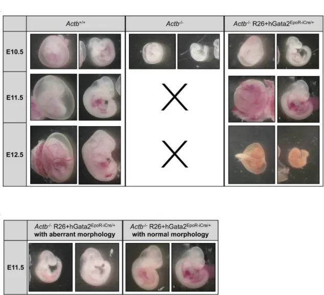
, breeding scheme in Figure 6A). Unlike their Actb2/2 embryo littermates we could obtain viable normal looking Actb2/2 R26+hGata2EpoR-iCre/+
embryos until E11.5, providing strong genetic evidence that erythroid-restricted transgene expression ofGata2is capable of rescuing the lethality of theActb2/2embryos at E10.5. However, despite this partial rescue allActb2/2R26+hGata2EpoR-iCre/+embryos were not surviving past
E12.5 (Figure 6B). Percentages of surviving embryos are summa-rized in Table S3. Figure 6B clearly illustrates that Actb2/2
R26+hGata2EpoR-iCre/+
embryos passed the primitive erythropoiesis phase and show prominent RBCs filled blood vessels at E11.5. We observed some heterogeneity in the rescue abilities of the embryos at E11.5 (Figure 6C). However, we found a direct correlation between the degree of normality and the overallGata2expression levels, with the most normal looking rescue embryos also expressing higher
Gata2 levels (corresponding to both endogenous and transgene transcripts). qRT-PCR experiments on the yolk sacs of Actb2/2
R26+hGata2EpoR-iCre/+
embryos demonstrate that the rescue embryos displaying higher Gata2 values (Figure 7A–C, light-grey bars) also have higherGata1, Hbb-y and Hbb-bh1 expression levels at both E10.25 and E11.5 (Figure 7A,C).Gata2target genes show variable expression profiles in the rescue embryos (Figure 7B). Interestingly, expression levels of gamma-actin did not decrease in the rescue embryos at E10.25 (Figure 7B) and at E11.5 (data not shown).
Discussion
This study has revealed an unexpected role for beta-actin during development. Beta-actin has been described in protrusive cellular structures and has traditionally been associated with a major role in cell migration [6,8,38]. TheActb2/2embryos appear normal until E8.5 and they undergo gastrulation without any obvious defects. Cell migration processes at these developmental stages therefore seem to be intact. We investigated other events requiring cell migration processes such as formation of the vasculature and hematopoietic system in the embryo. PECAM-1 staining suggests some vessel disorganization in the embryo proper and we also found vascular defects in the yolk sac. This could indicate a cell migration defect of hemangioblast cells, a progenitor population with both hematopoietic and vascular potential. These cells are thought to migrate from the embryo proper to the blood islands in the yolk sac where cells differentiate into primitive erythrocytes and endothelial cells. However, by tracking heman-gioblast cells expressing LacZ under theFlk1promotor [39,40], we observed no dramatic block in migration of hemangioblast cells at E9.5. This strongly suggests that a cell migration problem of hemangioblast cells per se is not causing the hematopoietic problems in the embryo and consequently has no major contribution to the observed lethality.
Our results however strongly suggest that an erythroid differentiation defect is most likely responsible for the observed embryonic lethality at E10.5 and one of the putative downstream players involved is Gata2. Indeed, the expression of this key erythroid transcription factor was dramatically downregulated at E8.5. Similar to theActb2/2embryos in this study,Gata2knockout embryos show marked anemia and fail to survive beyond the stage of primitive hematopoiesis [41]. Transgenic expression of Gata2
specifically in erythroid cells rescued the embryos from lethality at E10.5. The Actb2/2 R26+hGata2EpoR-iCre/+ embryos displayed
obvious remodeled RBC containing blood vessels in the yolk sac and embryo proper and the slight variations amongst those embryos could be correlated with the levels of transgenic Gata2
expression. After rescue the Actb2/2 R26+hGata2EpoR-iCre/+
embryos displayed similar levels of embryonic hemoglobins Hbb-y and Hbb-bh1, in line with restoration of primitive erythropoiesis. The vasculature of yolk sac and embryo proper appears intact at E11.5 in Actb2/2 R26+hGata2EpoR-iCre/+
embryos. This highly suggests that the vascular malformations observed in the Actb2/2 embryo proper immediately before lethality and the vascular remodeling defects in the yolk sac from E9.5 onwards are secondary defects, whereas downregulation of
Gata2and the associated block in primitive erythropoiesis is the primary defect causing the observed lethality at E10.5. The lack of full genetic rescue by erythroid-restricted GATA2 transgene expression could imply important roles ofGata2in later stages of cardiovascular development [42,43] that are not rescued using this approach.
One scenario whereby absence of beta-actin leads to reduced
Gata2mRNA levels is thatGata2expression is modulated by beta-actin in the nucleus. The nuclear role of beta-beta-actin has recently been consolidated and it appears to have a potent role in the regulation of transcription and gene expression by various molecular means [10,44,45]. Our ChIP analysis supports a function of beta-actin in the nucleus since we detected beta-actin binding to specific regions of the Gata2 promotor. One of the amplicons that was picked up by the ChIP experiment partly overlapped with a highly conserved region in theGata2promoter, suggesting the presence of important regulatory sequences in this region of the promoter. Indeed, the detailed analyses revealed multiple consensus sites for transcription factors with known roles in erythropoiesis and/or hematopoiesis. However, it is not clear whether these transcription factors bind to theGata2promotorin vivonor is it evident from literature that they associate with the actin protein. It also remains to be clarified whether beta-actin itself binds to the promotor as a transcription factor or whether it is part of a transcriptional complex. However, taking the previously described roles of beta-actin into account [10,11,12,44,45], we favor the possibility whereby beta-actin binds the promotor ofGata2in complex with other proteins.
Gamma-actin is, together with beta-actin, one of the main actin isoforms during embryonic development. It is approximately 2-fold upregulated in the embryo proper and in MEFs isolated from theActb2/2 embryos [13]. We observed similar compensatory mechanisms inActb2/2yolk sacs (Figure S1A–C). It remains to be clarified whether such an upregulation also occurs in the
Actb2/2 erythroid precursors but in two independent studies usingActb2/2MEFs and T-cells, an upregulation of the other cytoplasmic actin isoform has been observed [13,18]. Therefore, in addition to beta-actin specific nuclear effects onGata2expression, lack of cytoskeletal integrity which is important in erythrocytes, cannot completely be excluded.
Figure 6.Actb2/2R26+hGata2EpoR-iCre/+embryos remain viable at E11.5.(A) Schematic view of the breeding scheme used to generate Gata2expression in the erythroid lineage inActb2/2mice. (B) Pictures from freshly dissected embryos at developmental stages E10.5, E11.5 and E12.5. Black crosses mark the absence ofActb2/2embryos at stages E11.5 and E12.5. Note the presence of RBCs in theActb2/2R26+ hGata2EpoR-iCre/+embryos. Statistics on embryo recovery in function of genotype are in Table S3. E10.5: 156magnification, E11.5: 126magnification; E12.5: 106 magnification. (C) Morphology variation ofActb2/2R26+hGata2EpoR-iCre/+embryos at E11.5. Aberrant lookingActb2/2R26+hGata2EpoR-iCre/+ embryos (left panel) compared to normal lookingActb2/2R26+hGata2EpoR-iCre/+embryos (right panel), 126magnification. Embryos were imaged on a Leica MS5 (Leica Microsystems) stereomicroscope. Digital images were acquired using a Leica camera.
doi:10.1371/journal.pone.0067855.g006
Supporting Information
Figure S1 Compensatory up regulation of gamma-actin
occurs in Actb2/2 yolk sacs. (A) Relative gamma-actin
mRNA levels measured by qRT-PCR in yolk sacs of E8.5 and E10.25 embryos. Both embryonic stages show an approximately two fold increase in gamma-actin expression. Bars represent mean 6SEM; **P,.01, ***P,.001. (B) Western Blot analysis of gamma-actin protein level in yolk sacs of E8.5 embryos. A slight increase of gamma-actin can be detected in Actb2/2yolk sacs versus Actb+/+ yolk sacs. (C) Western Blot analysis of gamma-actin protein level in yolk sacs of E10.25 embryos. The increase of gamma-actin protein level in Actb2/2 yolk sacs is more pronounced. (D) Comparison of expression of mouse beta- and gamma actin at gastrula and organogenesis stage, data are from http://www.ncbi.nlm.nih.gov/unigene.
(TIFF)
Figure S2 Consensus sites possibly important for erythropoiesis in amplicon 3 and 8. Detail analysis of the large amplicon 3 (amplicon 3+regions between amplicon 2–3 and 3–4) and amplicon 8 reveal multiple DNA consensus sites for
binding of transcription factors possible involved in erythropoiesis in mouse.
(TIFF)
Table S1 Overview of the primer sets used in this study. (TIFF)
Table S2 Overview of the primer sets used for the ChIP analysis and gene location of the corresponding ampli-con.
(TIFF)
Table S3 Percentages of surviving embryos. Mendelian ratios are given in bold under the genotype.
(TIFF)
Author Contributions
Conceived and designed the experiments: DT BD. Performed the experiments: DT BD KB SB KS SG. Analyzed the data: DT. Contributed reagents/materials/analysis tools: JJH CA. Wrote the paper: DT SG JJH CA.
References
1. Vandekerckhove J, Weber K (1978) At least six different actins are expressed in a higher mammal: an analysis based on the amino acid sequence of the amino-terminal tryptic peptide. J Mol Biol 126: 783–802.
2. Ponte P, Gunning P, Blau H, Kedes L (1983) Human actin genes are single copy for alpha-skeletal and alpha-cardiac actin but multicopy for beta- and gamma-cytoskeletal genes: 39untranslated regions are isotype specific but are conserved in evolution. Mol Cell Biol 3: 1783–1791.
3. Moos M, Gallwitz D (1983) Structure of two human beta-actin-related processed genes one of which is located next to a simple repetitive sequence. EMBO J 2: 757–761.
4. McHugh KM, Crawford K, Lessard JL (1991) A comprehensive analysis of the developmental and tissue-specific expression of the isoactin multigene family in the rat. Dev Biol 148: 442–458.
5. Khaitlina SY (2001) Functional specificity of actin isoforms. Int Rev Cytol 202: 35–98.
6. Hoock TC, Newcomb PM, Herman IM (1991) Beta actin and its mRNA are localized at the plasma membrane and the regions of moving cytoplasm during the cellular response to injury. J Cell Biol 112: 653–664.
7. Watanabe H, Kislauskis EH, Mackay CA, Mason-Savas A, Marks SC Jr (1998) Actin mRNA isoforms are differentially sorted in normal osteoblasts and sorting is altered in osteoblasts from a skeletal mutation in the rat. J Cell Sci 111 (Pt 9): 1287–1292.
8. Hill MA, Gunning P (1993) Beta and gamma actin mRNAs are differentially located within myoblasts. J Cell Biol 122: 825–832.
9. Dugina V, Zwaenepoel I, Gabbiani G, Clement S, Chaponnier C (2009) Beta and gamma-cytoplasmic actins display distinct distribution and functional diversity. J Cell Sci 122: 2980–2988.
10. Visa N, Percipalle P (2010) Nuclear functions of actin. Cold Spring Harb Perspect Biol 2: a000620.
11. Hofmann WA, Stojiljkovic L, Fuchsova B, Vargas GM, Mavrommatis E, et al. (2004) Actin is part of pre-initiation complexes and is necessary for transcription by RNA polymerase II. Nat Cell Biol 6: 1094–1101.
12. Xu YZ, Thuraisingam T, Morais DA, Rola-Pleszczynski M, Radzioch D (2010) Nuclear translocation of beta-actin is involved in transcriptional regulation during macrophage differentiation of HL-60 cells. Mol Biol Cell 21: 811–820. 13. Tondeleir D, Lambrechts A, Mueller M, Jonckheere V, Doll T, et al. (2012)
Cells lacking beta-actin are genetically reprogrammed and maintain conditional migratory capacity. Mol Cell Proteomics.
14. Bunnell TM, Burbach BJ, Shimizu Y, Ervasti JM (2011) beta-Actin specifically controls cell growth, migration, and the G-actin pool. Mol Biol Cell 22: 4047– 4058.
15. Crawford K, Flick R, Close L, Shelly D, Paul R, et al. (2002) Mice lacking skeletal muscle actin show reduced muscle strength and growth deficits and die during the neonatal period. Mol Cell Biol 22: 5887–5896.
16. Kumar A, Crawford K, Close L, Madison M, Lorenz J, et al. (1997) Rescue of cardiac alpha-actin-deficient mice by enteric smooth muscle gamma-actin. Proc Natl Acad Sci U S A 94: 4406–4411.
17. Schildmeyer LA, Braun R, Taffet G, Debiasi M, Burns AE, et al. (2000) Impaired vascular contractility and blood pressure homeostasis in the smooth muscle alpha-actin null mouse. FASEB J 14: 2213–2220.
18. Bunnell TM, Ervasti JM (2010) Delayed embryonic development and impaired cell growth and survival in Actg1 null mice. Cytoskeleton (Hoboken) 67: 564– 572.
19. Shmerling D, Danzer CP, Mao X, Boisclair J, Haffner M, et al. (2005) Strong and ubiquitous expression of transgenes targeted into the beta-actin locus by Cre/lox cassette replacement. Genesis 42: 229–235.
20. Ema M, Takahashi S, Rossant J (2006) Deletion of the selection cassette, but not cis-acting elements, in targeted Flk1-lacZ allele reveals Flk1 expression in multipotent mesodermal progenitors. Blood 107: 111–117.
21. Drogat B, Kalucka J, Gutierrez L, Hammad H, Goossens S, et al. (2010) Vegf regulates embryonic erythroid development through Gata1 modulation. Blood 116: 2141–2151.
22. Nagy A (2003) Manipulating the Mouse Embryo: a laboratory manual. Cold Spring Harbor: Laboratoty Cold Spring Harbor. IX, 764.
23. Tsai FY, Orkin SH (1997) Transcription factor GATA-2 is required for proliferation/survival of early hematopoietic cells and mast cell formation, but not for erythroid and myeloid terminal differentiation. Blood 89: 3636–3643. 24. Khandekar M, Brandt W, Zhou Y, Dagenais S, Glover TW, et al. (2007) A
Gata2intronic enhancer confers its pan-endothelia-specific regulation. Develop-ment 134: 1703–1712.
25. Liu C, Levenstein M, Chen J, Tsifrina E, Yonescu R, et al. (1999) SZF1: a novel KRAB-zinc finger gene expressed in CD34+ stem/progenitor cells. Exp Hematol 27: 313–325.
26. Morris JF, Rauscher FJ 3rd, Davis B, Klemsz M, Xu D, et al. (1995) The myeloid zinc finger gene, MZF-1, regulates the CD34 promoter in vitro. Blood 86: 3640–3647.
27. Lacaud G, Gore L, Kennedy M, Kouskoff V, Kingsley P, et al. (2002) Runx1 is essential for hematopoietic commitment at the hemangioblast stage of development in vitro. Blood 100: 458–466.
28. Liao W, Ho CY, Yan YL, Postlethwait J, Stainier DY (2000) Hhex and scl function in parallel to regulate early endothelial and blood differentiation in zebrafish. Development 127: 4303–4313.
Figure 7. Morphologically normalActb2/2R26+hGata2EpoR-iCre/+embryos show improved Gata2 mRNA levels compared to Actb2/2embryos.(A) Relative E10.25 mRNA levels measured by qRT-PCR ofGata1,Gata2, Hbb-y, and Hbb-bh1 in yolk sacs ofActb+/+,Actb2/2
R26+hGata2EpoR-iCre/+embryos with aberrant morphology andActb2/2R26+hGata2EpoR-iCre/+embryos with normal morphology. (B) Relative E10.25 mRNA levels measured by qRT-PCR of Actg1 andGata2targets:c-kit, andEpoRin yolk sacs ofActb+/+,Actb2/2and the two groups ofActb2/
2R26+hGata2EpoR-iCre/+embryos. (C) Relative E11.5 mRNA levels measured by qRT-PCR ofGata1,Gata2, Hbb-y, and Hbb-bh1 in yolk sacs ofActb+/ +, Actb2/2 R26+hGata2EpoR-iCre/+ embryos with aberrant morphology (see Figure 6C) and Actb2/2 R26+hGata2EpoR-iCre/+with normal morphology. White bars =Actb+/+, dark-grey bars =Actb2/2 R26+hGata2EpoR-iCre/+ with aberrant morphology, light-grey bars =Actb2/2
R26+hGata2EpoR-iCre/+with normal morphology. doi:10.1371/journal.pone.0067855.g007
29. Tewari R, Gillemans N, Wijgerde M, Nuez B, von Lindern M, et al. (1998) Erythroid Kruppel-like factor (EKLF) is active in primitive and definitive erythroid cells and is required for the function of 5’HS3 of the beta-globin locus control region. EMBO J 17: 2334–2341.
30. Bose F, Fugazza C, Casalgrandi M, Capelli A, Cunningham JM, et al. (2006) Functional interaction of CP2 with GATA-1 in the regulation of erythroid promoters. Mol Cell Biol 26: 3942–3954.
31. Woo AJ, Moran TB, Schindler YL, Choe SK, Langer NB, et al. (2008) Identification of ZBP-89 as a novel GATA-1-associated transcription factor involved in megakaryocytic and erythroid development. Mol Cell Biol 28: 2675– 2689.
32. Dolcet X, Llobet D, Pallares J, Matias-Guiu X (2005) NF-kB in development and progression of human cancer. Virchows Arch 446: 475–482.
33. Semenza GL (2001) HIF-1 and mechanisms of hypoxia sensing. Curr Opin Cell Biol 13: 167–171.
34. Lee BK, Iyer VR (2012) Genome-wide studies of CCCTC-binding factor (CTCF) and cohesin provide insight into chromatin structure and regulation. J Biol Chem 287: 30906–30913.
35. Kaplan J, Calame K (1997) The ZiN/POZ domain of ZF5 is required for both transcriptional activation and repression. Nucleic Acids Res 25: 1108–1116. 36. Orford RL, Robinson C, Haydon JM, Patient RK, Guille MJ (1998) The
maternal CCAAT box transcription factor which controls GATA-2 expression is novel and developmentally regulated and contains a double-stranded-RNA-binding subunit. Mol Cell Biol 18: 5557–5566.
37. Nyabi O, Naessens M, Haigh K, Gembarska A, Goossens S, et al. (2009) Efficient mouse transgenesis using Gateway-compatible ROSA26 locus targeting vectors and F1 hybrid ES cells. Nucleic Acids Res 37: e55.
38. Peckham M, Miller G, Wells C, Zicha D, Dunn GA (2001) Specific changes to the mechanism of cell locomotion induced by overexpression of beta-actin. J Cell Sci 114: 1367–1377.
39. Huber TL, Kouskoff V, Fehling HJ, Palis J, Keller G (2004) Haemangioblast commitment is initiated in the primitive streak of the mouse embryo. Nature 432: 625–630.
40. Medvinsky A, Rybtsov S, Taoudi S (2011) Embryonic origin of the adult hematopoietic system: advances and questions. Development 138: 1017–1031. 41. Tsai FY, Keller G, Kuo FC, Weiss M, Chen J, et al. (1994) An early
haematopoietic defect in mice lacking the transcription factor GATA-2. Nature 371: 221–226.
42. Lim KC, Hosoya T, Brandt W, Ku CJ, Hosoya-Ohmura S, et al. (2012) ConditionalGata2inactivation results in HSC loss and lymphatic mispatterning. J Clin Invest 122: 3705–3717.
43. Huang Z, Dore LC, Li Z, Orkin SH, Feng G, et al. (2009) GATA-2 reinforces megakaryocyte development in the absence of GATA-1. Mol Cell Biol 29: 5168–5180.
44. Olson EN, Nordheim A (2010) Linking actin dynamics and gene transcription to drive cellular motile functions. Nat Rev Mol Cell Biol 11: 353–365. 45. Zheng B, Han M, Bernier M, Wen JK (2009) Nuclear actin and actin-binding