Administration of
Mycobacterium leprae
rHsp65
Aggravates Experimental Autoimmune Uveitis in Mice
Eliana B. Marengo1,3., Alessandra Gonc¸alves Commodaro2., Jean Pierre S. Peron1
, Luciana V. de Moraes1, Fernanda C. V. Portaro3, Rubens Belfort, Jr.2, Luiz Vicente Rizzo4, Osvaldo Augusto Sant’Anna3*
1Department of Immunology, Institute of Biomedical Sciences, University of Sa˜o Paulo, Sa˜o Paulo, Brazil,2Vision Institute, Federal University of Sa˜o Paulo, Sa˜o Paulo, Brazil,3Immunochemistry Laboratory, Instituto Butantan, Sa˜o Paulo, Brazil,4Albert Einstein Jewish Institute for Education and Research, Sa˜o Paulo, Brazil
Abstract
The 60kDa heat shock protein family, Hsp60, constitutes an abundant and highly conserved class of molecules that are highly expressed in chronic-inflammatory and autoimmune processes. Experimental autoimmune uveitis [EAU] is a T cell mediated intraocular inflammatory disease that resembles human uveitis. Mycobacterial and homologous Hsp60 peptides induces uveitis in rats, however their participation in aggravating the disease is poorly known. We here evaluate the effects of theMycobacterium lepraeHsp65 in the development/progression of EAU and the autoimmune response against the eye through the induction of the endogenous disequilibrium by enhancing the entropy of the immunobiological system with the addition of homologous Hsp. B10.RIII mice were immunized subcutaneously with interphotoreceptor retinoid-binding protein [IRBP], followed by intraperitoneally inoculation of M. leprae recombinant Hsp65 [rHsp65]. We evaluated the proliferative response, cytokine production and the percentage of CD4+IL-17+, CD4+
IFN-c+and CD4+Foxp3+cellsex vivo, by
flow cytometry. Disease severity was determined by eye histological examination and serum levels of IRBP and anti-Hsp60/65 measured by ELISA. EAU scores increased in the Hsp65 group and were associated with an expansion of CD4+ IFN-c+and CD4+IL-17+T cells, corroborating with higher levels of IFN-c. Our data indicate that rHsp65 is one of the managers
with a significant impact over the immune response during autoimmunity, skewing it to a pathogenic state, promoting both Th1 and Th17 commitment. It seems comprehensible that the specificity and primary function of Hsp60 molecules can be considered as a potential pathogenic factor acting as a whistleblower announcing chronic-inflammatory diseases progression.
Citation:Marengo EB, Commodaro AG, Peron JPS, de Moraes LV, Portaro FCV, et al. (2009) Administration ofMycobacterium lepraerHsp65 Aggravates Experimental Autoimmune Uveitis in Mice. PLoS ONE 4(11): e7912. doi:10.1371/journal.pone.0007912
Editor:Eshel Ben-Jacob, Tel Aviv University, Israel
ReceivedJuly 15, 2009;AcceptedOctober 23, 2009;PublishedNovember 19, 2009
Copyright:ß2009 Marengo et al. This is an open-access article distributed under the terms of the Creative Commons Attribution License, which permits unrestricted use, distribution, and reproduction in any medium, provided the original author and source are credited.
Funding:This work was supported by funds of Fundac¸a˜o de Amparo a Pesquisa do Estado de Sa˜o Paulo, Brazil [FAPESP], Conselho Nacional de Pesquisa, Brazil [CNPq], FADA-FAPESP and Pan-American Ophthalmological Foundation and INCTTOX - CNPq/FAPESP program. E. B. Marengo and A. G. Commodaro are recipients of FAPESP fellowships. R. Belfort Jr, L. V. Rizzo and O.A. Sant’Anna are researchers of CNPq-Brazil. The funders had no role in study design, data collection and analysis, decision to publish, or preparation of the manuscript.
Competing Interests:The authors have declared that no competing interests exist.
* E-mail: [email protected]
.These authors contributed equally to this work.
Introduction
Uveitis is an inflammation of the uvea, a layer of the eye located between the sclera and the retina. Uvea includes the iris, ciliary body, and choroid [1]. The animal model for human uveitis has experimental autoimmune uveitis [EAU] that also resembles that seen in diseases such as sympathetic ophthalmia, birdshot retino-choroidopathy, Vogt-Koyanagi-Harada’s disease, and Behcet’s disease [BD] [2]. EAU is an organ-specific T-cell-mediated disease that can be induced in susceptible animals such as non-human primates and rodents. Induction is done with immunization using retinal antigens like interphotoreceptor retinoid-binding protein [IRBP] or with S-antigen [arrestin]. It can also be done by adoptive transfer of IRBP or arrestin-specific T-cells [2,3,4]. The disease is characterized by vasculitis and granuloma formation in the neural retina, destruction of photoreceptor cells, and blindness [2,4].
The 60kDa heat shock protein family, HSP60, constitutes an abundant and highly conserved class of molecules. They prevent misfolding and promote the refolding and proper assembly of unfolded polypeptides generated under physiologic and stress
conditions. TheMycobacterium lepraeHsp65 [M. lepraeHsp65] is part of the HSP60 family denominated chaperonins. It is considered as one of the major immuno-reactive proteins in mycobacteria [5]. Mycobacterial Hsp65 shares approximately 50% amino acid identity with its mammalian homologue. The Hsp60 family comprises molecules that are immunodominant in several infectious processes [6], and which participate in pro-inflamma-tory events associated to pathologies such as arthritis, atheroscle-rosis, type I diabetes, and autoimmune demyelination [7,8,9]. As for its association with eye disease more specifically, higher anti-Hsp65 antibody titers were correlated to the retinopathy seen in type I diabetes patients [10].
Mycobacterial and homologous Hsp60 peptides can induce experimental uveitis in rats [11,12] and Hsp60 and anti-Hsp60 may be involved in the pathogenesis of the ocular manifestations of Behcet’s disease and acute anterior uveitis [13,14]. However, their participation in aggravating the disease is poorly known.
reactive to Hsp60 or Hsp65 in pathophysiological conditions suggests that these proteins play an important role as cellular targets in autoimmunity due to an existing similarity between the bacterial antigen and the autologous protein [16]. On the other hand, high anti-Hsp60/65 antibody titers were not found to be restricted to disease conditions and could also be detected in supposedly healthy individuals as they aged [17,18]. Indeed, autoantibodies can be indicators of susceptibility to the future development of various autoimmune diseases [19,20,21,22].
We previously showed that passive administration of wild typeM. lepraeHsp65 interfered with endogenous equilibrium by enhancing the entropy of the immunobiological system, as expressed by the early death of the systemic lupus erythematosus [NZBxNZW]F1
experimental mice [23]. We here applied a similar approach of inducing disequilibrium of physiological and immune states by adding homologous Hsp to evaluate the effects of the M. leprae Hsp65 in the development/progression of EAU by evaluating the autoimmune response against the eye. For that we analyzed the proliferative response, cytokine and antibody production in the B10.RIII experimental mice submitted to immunization and administration of Hsp65. The findings were compared to the histopathological analysis of the eye.
Results
Administration ofM. lepraerHsp65 Increased EAU Scores
To evaluate the influence of rHsp65 in the progression of EAU, B10.RIII mice were induced to develop uveitis by immunization with IRBP followed by inoculation with rHsp65, which occurred in the same day. Control mice showed moderate to severe signs of EAU according to histopathologic examination done on day 21. These signs were retinal disorganization, presence of inflamma-tory cells in the vitreous, vasculitis and granuloma formation [Figures 1A and 1B,control group]. Conversely, rHsp65-inoculated animals presented a more pronounced inflammatory response, with total disorganization of the retinal layers associated to detachment of the retina. Intense retinal folds, inflammatory infiltrating cells in the vitreous, vasculitis and granuloma formation were also observed [Figures 1A and 1B,rHsp65 group]. B10.RIII mice immunized with IRBP emulsified in incomplete Freund adjuvant plus PTX or immunized in CFA without PTX, even receiving rHsp65, do not express EAU [data not shown]. These indicate that Hsp have any commitment in the initiation process of this organ-specific autoimmune disease.
Analysis of Cytokine Levels
Draining lymph node cells from either control or rHsp65-inoculated mice were harvested on day 21 after immunization and assayed for cytokine secretion under IRBP and rHsp65 stimulating conditions. We observed an increased IFN-csecretion by IRBP
re-stimulated cells, which was associated with a higher expansion of CD4+
IFN-c+T cells [Figures 2A and 3A,p,0.05] in the rHsp65 group. These findings corroborate with our clinical findings, in which an increase in the EAU score was observed. On the other hand, no changes were observed in IFN-cafter re-stimulation with rHsp65 [Figure 2B].
Surprisingly, rHsp65 administration resulted in an increased IL-10 production after stimulation with IRBP and with rHsp65 [Figures 2C and 2D, p,0.05]. No alterations in IL-6 production levels were observed after IRBP and rHsp65 stimulation [Figures 2E and 2F].
rHsp65 Expanded IL-17 and IFN-c CD4+T Cell Population
Because the Th17 cells recently described are believed to play a relevant role in the pathogenesis of autoimmunity [24,25,26] and
the expression of Th1 is a characteristic phenotype of EAU, we evaluated the percentage of both Th1 and Th17 cells from lymph nodes of EAU mice inoculated with rHsp65 and compared to controls. Intracellular staining for IFN-cand IL-17 revealed that rHsp65 injection induced expansion of both CD4+ T cell populations [Figure 3]. Concerning T cell subsets, it is possible that the higher disease scores observed in the rHsp65 group would lead to an impaired percentage of T regulatory cells in the periphery. However, this was not the case, as no differences in the percentages of CD4+Foxp3+T cells were observed [Figure 4].
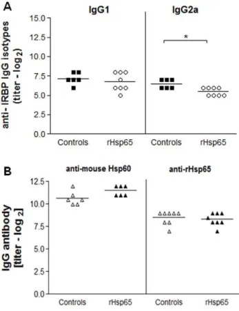
Anti-IRBP IgG2a Levels Decreased after rHsp65 Administration
We examined the effects of rHsp65 inoculation on IRBP and Hsp60/65 specific IgG antibody production. As shown in Figure 5A, anti-IRBP IgG2a levels decreased in the sera of the rHsp65 mice group when compared to control [p,0.05]. No significant dif-ferences in the levels of anti-IRBP IgG1 antibodies were observed.
Production of anti-mouse Hsp60 and anti-Hsp65 antibody showed no difference between test and control in the mice sera [Figure 5B]. However, production of anti-Hsp60 antibody [both in the test group and in controls] presented titers that were approximately 5.6-fold higher than those of anti-Hsp65 antibodies.
Discussion
Hsp60 are highly conserved molecules inducible by all forms of cellular stress. In spite of the best known function they have as Figure 1. Administration ofM. lepraerHsp65 increased EAU scores.
[A]: B10.RIII mice were immunized with 161–180 IRBP peptide followed by injection with rHsp65 [i.p.]. Eyes were collected for histopathology 21 days later. EAU scores were assigned on a 0 to 4 scale. [B]: Histopathological findings: a normal retinal architecture corresponding to non immunized
naı¨vemice [normal]; IRBP immunized mice [average score 3;control]; mice immunized with IRBP followed by rHsp65 inoculation [average score 4;
chaperonin, Hsp are also important players in the control of the immune response, being target by Hsp-specific T cells and antibodies in healthy subjects and also in chronic-inflammatory processes including autoimmune diseases [6,27,28,29]. Hsp have shown a dual role in immune-mediated disorders, being involved in the induction and propagation of autoimmune diseases as well as in suppressing them [30]. The precise mechanisms underlying these divergent Hsp60-mediated responses are not defined. It has been suggested that the increased expression of endogenous Hsp molecules in different stress conditions can propagate the ongoing inflammation and also constitutes an attractive target for T cells
and antibodies that are induced by extracellular [self or foreign] Hsp. In addition, inflammation can alter the antigen processing for Hsp and reveal new epitopes which can prime an immune response and lead to pathological status [31]. Furthermore, these molecules activate innate immunity through macrophages and dendritic cells, which in turn trigger the development of adaptive
Figure 2. rHsp65 inoculation increased IFN-cand IL-10 levels.
Draining lymph nodes cells from either rHsp65 inoculated or control mice were harvested at day 21 and stimulatedin vitro[106cells/ml] with
30mg/ml IRBP [A, C, E] and 20mg/mlM. lepraerHsp65 [B, D, F]. After 48 hours, IFN-c[A, B], IL-10 [C, D] and IL-6 [E, F] levels were determined
by ELISA. Data represent three independent experiments with n = 5 mice/group [*p,0.05 versus control group;t-test analysis].
doi:10.1371/journal.pone.0007912.g002
Figure 3. Expansion of CD4+
IFN-c+and CD4+IL-17+T cells after
rHsp65 inoculation.Cells from draining lymph nodes collected at day 21 after immunization were labeled with anti-CD4 mAb, fixed and permeabilized, stained intracellularly and analyzed by flow-cytometry evaluating the CD4, IFN-cand IL-17 expression. Results are expressed in
mean6SD. Data represent three independent experiments with n = 5 mice/group [*p,0.05; **p,0.01 versus control group;t-test analysis]. doi:10.1371/journal.pone.0007912.g003
Figure 4. rHsp65 showed no effect on CD4+Foxp3+T cells.Cells from draining lymph nodes were harvested at day 21 after immuniza-tion, stained and analyzed by flow-cytometry evaluating the CD4 and Foxp3 expressions. Results are expressed in mean6SD. Data represent three independent experiments with n = 5 mice/group.
doi:10.1371/journal.pone.0007912.g004
Figure 5. rHsp65 modulated anti-IRBP IgG2a levels.Serum levels of anti-IRBP IgG1 and IgG2a isotypes [A] and IgG anti-mouse Hsp60 and anti-rHsp65 [B] antibody production in IRBP-immunized mice either or not inoculated with rHsp65. Antibody levels were determined by ELISA at day 21 after immunization. Results are expressed in titers6SD in each experimental group. Data represent three independent experi-ments with n = 6–8 mice/group [*p,0.05 versus control group;t-test analysis].
immune response to self Hsp60 by extracellular Hsp60/65. Besides that, anti-Hsp65 antibodies can also be a potent pathological trigger [7,32]. The immune response to Hsp can be mined for information on the state of the immune system [33,34]; in altered conditions, Hsp and their antibodies can act as biomarkers during postnatal life [20,35].
In this study we showed that a single administration ofM. leprae rHsp65 in the highly susceptible B10.RIII immunized mouse was capable to increase the scores of uveitis when compared to controls [p,0.05]. Also, rHsp65-inoculated animals presented a more pronounced inflammatory response, with total disorganization of the retinal layers associated to its detachment. Intense retinal folds, inflammatory infiltrating cells in the vitreous, vasculitis and granuloma formation were also observed.
Most reports that attempted to correlate Hsp60 with uveitis were limited to studies done with systemic Behcet’s disease [11,36,37,38]; increased T and B cell activity against Hsp60 and Hsp65 has been documented in these patients [11,39,40,41]. The inoculation of bacterial Hsp65 peptides either subcutaneously or orally, or their human analogues, induced uveitis in Lewis rats by self-reactive T cells specific to these peptides. Although the Hsp60 family members share a common basic antigenicity, they differ in the reactivity to the anti-Hsp60 antibodies present in the sera of BD patients [42]. Cancino-Diaz and collaborators hypothesized that the high levels of anti-bacterial Hsp60 antibodies found in patients with uveitis was produced during episodes of bacterial infection that may have triggered an autoimmune reaction through a mechanism of molecular mimicry between the human and microbial Hsp. This suggests also that an immune cross-reaction between retinal and Hsp molecules and a related autoimmune response may be involved in the development of BD [41].
Many studies showed Hsp60 as a ligand for TLR-2 and TLR-4, suggesting a role for Hsp60 as an immunological and endogenous sign of danger that leads to rapid inflammatory cytokine release and enhancement of adaptive Th1-type responses [43,44,45,46]. Genetic susceptibility to EAU is associated to a dominant Th1-type response to the uveitogenic retinal antigen; the effector T cells possess a Th1-like phenotype that is expressed as high IFN-c and low IL-4
cytokines production [47,48]. In our study, the levels of IFN-c
secretion and the percentage of CD4+
IFN-c+and CD4+ IL-17+
cells were significantly higher in the rHsp65 group than in control mice. IL-17-producing CD4+
T cells or Th17 constitute a newly identified inflammatory cell population, which is critically involved in EAU [24,25,49] suggesting that rHsp65 aggravates EAU pathology by expanding antigen-specific Th1 and Th17 cells. Knowing that IL-6 is associated with the production of the Th17 effector phenotype [50] and that the cytokine is responsible, at least partially, for an ocular inflammation in which differentiation of Th17 is IL-6-dependent [26], we expected to see an increased secretion of IL-6 in rHsp65-inoculated mice. Expansion of Th17 did not reflect in a higher production of IL-6 in the rHsp65 group, when compared to controls. The analysis of the kinetics of cytokine secretion and other time-points may have showed otherwise and explain the lack of differential IL-6 production, in the rHsp65 group and emerge an increase of IL-10 production by IRBP and Hsp65-specific cells. Although IL-10 has been implicated in the progression control of immune-mediated diseases and in the maintenance of tolerance [51,52], in our study it did not correlate with suppression, since the EAU score was higher in animals that received Hsp65. Concerning T cell subsets, it is possible that the higher disease scores observed in the rHsp65 group would lead to an impaired percentage of T regulatory cells in the periphery. However, this was not the case, as no differences in the percentages of
CD4+ Foxp3+
T cells were observed. The elevated IL-10 production in this context may works as an attempt to control the balance Th2/Th1 during the intense inflammatory response caused by the administration of rHsp65, other than being an attempt to regulate the response to self antigens. The higher IL-10 cytokine production and the decrease in IgG2a anti-IRBP levels in rHsp65-inoculated mice reinforce the notion of reacquired homeostasis states.
No significant differences in the production of anti-mouse Hsp60 and anti-Hsp65 antibody levels in both rHsp65 and control groups were observed [Figure 5B]. However, the anti-Hsp65 antibody production was higher in B10.RIII immunized than naı¨ve mice, which showed basal production [data not shown]. The high anti-Hsp60/65 antibody titers were not found to be restricted to disease conditions. That they were detected in supposedly healthy individuals [17,18] may be due to the fact that the pathophysiologic status of the individuals were not deeply investigated. However, we cannot rule out that anti-Hsp65 antibodies are involved in vascular damage and consequently aggravate ocular disease in Hsp65-inoculated mice, since EAU can present vasculitis and granuloma formation in the neural retina [2,4]. Indeed, it was reported that the anti-Hsp60 autoantibodies mediates endothelial cell cytotoxicity [53]. The possible correla-tion between anti-Hsp65 antibodies and vascular lesions and the physiopathological mechanisms involving pro-inflammatory cyto-kines in the serum of inoculated and control mice may clarify the role of these soluble molecules in the severity of uveitis.
Hsp have been implicated in the induction and propagation of autoimmunity in several diseases. Our findings demonstrate that this may also be true in uveitis and it opens the possibility that the blockage of endogenous produced Hsp, whose expression should be enhanced during autoimmunity, may present an interesting target for immunotherapy of this syndrome of the eye. Furthermore, in infectious diseases of the eye, such as toxoplas-mosis, recurrence of lesions often go unexplained and because Hsp are highly conserved in nature it is entirely possible that the pathogen’s Hsp may contribute to disease flare up.
Through an original approach based in the Prigogine’s principle [54,55], the pathophysiological role of the rHsp65 ofM. lepraewas also evaluated in the in vivo spontaneous Systemic Lupus Erythematosus [SLE] model. It is assumed that the immune system keeps the order state at the cost of irreversibility and that in faster reiterated rounds of stimulation the system is not able to come back to its original state [23]. Both in the organ-specific EAU and in the systemic SLE, these findings strongly support that the over expression of the Hsp family and the altered microenvironmental state in pathologic conditions can modify the antigen processing of Hsps molecules, providing it a toxin function.
acuteness, most likely because of a cumulative and irreversible effect of the immune system.
Our data suggest that Hsp65 has pathological significance during the ocular autoimmune disease by increasing the immune response associated with an expansion of CD4+
IFN-c+ and
CD4+ IL-17+
T cells, probably by skewing it to a pathogenic state, promoting both Th1 and Th17 commitment.
These findings can be used for a better understanding of the relationship between self molecules, such as Hsp, and the immune response under physiological and pathological conditions. The specificity and primary function of Hsp60 can be considered as a potential pathogenic factor that also may act as a whistleblower announcing chronic-inflammatory diseases progression. It may be consider the immune system as a broken mirror in which identical or similar images, or their own reflections, distinctly recognized or blurred. In this sense, depending on the pathophysiological conditions, unpredictable immune responsiveness will be evoked by cumulative and temporal effects.
Materials and Methods
Antigens and Reagents
Peptide SGIPYIISYLHPGNTILHVD representing residues 161–180 of IRBP was synthesized using the Fmoc procedure [60] in a Shimadzu PSSM8 peptide synthesizer [Shimadzu, Tokyo, Japan]. The Fmoc-amino acids were purchased from Novabiochem [Nottingham, UK]. Bordetella pertussis toxin and Complete Freund Adjuvant [CFA] were purchased from Sigma-Aldrich [St. Louis, MO, USA]. Purified mouse Hsp60 [ESP-741] was obtained from Stressgen Bioreagents [San Diego, CA, EUA].
Expression and Purification of the Recombinant
Mycobaterium lepraeHsp65 inEscherichia coli
Clone pIL161 containing the coding sequence of theM. leprae Hsp65 was kindly given by Prof. Ce´lio L. Silva from University of Sao Paulo, Ribeira˜o Preto, Brazil. The clone was amplified inE. coliDH5acells. Expression of the recombinant wild-type Hsp65 protein [rHsp65] was performed as described in [61]. The rHsp65 present in the bacterial pellet was solubilized with 6 M urea and submitted to preparative SDS-PAGE gel. Elution of the Hsp65 band from the polyacrylamide gel slice was performed to obtain the purified protein [23]. Concentration of the recombinant Hsp65 protein was determined using the Bradford assay [62].
Animals
Six to eight weeks old B10.RIII mice were obtained from the animal facilities at the University of Sao Paulo, Brazil. All animals were housed under specific pathogen-free conditions and handled under ethical conditions. The Animal Care Committee of the Institute of Biomedical Sciences at the University of Sao Paulo approved all the procedures used in this study.
Induction of EAU and rHsp65 Treatment
Mice were immunized subcutaneously [s.c.] at the base of the tail with 30mg of 161–180 IRBP peptide emulsified in 0.2 ml CFA [v/v] and injected intraperitoneally [i.p.] with 0.5mg ofBordetella pertussistoxin [PTX] in 0.1 ml, as additional adjuvant. Following immunization, a group of 6 to 8 animals was inoculated with 2.5mg of M. leprae rHsp65 in 0.2 ml of phosphate buffer saline
pH 7.4 [PBS] by i.p. route. At day 21 after immunization, rHsp65 inoculated and control mice were sacrificed. The histopathology of the eye, cellular phenotype, cytokines and anti–IRBP and anti-Hsp60/Hsp65 antibody production were then analyzed.
Histopathology EAU
Eyes were collected and prepared for histopathological evalua-tion at day 21 after immunizaevalua-tion. The eyes were immersed for 1 h in phosphate-buffered glutaraldehyde 4%, transferred into phos-phate-buffered formaldehyde 10% for 24 h, and to ethanol 70% until processing. Fixed and dehydrated tissues were embedded in paraffin wax; 4–6mm sections were cut through the papillary-optic nerve plane. Sections were stained by hematoxylin and eosin.
Presence of disease was evaluated in a double-blinded fashion by examining six sections cut at different levels in each eye. Severity of EAU was scored on a scale of 0 [no disease] to 4 [maximum disease] in half-point increments, according to a semi quantitative system described previously [2] and which considers the lesion type, size, and number. The minimal EAU score is characterized by inflammatory cell infiltration of the ciliary body, choroids, or retina [EAU score 0.5]. Progressively higher scores were assigned according to the amount, in the tissues, of discrete lesions such as vasculitis, granuloma formation, retinal folding and/or detachment and photoreceptor damage.
Determination of Cytokine Production
Draining lymph node cells harvested at day 21 after immunization were cultured in 24-well plates [106 cells/ well] and stimulated with 30mg/ml IRBP or 20mg/ml M. leprae
rHsp65. Supernatants were collected for cytokine analysis after 48 h and stored at280uC until assayed. The IFN-c, 10 and IL-6 cytokines were detected with the ELISA kit from eBiosciences [San Diego, CA, USA] or BD Pharmingen [La Jolla, CA, USA] according to the manufacturer’s instructions.
Flow Cytometry
Lymph node cells were incubated for 20 min on ice with Fc block [hamster anti-mouse CD16/CD32, clone 24G2; BD Pharmingen, San Diego, CA, USA] and labeled with anti-CD4 PE-Cy5 [BD Pharmingen, San Diego, CA, USA], anti-CD25 PE [BD Pharmin-gen, San Diego, CA, USA] or matched isotype controls for an additional 30 minutes. All antibodies were obtained from BD Pharmingen [San Diego, CA, USA]. For intracellular Foxp3 staining, cells were fixed and permeabilized according to the manufacturer’s procedure for the staining set [eBioscience, San Diego, CA, USA] and labeled with PE-conjugated anti-mouse Foxp3 antibody. Cells were washed and then analyzed by flow-cytometry.
For intracellular detection of IFN-c and IL-17, cells were
collected and stimulated for 5 h with 50 ng/ml phorbol myristate acetate [PMA] [Sigma-Aldrich, St. Louis, MO, USA] and 750 ng/ml ionomycin [Calbiochem, La Jolla, CA, USA] in the presence of Golgistop at the recommended concentrations [BD Pharmingen, San Diego, CA, USA]. Another group of cells was not stimulated and used as a control. Both groups of cells [stimulated and control] were stained with fluorescein conjugated anti-CD4+, fixed and permeabilized with Cytofix/Cytoperm solution [BD Pharmingen, San Diego, CA, USA] and then labeled with FITC-conjugated anti-IFN-c and PE-conjugated
anti-IL-17 [BD Pharmingen, San Diego, CA, USA]. Samples were acquired on a FACSCalibur [BD Pharmingen, San Diego, CA, USA] and data were analyzed with CellQuest-Pro software [BD Pharmingen, San Diego, CA, USA].
Titration of Anti-IRBP, Anti-mouse Hsp60 and Anti-Hsp65 Antibodies
[Nunc MaxiSorp, Roskilde, Denmark] were coated with 1mg/well of one of the following: IRBP, Mycobacterium leprae rHsp65, or mouse Hsp60, diluted in 0.1 M NaHCO3-pH 9.6, overnight at
4uC. Plates were washed three times with PBS containing 0.05% Tween 20 [PBS–T]. Further steps were performed as described in [23]. Serum was serially diluted starting at 1:16. Peroxidase– labeled anti-mouse IgG [1:2500], IgG1 [1:1000] or IgG2a [1:1000] were obtained from BD Pharmingen [San Diego, CA, USA]. The antibody titers were expressed as log2of the reciprocal
serum dilution giving an absorbance value of 20% of the saturation level.
Statistical Analysis
Results are expressed as mean values6standard deviation. The statistical significance was determined by unpaired t-test and set at
p,0.05 using the GraphPad Prism 4.0 software [La Jolla, CA, USA].
Acknowledgments
We are grateful to Paulo Albe for technical support in performing histological sections and Dr. Robson L. Melo for the 161–180 IRBP peptide synthesis. We would like to thank the assistance of Marcia Triunfol of Publicase.
Author Contributions
Conceived and designed the experiments: EBM AGC OAS. Performed the experiments: EBM AGC JPSP OAS. Analyzed the data: EBM AGC FCVP RBJ LVR OAS. Contributed reagents/materials/analysis tools: LVR OAS. Wrote the paper: EBM AGC LVdM LVR OAS.
References
1. Whitcup SM, Nussenblatt RB (1997) Immunologic mechanisms of uveitis. New targets for immunomodulation. Arch Ophthalmol 115: 520–525.
2. Caspi RR, Roberge FG, Chan CC, Wiggert B, Chader GJ, et al. (1988) A new model of autoimmune disease. Experimental autoimmune uveoretinitis induced in mice with two different retinal antigens. J Immunol 140: 1490–1495. 3. Mochizuki M, Kuwabara T, McAllister C, Nussenblatt RB, Gery I (1985)
Adoptive transfer of experimental autoimmune uveoretinitis in rats. Immuno-pathogenic mechanisms and histologic features. Invest Ophthalmol Vis Sci 26: 1–9.
4. Rizzo LV, Silver P, Wiggert B, Hakim F, Gazzinelli RT, et al. (1996) Establishment and characterization of a murine CD4+T cell line and clone that induce experimental autoimmune uveoretinitis in B10.A mice. J Immunol 156: 1654–1660.
5. Thole JE, Hindersson P, de Bruyn J, Cremers F, van der Zee J, et al. (1988) Antigenic relatedness of a strongly immunogenic 65 kDA mycobacterial protein antigen with a similarly sized ubiquitous bacterial common antigen. Microb Pathog 4: 71–83.
6. Kaufmann SH (1990) Heat shock proteins and the immune response. Immunol Today 11: 129–136.
7. Perschinka H, Mayr M, Millonig G, Mayerl C, van der Zee R, et al. (2003) Cross-reactive B-cell epitopes of microbial and human heat shock protein 60/65 in atherosclerosis. Arterioscler Thromb Vasc Biol 23: 1060–1065.
8. George J, Afek A, Gilburd B, Shoenfeld Y, Harats D (2001) Cellular and humoral immune responses to heat shock protein 65 are both involved in promoting fatty-streak formation in LDL-receptor deficient mice. J Am Coll Cardiol 38: 900–905.
9. Mor F, Cohen IR (1992) T cells in the lesion of experimental autoimmune encephalomyelitis. Enrichment for reactivities to myelin basic protein and to heat shock proteins. J Clin Invest 90: 2447–2455.
10. Weitgasser R, Lechleitner M, Koch T, Galvan G, Muhlmann J, et al. (2003) Antibodies to heat-shock protein 65 and neopterin levels in patients with type 1 diabetes mellitus. Exp Clin Endocrinol Diabetes 111: 127–131.
11. Stanford MR, Kasp E, Whiston R, Hasan A, Todryk S, et al. (1994) Heat shock protein peptides reactive in patients with Behcet’s disease are uveitogenic in Lewis rats. Clin Exp Immunol 97: 226–231.
12. Pervin K, Childerstone A, Shinnick T, Mizushima Y, van der Zee R, et al. (1993) T cell epitope expression of mycobacterial and homologous human 65-kilodalton heat shock protein peptides in short term cell lines from patients with Behcet’s disease. J Immunol 151: 2273–2282.
13. Direskeneli H, Hasan A, Shinnick T, Mizushima R, van der Zee R, et al. (1996) Recognition of B-cell epitopes of the 65 kDa HSP in Behcet’s disease. Scand J Immunol 43: 464–471.
14. Huhtinen M, Puolakkainen M, Laasila K, Sarvas M, Karma A, et al. (2001) Chlamydial antibodies in patients with previous acute anterior uveitis. Invest Ophthalmol Vis Sci 42: 1816–1819.
15. Kohm AP, Fuller KG, Miller SD (2003) Mimicking the way to autoimmunity: an evolving theory of sequence and structural homology. Trends Microbiol 11: 101–105.
16. Dudani AK, Gupta RS (1989) Immunological characterization of a human homolog of the 65-kilodalton mycobacterial antigen. Infect Immun 57: 2786–2793.
17. Pockley AG, Bulmer J, Hanks BM, Wright BH (1999) Identification of human heat shock protein 60 (Hsp60) and anti-Hsp60 antibodies in the peripheral circulation of normal individuals. Cell Stress Chaperones 4: 29–35. 18. Xu Q, Schett G, Perschinka H, Mayr M, Egger G, et al. (2000) Serum soluble
heat shock protein 60 is elevated in subjects with atherosclerosis in a general population. Circulation 102: 14–20.
19. Quintana FJ, Buzas E, Prohaszka Z, Biro A, Kocsis J, et al. (2004) Knock-out of the histidine decarboxylase gene modifies the repertoire of natural autoantibod-ies. J Autoimmun 22: 297–305.
20. Quintana FJ, Hagedorn PH, Elizur G, Merbl Y, Domany E, et al. (2004) Functional immunomics: microarray analysis of IgG autoantibody repertoires predicts the future response of mice to induced diabetes. Proc Natl Acad Sci U S A 101 Suppl 2: 14615–14621.
21. Quintana FJ, Cohen IR (2001) Autoantibody patterns in diabetes-prone NOD mice and in standard C57BL/6 mice. J Autoimmun 17: 191–197.
22. Quintana FJ, Cohen IR (2004) The natural autoantibody repertoire and autoimmune disease. Biomed Pharmacother 58: 276–281.
23. Marengo EB, de Moraes LV, Faria M, Fernandes BL, Carvalho LV, et al. (2008) Administration of M. leprae Hsp65 interferes with the murine lupus progression. PLoS ONE 3: e3025.
24. Luger D, Silver PB, Tang J, Cua D, Chen Z, et al. (2008) Either a Th17 or a Th1 effector response can drive autoimmunity: conditions of disease induction affect dominant effector category. J Exp Med 205: 799–810.
25. Yoshimura T, Sonoda KH, Miyazaki Y, Iwakura Y, Ishibashi T, et al. (2008) Differential roles for IFN-gamma and IL-17 in experimental autoimmune uveoretinitis. Int Immunol 20: 209–214.
26. Yoshimura T, Sonoda KH, Ohguro N, Ohsugi Y, Ishibashi T, et al. (2009) Involvement of Th17 cells and the effect of anti-IL-6 therapy in autoimmune uveitis. Rheumatology (Oxford) 48: 347–354.
27. Young RA (1990) Stress proteins and immunology. Annu Rev Immunol 8: 401–420.
28. Zugel U, Kaufmann SH (1999) Role of heat shock proteins in protection from and pathogenesis of infectious diseases. Clin Microbiol Rev 12: 19–39. 29. Van Eden W, Wick G, Albani S, Cohen I (2007) Stress, heat shock proteins, and
autoimmunity: how immune responses to heat shock proteins are to be used for the control of chronic inflammatory diseases. Ann N Y Acad Sci 1113: 217–237. 30. Rajaiah R, Moudgil KD (2009) Heat-shock proteins can promote as well as
regulate autoimmunity. Autoimmun Rev 8: 388–393.
31. Moudgil KD, Durai M (2008) Regulation of autoimmune arthritis by self-heat-shock proteins. Trends Immunol 29: 412–418.
32. Rossmann A, Henderson B, Heidecker B, Seiler R, Fraedrich G, et al. (2008) T-cells from advanced atherosclerotic lesions recognize hHSP60 and have a restricted T-cell receptor repertoire. Exp Gerontol 43: 229–237.
33. Cohen IR (2007) Biomarkers, self-antigens and the immunological homunculus. J Autoimmun 29: 246–249.
34. Cohen IR (2007) Real and artificial immune systems: computing the state of the body. Nat Rev Immunol 7: 569–574.
35. Quintana FJ, Farez MF, Viglietta V, Iglesias AH, Merbl Y, et al. (2008) Antigen microarrays identify unique serum autoantibody signatures in clinical and pathologic subtypes of multiple sclerosis. Proc Natl Acad Sci U S A 105: 18889–18894.
36. Kaneko S, Suzuki N, Yamashita N, Nagafuchi H, Nakajima T, et al. (1997) Characterization of T cells specific for an epitope of human 60-kD heat shock protein (hsp) in patients with Behcet’s disease (BD) in Japan. Clin Exp Immunol 108: 204–212.
37. Lehner T (1997) The role of heat shock protein, microbial and autoimmune agents in the aetiology of Behcet’s disease. Int Rev Immunol 14: 21–32. 38. Stanford M, Whittall T, Bergmeier LA, Lindblad M, Lundin S, et al. (2004) Oral
tolerization with peptide 336–351 linked to cholera toxin B subunit in preventing relapses of uveitis in Behcet’s disease. Clin Exp Immunol 137: 201–208. 39. Hu W, Hasan A, Wilson A, Stanford MR, Li-Yang Y, et al. (1998) Experimental
mucosal induction of uveitis with the 60-kDa heat shock protein-derived peptide 336–351. Eur J Immunol 28: 2444–2455.
40. Uchio E, Stanford M, Hasan A, Satoh S, Ohno S, et al. (1998) HSP-derived peptides inducing uveitis and IgG and IgA antibodies. Exp Eye Res 67: 719–727.
42. Tanaka T, Yamakawa N, Koike N, Suzuki J, Mizuno F, et al. (1999) Behcet’s disease and antibody titers to various heat-shock protein 60 s. Ocul Immunol Inflamm 7: 69–74.
43. Hightower LE (1991) Heat shock, stress proteins, chaperones, and proteotoxi-city. Cell 66: 191–197.
44. Kol A, Lichtman AH, Finberg RW, Libby P, Kurt-Jones EA (2000) Cutting edge: heat shock protein (HSP) 60 activates the innate immune response: CD14 is an essential receptor for HSP60 activation of mononuclear cells. J Immunol 164: 13–17.
45. Ohashi K, Burkart V, Flohe S, Kolb H (2000) Cutting edge: heat shock protein 60 is a putative endogenous ligand of the toll-like receptor-4 complex. J Immunol 164: 558–561.
46. Zanin-Zhorov A, Nussbaum G, Franitza S, Cohen IR, Lider O (2003) T cells respond to heat shock protein 60 via TLR2: activation of adhesion and inhibition of chemokine receptors. Faseb J 17: 1567–1569.
47. Sun B, Rizzo LV, Sun SH, Chan CC, Wiggert B, et al. (1997) Genetic susceptibility to experimental autoimmune uveitis involves more than a predisposition to generate a T helper-1-like or a T helper-2-like response. J Immunol 159: 1004–1011.
48. Caspi RR, Silver PB, Chan CC, Sun B, Agarwal RK, et al. (1996) Genetic susceptibility to experimental autoimmune uveoretinitis in the rat is associated with an elevated Th1 response. J Immunol 157: 2668–2675.
49. Peng Y, Han G, Shao H, Wang Y, Kaplan HJ, et al. (2007) Characterization of IL-17+interphotoreceptor retinoid-binding protein-specific T cells in experi-mental autoimmune uveitis. Invest Ophthalmol Vis Sci 48: 4153–4161. 50. Ogura H, Murakami M, Okuyama Y, Tsuruoka M, Kitabayashi C, et al. (2008)
Interleukin-17 promotes autoimmunity by triggering a positive-feedback loop via interleukin-6 induction. Immunity 29: 628–636.
51. Rizzo LV, Xu H, Chan CC, Wiggert B, Caspi RR (1998) IL-10 has a protective role in experimental autoimmune uveoretinitis. Int Immunol 10: 807–814.
52. Rizzo LV, Morawetz RA, Miller-Rivero NE, Choi R, Wiggert B, et al. (1999) IL-4 and IL-10 are both required for the induction of oral tolerance. J Immunol 162: 2613–2622.
53. Schett G, Xu Q, Amberger A, Van der Zee R, Recheis H, et al. (1995) Autoantibodies against heat shock protein 60 mediate endothelial cytotoxicity. J Clin Invest 96: 2569–2577.
54. Prigogine I (1978) Time, Structure, and Fluctuations. Science 201: 777–785. 55. Prigogine I (2003) Chemical kinetics and dynamics. Ann N Y Acad Sci 988:
128–132.
56. Biozzi G, Mouton D, Sant’Anna OA, Passos HC, Gennari M, et al. (1979) Genetics of immunoresponsiveness to natural antigens in the mouse. Curr Top Microbiol Immunol 85: 31–98.
57. Sant’anna OA, Ferreira VC, Reis MH, Gennari M, Ibanez OM, et al. (1982) Genetic parameters of the polygenic regulation of antibody responsiveness to flagellar and somatic antigens of salmonellae. J Immunogenet 9: 191–205. 58. Ibanez OM, Stiffel C, Ribeiro OG, Cabrera WK, Massa S, et al. (1992) Genetics
of nonspecific immunity: I. Bidirectional selective breeding of lines of mice endowed with maximal or minimal inflammatory responsiveness. Eur J Immunol 22: 2555–2563.
59. Da Silva AC, de Souza KW, Machado RC, da Silva MF, Sant’anna OA (1998) Genetics of immunological tolerance: I. Bidirectional selective breeding of mice for oral tolerance. Res Immunol 149: 151–161.
60. Atherton E, Sheppard R (1989) Solid Phase Peptide Synthesis - a practical approach. Oxford: IRL Press.
61. Portaro FC, Hayashi MA, De Arauz LJ, Palma MS, Assakura MT, et al. (2002) The Mycobacterium leprae hsp65 displays proteolytic activity. Mutagenesis studies indicate that the M. leprae hsp65 proteolytic activity is catalytically related to the HslVU protease. Biochemistry 41: 7400–7406.