Volume 2012, Article ID 697541,10pages doi:10.1155/2012/697541
Research Article
Monocrotaline: Histological Damage and Oxidant Activity in
Brain Areas of Mice
Jos´e Eduardo Ribeiro Hon ´orio Junior,
1Germana Silva Vasconcelos,
1Francisca Taciana Sousa Rodrigues,
1Jos´e Guedes Sena Filho,
2Jos´e Maria Barbosa-Filho,
2Carlos Clayton Torres Aguiar,
3Luzia Kalyne Almeida Moreira Leal,
4Pedro Marcos Gomes Soares,
5David John Woods,
6Marta Maria de Franc¸a Fonteles,
4and Silvˆania Maria Mendes Vasconcelos
11Department of Physiology and Pharmacology, Faculty of Medicine, Federal University of Cear´a, Rua Coronel Nunes de Melo,
1127-60430-270 Fortaleza, CE, Brazil
2Department of Pharmaceutical Sciences, Federal University of Paraiba, Jo˜ao Pessoa 58051-970, PB, Brazil
3School of Medicine, University of Fortaleza (UNIFOR), Rua Desembargador Floriano Benevides Magalh˜aes, 221 3◦Andar,
60811-690 Fortaleza, CE, Brazil
4Department of Pharmacy, Faculty of Pharmacy, Odontology and Nursing, Federal University of Cear´a, Rua Capit˜ao Francisco Pedro
1210, 60431-327 Fortaleza, CE, Brazil
5Department of Morphology, School of Medicine, Federal University of Cear´a, Rua Delmiro de Farias s/n, Rodolfo Te´ofilo,
Fortaleza, 60416-030, CE, Brazil
6School of Pharmacy, University of Otago, P.O. Box 913, Dunedin 9016, New Zealand
Correspondence should be addressed to Silvˆania Maria Mendes Vasconcelos,silvania [email protected]
Received 3 September 2012; Revised 30 October 2012; Accepted 31 October 2012
Academic Editor: Christopher Horst Lillig
Copyright © 2012 Jos´e Eduardo Ribeiro Hon ´orio Junior et al. This is an open access article distributed under the Creative Commons Attribution License, which permits unrestricted use, distribution, and reproduction in any medium, provided the original work is properly cited.
This work was designed to study MCT effect in histopathological analysis of hippocampus (HC) and parahippocampal cortex (PHC) and in oxidative stress (OS) parameters in brain areas such as hippocampus (HC), prefrontal cortex (PFC), and striatum (ST). Swiss mice (25–30 g) were administered a single i.p. dose of MCT (5, 50, or 100 mg/kg) or 4% Tween 80 in saline (control group). After 30 minutes, the animals were sacrificed by decapitation and the brain areas (HC, PHC, PFC, or ST) were removed for histopathological analysis or dissected and homogenized for measurement of OS parameters (lipid peroxidation, nitrite, and catalase) by spectrophotometry. Histological evaluation of brain structures of rats treated with MCT (50 and 100 mg/kg) revealed lesions in the hippocampus and parahippocampal cortex compared to control. Lipid peroxidation was evident in all brain areas after administration of MCT. Nitrite/nitrate content decreased in all doses administered in HC, PFC, and ST. Catalase activity was increased in the MCT group only in HC. In conclusion, monocrotaline caused cell lesions in the hippocampus and parahippocampal cortex regions and produced oxidative stress in the HC, PFC, and ST in mice. These findings may contribute to the neurological effects associated with this compound.
1. Introduction
Monocrotaline (MCT) is a pyrrolizidine alkaloid produced by Crotalaria genus, which causes hepatotoxic effects in animals and man [1,2]. Pyrrolizidine alkaloids are activated
NADH oxidase. The probable mechanism is a change in the conformation of complex I resulting from modification of cysteine thiol groups by the metabolite [5].
Humans are exposed to MCT following consumption of herbal teas or contaminated food grains [1,6]. Lethal toxicity has been reported in sheep 38–120 h after administration ofCrotalaria retusa5–40 g/kg [7,8]. Horses are particularly sensitive to intoxication by MCT and show classic picture of liver fibrosis and neurological symptoms (Kimberley horse disease) with chronic exposure to the alkaloid [9, 10]. Preliminary studies show that MCT causes lesions in some organs, such as liver [11] and lungs [12]. There are also reports of histological changes in these organs and MCT neurotoxicity is associated with its lipophilicity [9,13], but there is a little information about the associated histological changes in the central nervous system (CNS).
Oxidative stress (OS) is classically defined as a redox imbalance caused by an excess of oxidants or a lack of natural antioxidants in the system [14, 15]. The normal brain consumes higher rate of oxygen per unit mass of tissue and contains low levels of antioxidants. There are excitatory amino acids in normal brain, and these amino acids are important to normal function. Also, neurotransmitters are molecules involved with oxidation process. For example, dopamine and noradrenaline react with molecular oxygen to form quinines and semiquinone, which can deplete glutathione [16, 17]. Likewise, in brain, the oxidation of dopamine by monoamine oxidase releases hydrogen per-oxide as a metabolic product that can cause tissue injury, including lipid peroxidation, DNA damage, and enzyme inactivation [18]. Brain lipids are highly enriched with polyunsaturated fatty acids [16, 19]. In addition, certain regions of the central nervous system, such as striatum and hippocampus, may be particularly sensitive to OS because of their low endogenous level of vitamin E, an important biochemical antioxidant, relative to other brain regions [20]. Some studies with MCT observed that the levels of glutathione (GSH) are more than 50% increase in rat liver after MCT (65 mg/kg, i.p.) administration [4]. On the other hand, MCT increases the intensity of cellular OS in pulmonary artery endothelial cells [1,12]. Based on the facts cited above and that MCT administration to mice caused CNS alterations [21], we decided to assess the effect of MCT administration in histopathological analysis as well as in tests of oxidative stress through thiobarbituric acid reactive substances (TBARS), as an indicator of lipid peroxidation, as well as catalase activity and nitrite/nitrate content in the hippocampus, prefrontal cortex, and striatum. The idea is to study the relation of ROS and reactive nitrogen species (RNS) with MCT neurotoxic effects.
2. Materials and Methods
2.1. Plant Material. Crotalaria retusaleaves were collected in the district of Patos-Paraiba (Brazil), in November 2009, and identified by the botanist Maria de Fatima Agra. A voucher specimenAgra e Gois 3607is kept at the Professor Lauro Pires Xavier herbarium (Joao Pessoa, PB, Brazil).
2.2. Extraction and Isolation of Monocrotaline. The ground seeds (640 g) of C. retusa were extracted three times with 95% ethanol for 72 h. The ethanolic extracts were evaporated under reduced pressure to yield a brown viscous residue (53.4 g), which was extracted with 3% hydrochloric acid until the washing was negative to Dragendorff’s reagent. The acidic aqueous solution was made alkaline (pH 9) with ammonia (25%) and repeatedly extracted with chlo-roform. The chloroformic solution was dried with Na2SO4
anhydrous, filtered, and evaporated until dryness to obtain the total alkaloid fraction (10.48 g). Analytical thin layer chromatography showed the presence of a single alkaloid. The dried residue was recrystallized from ethanol to give colorless prismatic crystals (yield ca. 1.4% based on the dry weight of the seeds material), which melted at 197-198◦C. It was identified as monocrotaline based on 1H NMR,
13C NMR, HMQC, and HMBC spectroscopic data and comparison with those reported by Barreiro et al. (1980) and Cheng et al. (1986) [22,23].
2.3. Animals. Male Swiss mice, 25–35 g, from the Animal House of the Federal University of Cear´a were used. Experi-ments were carried out according to the Guide for the Care and Use of Laboratory Animals of the US Department of Health and Human Services, Washington, DC, USA, (1985) and were performed under the consent and surveillance of the Department of Physiology and Pharmacology of Federal University of Cear´a Ethics Committee for the use of animals in research (CEPA N◦10/08).
2.4. Drugs and Animal Treatment. Purified MCT was pro-vided by Dr. Jos´e Maria Barbosa Filho from Pharmaceu-tical Technology Laboratory, Federal University of Para´ıba (Brazil). MCT was emulsified with 4% Tween 80 (SIGMA, USA) in saline 0.9% [24].
Animals were treated once with MTC at the doses of 5, 50, or 100 mg/kg i.p. and divided into 3 groups of 10 animals each. Control groups (vehicle) received 4% Tween 80 dissolved in saline 0.9% at the same volume as treated animals (10 mL/kg). Twenty-four hours after the drug administration the animals were sacrificed and their brain areas (hippocampus-HC, parahippocampal cortex-PHC, prefrontal cortex-PFC, or striatum-ST) were removed for histological analysis or dissected and homogenized with 10% phosphate buffer 0.05 M pH 7.4 for OS (HC, PFC, or ST) parameters determinations.
Control
Parahippocampal cortex Hippocampus
(a) (d)
(b) (e)
(c) (f)
CA1
MCT 50 mg
MCT 100 mg
∗
∗
∗
∗
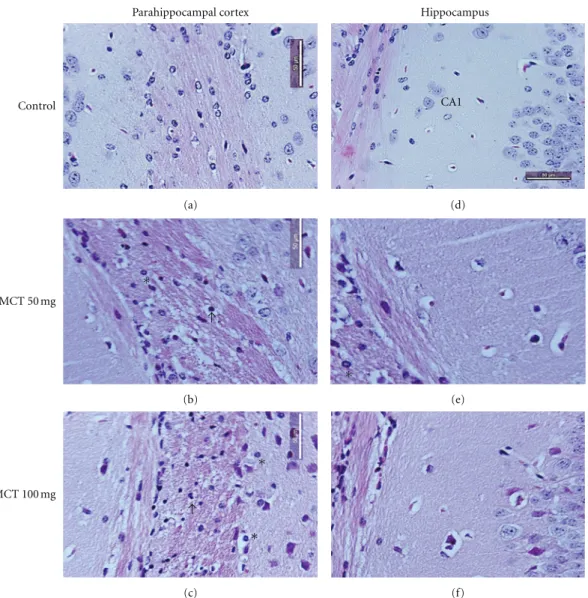
Figure1: Histologic analysis of representative parahippocampal cortex and hippocampus sections. Control mice showing normal brain
tissue ((a) and (d)). MCT-induced severe structural disorganization, edema, moderate pyknotic cells (arrow), vacuolization (arrowhead), inflammatory cell infiltration (∗) in parahippocampal ((b) and (c)), and hippocampus ((e) and (f)) regions in 50 and 100 mg/kg
concentrations, respectively. H&E staining ((a), (b), (c), (d), (e), and (f) 400x).
(0) Normal morphology.
(1) Minor damage (edema, few pyknotic cells).
(2) Moderate damage (structural disorganization, edema, moderate pyknotic cells, vacuolization, inflammatory cell infiltration).
(3) Intense damage (structural disorganization, edema, intense pyknotic cells, vacuolization, inflammatory cell infiltration).
We included immunohistochemistry analysis in our results. Immunohistochemistry for caspase-3 was performed using the streptavidin-biotin-peroxidase method [26] in formalin-fixed, paraffin-embedded tissue sections (4µm thick), mounted on poly-L-lysine-coated microscope slides. The sections were deparaffinized and rehydrated through xylene and graded alcohols. After antigen retrieval, endoge-nous peroxidase was blocked (15 min) with 3% (v/v)
hydrogen peroxide and washed in phosphate-buffered saline (PBS). Sections were incubated overnight (4◦C) with
pri-mary anti-caspase-3 antibody (polyclonal goat anti-mouse) diluted 1 : 200 in PBS plus bovine serum albumin (PBS-BSA). The slides were then incubated with biotinylated goat anti-IgG, diluted 1 : 200 in PBS-BSA. After washing, the slides were incubated with avidin-biotin-horseradish peroxidase conjugate (Strep ABC complex by Vectastain ABC reagent and peroxidase substrate solution) for 30 min, according to the Vectastain protocol. Caspase-3 was visualized with the chromogen 3,3′ diaminobenzidine (DAB). Negative
P
ar
ahippocampal c
o
rt
ex
Control
(a)
MCT 50 mg
(b)
MCT 100 mg
(c)
H
ippocampus
CA1
(d) (e) (f)
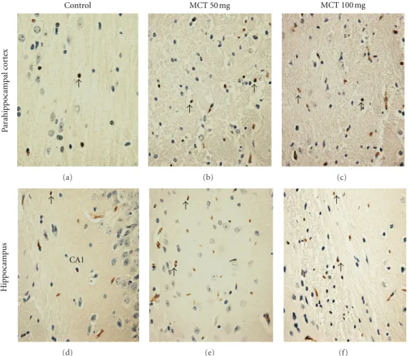
Figure2: Representative examples of caspase-3 immunohistochemistry in the parahippocampal cortex and hippocampus of mice (400x).
(a) and (d) Control groups. (b) and (e) Presented is an increase of the immunostaining for caspase-3 (arrow) in parahippocampal cortex and hippocampus of mice treated MCT (50 mg/kg). (c) and (f) Presented is an increase of the immunostaining for caspase-3 (arrow) in parahippocampal cortex and hippocampus of mice treated MCT (100 mg/kg).
2.6. Measurement of Lipid Peroxidation. Lipid peroxides formation was analyzed by measuring the thiobarbituric-acid reacting substances (TBARS) in the homogenates [27]. The samples were added to a catalytic system of formation of free radicals (FeSO40.01 mM and ascorbic acid 0.1 mM) and
then incubated at 37◦C for 30 min. The reaction was stopped
with 0.5 mL of 10% trichloroacetic acid, then the samples were centrifuged (3000 rpm/15 min), and the supernatants were retrieved and mixed with 0.5 mL of 0.8% thiobarbituric acid then heated in a boiling water bath for 15 min and after this period, they were immediately kept cold in a bath of ice. Lipid peroxidation was determined by the absorbance at 532 nm and was expressed as µmol of malondialdehyde (MDA)/g tissue.
2.7. Nitrite-Nitrate Determination. For the assessment of nitrite, derived from nitric oxide (NO), 100µL of Griess reagent (1% sulfanilamide in 1% H3PO4/0.1%
N-(1-naphthyl)-ethylenediamine dihydrochloride/1% H3PO4/
distilled water, 1 : 1 : 1 : 1) were added to 100µL of brain homogenates or to 100µL of NaNO2 at concentrations
ranging from 0.75 to 100µL (standard curve). The absorbance was measured with a reader plate at 560 nm. The
standard curve was used for the determination of nitrite concentrations in samples [28]. Results were expressed as
µM.
2.8. Evaluation of Catalase Activity. The catalase activity was measured by the method that employs hydrogen peroxide to generate H2O and O2 [27]. The activity was measured
by the degree of this reaction. The assay mixture contained 0.3 mL of hydrogen peroxide in 50 mL of 0.05 M phosphate buffer, pH 7.0. The sample aliquot (20µL) was added to 980µL of the substrate mixture. Initial and final absorbances were recorded at 230 nm after 1 and 6 min, respectively. A standard curve was established using purified catalase (Sigma, MO, USA) under identical conditions. All samples were diluted with 0.1 mmol/L phosphate buffer (pH 7.0) to provoke a 50% inhibition of the diluent rate (i.e., the uninhibited reaction). Protein was determined using bovine serum albumin as standard [29]. Results were expressed as mM/min/mg protein.
Control MCT 5 MCT 50 MCT 100 0
1 2 3 4 5
a, b
a, b
TB
A
R
S
µ
mol of
MD
A/mg of
pr
ot
ein
(a)
Control MCT 5 MCT 50 MCT 100
0 1 2 3 4 5
a, b
a, b
TB
A
R
S
µ
mol of
MD
A/mg of
pr
ot
ein
(b)
Control MCT 5 MCT 50 MCT 100
0 1 2 3 4 5
a, b
a, b
TB
A
R
S
µ
mol of
MD
A/mg of
pr
ot
ein
(c)
Figure3: Levels of TBARS (lipid peroxidation level) in HC (a), PFC (b), or ST (c) of adult mice treated with vehicle or MCT (5, 50 or
1000 mg/kg). The results are presented as mean±S.E.M. In parentheses, there is the number of animals per group. ((a) and (b))P <0.05 when compared to controls or MCT 5 mg/kg, respectively (ANOVA and Student-Newman-Keuls test as a post hoc).
(post hoc) by GraphPad Prism 5.0 version for Windows, GraphPad Software, San Diego, CA USA. Differences were considered statistically significant atP <0.05.
3. Results
3.1. Histopathological Analysis. Histological evaluation of brain structures of the rat treated with MCT (50 or 100 mg/kg) revealed lesions in the hippocampus and parahippocampal cortex compared to control (score: 0 (0-1)). The scores show severe structural disorganization, edema, moderate pyknotic cells, vacuolization, and inflam-matory cell infiltration in both concentration 50 mg/kg (score: 2 (2-3)) and 100 mg/kg (score: 2 (1-3)) (Figure 1). Immunohistochemistry analysis in our results showed an increase of caspase 3, indicating apoptosis caused by MCT in relation to control animals (Figure 2).
3.2. Oxidative Stress Parameters
3.2.1. Lipid Peroxidation. The effect of MCT in lipid per-oxidation is presented inFigure 3. MCT administration, in both doses (50 or 100 mg/kg), increased MDA levels (a lipid peroxidation marker) in the HC (F(3, 27) = 6.353; P =
0.0025), PFC (F(3, 26) = 4.441; P = 0.0133), and ST
(F(3, 27)=8.143;P=0.0007) as compared to control (HC:
2.26±0.32; PFC: 1.39±0.30; ST: 1.26±0.23) and MCT
5 mg/kg (HC: 1.57±0.25; PFC: 1.46±0.43; ST: 1.13±0.16)
groups.
3.2.2. Nitrite Content. MCT in all doses (5, 50 or 100 mg/kg) resulted in low levels of nitrite in HC (F(3, 35)=5.447;P=
0.0039) and PFC (F(3, 28)=5.528;P=0.0047) as compared
to the control group (HC: 9.17±0.66; PFC: 7.25±0.41)
(Figures 4(a)and4(b)). Similar effect was observed in ST
(Figure 4(c)) in the levels of nitrite only with high doses of
MCT (50 or 100 mg/kg) as compared to control (F(3, 36)=
4.160;P=0.0132).
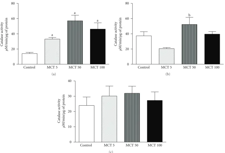
3.2.3. Catalase Activity. An increase in catalase activity was seen in the MCT group, in all doses, in HC (F(3, 21)=7.976;
P = 0.0014) as compared to the control group (control:
14.12±1.53) (Figure 5(a)). No alteration was observed in
catalase activity in PFC (F(3, 27) = 4.243;P = 0.0154) or
ST (F(3, 35) = 0.4684; P = 0.7064) after administration
Control MCT 5 MCT 50 MCT 100 0
5 10 15
a a a
N
it
rit
e c
o
nt
ent
µ
mol/g of
tissue
(a)
Control MCT 5 MCT 50 MCT 100
0 2 4 6 8 10
a
a
a
N
it
rit
e c
o
nt
ent
µ
mol/g of
tissue
(b)
Control MCT 5 MCT 50 MCT 100
0 5 10 15
a a
N
it
rit
e c
o
nt
ent
µ
mol/g of
tissue
(c)
Figure4: Content of nitrite HC (a), PFC (b), or ST (c) of adult mice treated with vehicle or MCT (5, 50 or 1000 mg/kg). The results are
presented as mean±S.E.M. In parentheses, there is the number of animals per group. (a)P <0.05 when compared to controls (ANOVA and Student-Newman-Keuls test as a post hoc).
4. Discussion
The alkaloids are molecules that may cause hepatotoxic, pneumotoxic, mutagenic, and neurotoxic effects [4]. Kim-berley horse disease is observed in horses intoxicated with monocrotaline (pyrrolizidine alkaloid) and it presents neu-rological symptoms which have been associated with neural alteration [30]. On this way, Nobre et al. observed that animals intoxicated with MCT showed clinical symptoms related to CNS such as depression, decreased locomotor activity, and motor incoordination [9,24].
Our histopathological results showed that 50 or 100 mg/kg of monocrotaline caused lesions in the hip-pocampus and parahippocampal cortex regions. Lesions observed in our work were severe structural disorganization, edema, moderate pyknotic cells, vacuolization, and inflam-matory cell infiltration. These results are according to previous studies that showed vacuolization and increase in cell body in cortical astrocyte/neuron primary co-ultures, referring to neuronal damage induced by MCT [31]. Similarly, another study showed that pyrrolizidine alkaloid monocrotaline induced cytotoxicity, morphological changes, and oxidative and genotoxic damages to glial cells, using the human glioblastoma cell line GL-15 as a model
[32]. The results of monocrotaline immunochemistry in two doses showed an increase of caspase 3 in brain areas compared to control, indicating apoptosis, so these results are according to histological data. Apoptosis is characterized by DNA fragmentation, membrane blebbing, cell shrinkage, and disassembly into membrane-enclosed vesicles, which prevents a host inflammatory response to intracellular components [33]. At molecular level, classical apoptosis is caused by the activation of a family of cysteine protease known as caspases that cleave their target protein at specific aspartic acids. Two major apoptotic pathways culminate in the activation of downstream executioner caspases 3 and 6 [34,35]. So caspase 3 is an immunohistochemistry marker to apoptosis.
Based on these histological alterations in hippocampus and parahippocampal cortex regions and behavioral studies that demonstrated that MCT is able to cause serious alterations in CNS [9, 13, 24], we decided to investigate oxidative stress in HC, PFC, and ST of mice treated with MCT. These brain areas were selected due to susceptibility to oxidative stress and they are involved with motor behavior.
Control MCT 5 MCT 50 MCT 100 0
20 40 60 80
a
a
a
Catalase acti
vi
ty
µ
M/min/
µ
g of
pr
ot
ein
(a)
Control MCT 5 MCT 50 MCT 100
0 20 40 60 80
b
Catalase acti
vi
ty
µ
M/min/
µ
g of
pr
ot
ein
(b)
Control MCT 5 MCT 50 MCT 100
0 10 20 30 40
Catalase acti
vi
ty
µ
M/min/
µ
g of
pr
ot
ein
(c)
Figure5: Catalase activity in HC (a), PFC (b), or ST (c) of adult mice treated with vehicle or MCT (5, 50 or 1000 mg/kg). The results are
presented as mean±S.E.M. In parentheses, there is the number of animals per group. ((a) and (b))P <0.05 when compared to controls or MCT 5 mg/kg, respectively, (ANOVA and Student-Newman-Keuls test as a post hoc).
may be a common pathogenic mechanism underlying many disorders in CNS, since the brain has comparatively greater vulnerability to oxidative damage [39]. The data presented in this study demonstrated that MCT (50 or 100 mg/kg) in the three brain areas increased TBARS, indicating an increase in lipid peroxidation. Membrane lipid derangements, includ-ing lipid peroxidation, have been reported to contribute significantly to paroxysmal membrane dysfunction in neu-rodegenerative diseases [40]. McEwen indicates that stressors were having neurotoxic effects on the hippocampus which predisposes to the development of depression [41]. Our results showed that the dose of MCT 50 or 100 mg/kg could contribute to alteration in CNS due to increased lipid peroxidation in the HP, PFC, and ST. In fact, besides the brain is more vulnerable to injury by lipid peroxidation products than other tissues, animals with high OS have an increase in prostaglandin levels and the overproduction of the compounds could release O2−and OH•[42].
Many studies have focused on the biochemical and molecular actions of nitric oxide (NO) in normal conditions, as well as its potential alteration in CNS [37,43,44]. We investigated MCT effects on nitrite levels and observed low levels of nitrite in all studied brain areas after MCT admin-istration. The low response in the levels of nitrite/nitrate
and in the values of HC, PFC, and ST may has occurred because the nitric oxide is an unstable molecule [45, 46]. In the CNS, the major reactive nitrogen species (RNS) mediator of oxidative stress is peroxynitrite ONOO− [47].
The participation of peroxynitrite in oxidative stress is difficult to determine due to their reduced life span and interaction with other molecules [48]. However, it has been demonstrated that stressful conditions produce changes in NO metabolism and glutamatergic receptors to produce part of its stimulatory action on the CNS [49]. NO, in physiological levels, participates in a variety of physiological processes consisting of neurotransmission and regulation of blood vessel wall [50].
mechanism of catalase against the oxidative effects caused by MCT administration related to TBARS increase in the brain regions studied.
Oxidative stress has been related to many neurodegen-erative diseases, such as ischemia and Parkinson disease [16]. Rotenone, a chemical that belongs to the family of isoflavones naturally found in the roots and stems of several plants, is used as a broad-spectrum pesticide [54]. It has characteristics alike MCT, such as potent inhibitor of mitochondrial complex-I and crosses the blood-brain barrier as well as the cell membrane easily because of its lipophilic structure [55]. The present results showed that MCT increases lipidic peroxidation and catalase activity, only in HC, while it decreases nitrite/nitrate contente. Hippocampus was probably more affected because it is a rich area in mitochondria. These are related to a production of antioxidant defense when the brain is attacked by a harmful agent as monocrotaline [56, 57]. Rotenone, a neurotoxin used as a Parkinson model, also augments lipidic peroxidation and nitric oxide content in chronically treated rats [54]. This substance also increases catalase activity after chronic administration to rats [55].
5. Conclusion
Our findings showed that monocrotaline caused cell lesions in the HC and PFC regions. Also, it produced oxidative stress in the HC, PFC, and ST in mice. These alterations may contribute to the neurological effects related to this compound.
Acknowledgment
The authors acknowledge support from the Brazilian National Research Council (CNPq).
References
[1] R. J. Huxtable, “Activation and pulmonary toxicity of pyrrolizidine alkaloids,”Pharmacology and Therapeutics, vol. 47, no. 3, pp. 371–389, 1990.
[2] S. M. M. Vasconcelos, J. E. R. Hon ´orio-J ´unior, R. N. D. C. Abreu, M. C. C. Silva, J. M. Barbosa-Filho, and R. F. G. Lobato, “Pharmacologic study of some plant species from the Brazilian Northeast: Calotropis procera Agava sisalana, Solanum paludosum, Dioscorea cayenensis and Crotalaria retusa,” in, Medicinal Plants: Classification, Biosynthesis and
Pharmacology, A. Varela and J. Jasiah Ibanez, Eds., vol. 4, pp.
189–202, Nova Science, Hauppauge, NY, USA, 2009.
[3] C. Kosogof, J. J. Tepe, and R. M. Williams, “DNA cross-linking by a phototriggered pyrrolic progenitor developed from monocrotaline,”Tetrahedron Letters, vol. 42, no. 38, pp. 6641–6643, 2001.
[4] C. C. Yan and R. J. Huxtable, “Effects of taurine and guanid-inoethane sulfonate on toxicity of the pyrrolizidine alkaloid monocrotaline,”Biochemical Pharmacology, vol. 51, no. 3, pp. 321–329, 1996.
[5] F. E. Mingatto, D. J. Dorta, A. B. dos Santos et al., “De-hydromonocrotaline inhibits mitochondrial complex I.
A potential mechanism accounting for hepatotoxicity of monocrotaline,”Toxicon, vol. 50, no. 5, pp. 724–730, 2007. [6] A. C. de Souza, M. R. Hatayde, and G. H. Bechara,
“Patholog-ical aspects of poisoning by Crotalaria spectabilis (Fabaceae) seeds in swine,”Pesquisa Veterinaria Brasileira, vol. 17, no. 1, pp. 12–18, 1997.
[7] T. W. Petry, G. T. Bowden, R. J. Huxtable, and I. G. Sipes, “Characterization of hepatic DNA damage induced in rats by the pyrrolizidine alkaloid monocrotaline,”Cancer Research, vol. 44, no. 4, pp. 1505–1509, 1984.
[8] A. L. Rose, C. A. Garden, J. D. McConnell, and L. B. Bull, “Field and experimental investigations of, “walk-about” disease of horses (Kimberly horse disease) in Northern Australia. Crotalaria poisoning in horses,”Australian Veterinary Journal, vol. 33, pp. 25–33, 1957.
[9] V. M. da Trindade Nobre, F. Riet-Correa, A. F. Dantas, J. M. Barbosa Filho, I. M. Tabosa, and J. S. Vasconcelos, “Poisoning by Crotalaria retusa (Fabaceae) in Equidae in the semiarid region of Para´ıba,”Pesquisa Veterinaria Brasileira, vol. 24, no. 3, pp. 132–143, 2004.
[10] H. Sies and E. Cadenas, “Oxidative stress: damage to intact cells and organs,” Philosophical Transactions of the Royal
Society, vol. 311, no. 1152, pp. 617–631, 1985.
[11] B. Joseph, V. Kumaran, E. Berishvili, K. K. Bhargava, C. J. Pale-stro, and S. Gupta, “Monocrotaline promotes transplanted cell engraftment and advances liver repopulation in rats via liver conditioning,”Hepatology, vol. 44, no. 6, pp. 1411–1420, 2006. [12] Y. Morimatsu, N. Sakashita, Y. Komohara et al., “Development and characterization of an animal model of severe pulmonary arterial hypertension,”Journal of Vascular Research, vol. 49, no. 1, pp. 33–42, 2011.
[13] R. J. Huxtable, C. C. Yan, S. Wild, S. Maxwell, and R. Cooper, “Physicochemical and metabolic basis for the differing neu-rotoxicity of the pyrrolizidine alkaloids, trichodesmine and monocrotaline,”Neurochemical Research, vol. 21, no. 2, pp. 141–146, 1996.
[14] D. G. Graham, “Oxidative pathways for catecholamines in the genesis of neuromelanin and cytotoxic quinones,”Molecular
Pharmacology, vol. 14, no. 4, pp. 633–643, 1978.
[15] B. Poljsak and I. Milisav, “The neglected significance of, ‘antioxidative stress’,” Oxidative Medicine and Cellular
Longevity, vol. 2012, Article ID 480895, 12 pages, 2012.
[16] B. Halliwell and J. M. C. Gutteridge,Free Radicals in Biology
and Medicine, 1999.
[17] L. L. Dugan and D. W. Choi, “Hypoxic-ischemic brain injury and oxidative stress,” in Basic Neurochemistry: Molecular,
Cellular and Medical Aspects, G. J. Siegel, B. W. Agranoff, E.
W. Alberts, S. K. Fisher, and M. D. Uhler, Eds., pp. 722–723, Williams & Wilkins, Baltimore, Md, USA, 6th edition, 1999. [18] C. M. Bergamini, S. Gambetti, A. Dondi, and C. Cervellati,
“Oxygen, reactive oxygen species and tissue damage,”Current
Pharmaceutical Design, vol. 10, no. 14, pp. 1611–1626, 2004.
[19] M. Gottlieb, R. Leal-Campanario, M. R. Campos-Esparza et al., “Neuroprotection by two polyphenols following excitotox-icity and experimental ischemia,”Neurobiology of Disease, vol. 23, no. 2, pp. 374–386, 2006.
[20] S. M. Aziz, M. Toborek, B. Hennig, E. Endean, and D. W. Lipke, “Polyamine regulatory processes and oxidative stress in monocrotaline-treated pulmonary artery endothelial cells,”
Cell Biology International, vol. 21, no. 12, pp. 801–812, 1997.
[21] N. T. T. Huong, K. Matsumoto, R. Kasai, K. Yamasaki, and H. Watanabe, “In vitro antioxidant activity of Vietnamese ginseng saponin and its components,”Biological and
[22] E. J. Barreiro, A. L. Pereira, L. N. L. F. Gomes, and J. R. Silva, “Carbon-13 nuclear magnetic resonance of pyrrolizidine alkaloids: a reassignment,”Journal of Chemical Research, pp. 330–331, 1980.
[23] D. L. Cheng, S. B. Tu, A. A. Enti, and E. Roder, “Occurrence of the pyrrolizidine alkaloid monocrotaline in Crotalaria
assamica Benth. and Crotalaria calycina Schrank,” Scientia
Pharmaceutica, vol. 54, no. 4, pp. 351–355, 1986.
[24] V. M. T. Nobre, A. F. M. Dantas, F. Riet-Correa, J. M. Barbosa Filho, I. M. Tabosa, and J. S. Vasconcelos, “Acute intoxication by Crotalaria retusa in sheep,”Toxicon, vol. 45, no. 3, pp. 347– 352, 2005.
[25] E. Gilat, T. Kadar, A. Levy et al., “Anticonvulsant treatment of sarin-induced seizures with nasal midazolam: an electro-graphic, behavioral, and histological study in freely moving rats,”Toxicology and Applied Pharmacology, vol. 209, no. 1, pp. 74–85, 2005.
[26] S. M. Hsu and L. Raine, “Protein A, avidin, and biotin in immunohistochemistry,” Journal of Histochemistry and
Cytochemistry, vol. 29, no. 11, pp. 1349–1353, 1981.
[27] A. C. Maehly and B. Chance, “The assay of catalases and peroxidases,”Methods of Biochemical Analysis, vol. 1, pp. 357– 424, 1954.
[28] L. C. Green, S. R. Tannenbaum, and P. Goldman, “Nitrate synthesis in the germfree and conventional rat,”Science, vol. 212, no. 4490, pp. 56–58, 1981.
[29] O. H. Lowry, N. J. Rosebrough, A. L. Farr, and R. J. Randall, “Protein measurement with the Folin phenol reagent,”The
Journal of Biological Chemistry, vol. 193, no. 1, pp. 265–275,
1951.
[30] M. R. Gardiner, R. Royce, and A. Bokora, “Studies on crotalaria crispata, a newly recognised cause of kimberley horse disease,”Journal of Pathology & Bacteriology, vol. 89, no. 1, pp. 43–55, 1965.
[31] B. P. S. Pitanga, V. D. A. Silva, C. S. Souza et al., “Assessment of neurotoxicity of monocrotaline, an alkaloid extracted from Crotalaria retusa in astrocyte/neuron co-culture system,”
NeuroToxicology, vol. 32, no. 6, pp. 776–784, 2011.
[32] J. P. Silva-Neto, R. A. Barreto, B. P. S. Pitanga et al., “Genotoxicity and morphological changes induced by the alkaloid monocrotaline, extracted from Crotalaria retusa, in a model of glial cells,”Toxicon, vol. 55, no. 1, pp. 105–117, 2010. [33] V. Crysns and J. Yuan, “The cutting edge: caspases in apoptosis and disease,” inWhen Cells Die, R. Lockshin, Z. zakeri, and J. Tilly, Eds., pp. 177–210, Wiley-Liss, New York, NY, USA, 1998. [34] J. J. Kang, M. D. Schaber, S. M. Srinivasula et al., “Cascades of mammalian caspase activation in the yeastSaccharomyces
cerevisiae,”Journal of Biological Chemistry, vol. 274, no. 5, pp.
3189–3198, 1999.
[35] G. A. C. Brito, J. Fujji, B. A. Carneiro-Filho, A. A. M. Lima, T. Obrig, and R. L. Guerrant, “Mechanism ofClostridium difficile toxin A-induced apoptosis in T84 cells,”Journal of Infectious
Diseases, vol. 186, no. 10, pp. 1438–1447, 2002.
[36] E. E. Essick and F. Sam, “Oxidative stress and autophagy in cardiac disease, neurological disorders, aging and cancer,”
Oxidative Medicine and Cellular Longevity, vol. 3, no. 3, pp.
168–177, 2010.
[37] C. C. Aguiar, A. B. Almeida, P. V. Araujo et al., “Oxidative stress and epilepsy: literature review,”Oxidative Medicine and
Cellular Longevity, vol. 2012, Article ID 795259, 12 pages,
2012.
[38] K. Takuma, A. Baba, and T. Matsuda, “Astrocyte apoptosis: implications for neuroprotection,” Progress in Neurobiology, vol. 72, no. 2, pp. 111–127, 2004.
[39] F. Ng, M. Berk, O. Dean, and A. I. Bush, “Oxidative stress in psychiatric disorders: evidence base and therapeutic implica-tions,”International Journal of Neuropsychopharmacology, vol. 11, no. 6, pp. 851–876, 2008.
[40] M. G. Naffah-Mazzacoratti, “Profile of prostaglandin levels in the rat hippocampus in pilocarpine model of epilepsy,”
Neurochemistry International, vol. 27, no. 6, pp. 461–466, 1995.
[41] B. S. McEwen and R. M. Sapolsky, “Stress and cognitive function,”Current Opinion in Neurobiology, vol. 5, no. 2, pp. 205–216, 1995.
[42] M. I. Bellissimo, D. Amado, D. S. P. Abdalla, E. C. Ferreira, E. A. Cavalheiro, and M. G. Naffah-Mazzacoratti, “Superoxide dismutase, glutathione peroxidase activities and the hydroper-oxide concentration are modified in the hippocampus of epileptic rats,”Epilepsy Research, vol. 46, no. 2, pp. 121–128, 2001.
[43] S. S. Zoroˇglu, H. Herken, M. Y¨urekli et al., “The pos-sible pathophysiological role of plasma nitric oxide and adrenomedullin in schizophrenia,” Journal of Psychiatric
Research, vol. 36, no. 5, pp. 309–315, 2002.
[44] R. Garg and A. Kumar, “Possible role of citalopram and desipramine against sleep deprivation-induced anxiety like-behavior alterations and oxidative damage in mice,”Indian
Journal of Experimental Biology, vol. 46, no. 11, pp. 770–776,
2008.
[45] F. T. Bonner and G. Stedman, “The chemistry of nitric oxide and redox-related species,” inMethods in Nitric Oxide
Research, pp. 3–18, John Wiley & Sons, New York, NY, USA,
1996.
[46] F. T. Bonner, “Nitric oxide gas,”Methods in Enzymology, vol. 268, pp. 50–57, 1996.
[47] J. S. Beckman, “The double-edged role of nitric oxide in brain function and superoxide-mediated injury,”Journal of
Developmental Physiology, vol. 15, no. 1, pp. 53–59, 1991.
[48] J. Fukuto and L. J. Ignarro, “In vivo aspects of nitric oxide (NO) chemistry: does peroxynitrite (-OONO) play a major role in cytotoxicity?”Accounts of Chemical Research, vol. 30, no. 4, pp. 149–152, 1997.
[49] L. J. Ignarro, G. M. Buga, K. S. Wood, R. E. Byrns, and G. Chaudhuri, “Endothelium-derived relaxing factor produced and released from artery and vein is nitric oxide,”Proceedings of the National Academy of Sciences of the United States of
America, vol. 84, no. 24, pp. 9265–9269, 1987.
[50] D. O. Barros, S. M. L. Xavier, C. O. Barbosa et al., “Effects of the vitamin E in catalase activities in hippocampus after status epilepticus induced by pilocarpine in Wistar rats,”
Neuroscience Letters, vol. 416, no. 3, pp. 227–230, 2007.
[51] B. D. Watson, “Evaluation of the concomitance of lipid peroxidation in experimental models of cerebral ischemia and stroke,”Progress in Brain Research, vol. 96, pp. 69–95, 1993. [52] M. Erecinska and I. A. Silver, “Calcium handling by
hip-pocampal neurons under physiologic and pathologic condi-tions,”Advances in Neurology, vol. 71, pp. 119–136, 1996. [53] A. M. Dymond and P. H. Crandall, “Oxygen availability and
blood flow in the temporal lobes during spontaneous epileptic seizures in man,”Brain Research, vol. 102, no. 1, pp. 191–196, 1976.
[54] R. Verma and B. Nehru, “Effect of centrophenoxine against rotenone-induced oxidative stress in an animal model of Parkinson’s disease,”Neurochemistry International, vol. 55, no. 6, pp. 369–375, 2009.
oxide and lipid peroxidation products in rat brain,”
Experi-mental Neurology, vol. 186, no. 2, pp. 235–241, 2004.
[56] K. H. Lauritzen, C. Cheng, H. Wiksen, L. H. Bergersen, and A. Klungland, “Mitochondrial DNA toxicity compromises mito-chondrial dynamics and induces hippocampal antioxidant defenses,”DNA Repair, vol. 10, no. 6, pp. 639–653, 2011. [57] L. L. Dugan and J. S. Kim-Han, “Hypoxic-ischemic brain
injury and oxidative stress,” inBasic Neurochemistry
Molec-ular, CellMolec-ular, and Medical Aspects, G. J. Siegel, Ed.,
Submit your manuscripts at
http://www.hindawi.com
Hindawi Publishing Corporation
http://www.hindawi.com Volume 2013 Oxidative Medicine and Cellular Longevity
Hindawi Publishing Corporation
http://www.hindawi.com Volume 2013 Hindawi Publishing Corporation
http://www.hindawi.com Volume 2013
The Scientiic
World Journal
International Journal of
Endocrinology
Hindawi Publishing Corporation
http://www.hindawi.com Volume 2013
ISRN
Anesthesiology
Hindawi Publishing Corporation
http://www.hindawi.com Volume 2013
Hindawi Publishing Corporation http://www.hindawi.com
Oncology
Volume 2013
PPAR
R e s e a r c h
Hindawi Publishing Corporation
http://www.hindawi.com Volume 2013
Ophthalmology
Journal ofHindawi Publishing Corporation
http://www.hindawi.com Volume 2013
ISRN
Allergy
Hindawi Publishing Corporation
http://www.hindawi.com Volume 2013
BioMed Research International
Hindawi Publishing Corporation
http://www.hindawi.com Volume 2013
Hindawi Publishing Corporation
http://www.hindawi.com Volume 2013
Obesity
ISRN
Addiction
Hindawi Publishing Corporation
http://www.hindawi.com Volume 2013
Hindawi Publishing Corporation
http://www.hindawi.com Volume 2013
Computational and Mathematical Methods in Medicine
ISRN
AIDS
Hindawi Publishing Corporation
http://www.hindawi.com Volume 2013
Clinical & Developmental Immunology
Hindawi Publishing Corporation
http://www.hindawi.com Volume 2013
Diabetes ResearchJournal of
Hindawi Publishing Corporation
http://www.hindawi.com Volume 2013
Evidence-Based Complementary and Alternative Medicine
Volume 2013 Hindawi Publishing Corporation
http://www.hindawi.com Hindawi Publishing Corporation
http://www.hindawi.com Volume 2013 Gastroenterology Research and Practice
Hindawi Publishing Corporation
http://www.hindawi.com Volume 2013 ISRN
Biomarkers
Hindawi Publishing Corporation
http://www.hindawi.com Volume 2013
MEDIATORS
INFLAMMATION