Canine Disorder Mirrors Human Disease:
Exonic Deletion in
HES7
Causes Autosomal
Recessive Spondylocostal Dysostosis in
Miniature Schnauzer Dogs
Cali E. Willet1*, Mariano Makara1, George Reppas2, George Tsoukalas1, Richard Malik3, Bianca Haase1, Claire M. Wade1*
1Faculty of Veterinary Science, University of Sydney, Sydney, NSW, Australia,2Vetnostics, North Ryde, NSW, Australia,3Centre for Veterinary Education, University of Sydney, Sydney, NSW, Australia
*cali.willet@sydney.edu.au(CEW);claire.wade@sydney.edu.au(CMW)
Abstract
Spondylocostal dysostosis is a congenital disorder of the axial skeleton documented in human families from diverse racial backgrounds. The condition is characterised by truncal shortening, extensive hemivertebrae and rib anomalies including malalignment, fusion and reduction in number. Mutations in the Notch signalling pathway genesDLL3,MESP2,
LFNG,HES7andTBX6have been associated with this defect. In this study, spondylocostal
dysostosis in an outbred family of miniature schnauzer dogs is described. Computed to-mography demonstrated that the condition mirrors the skeletal defects observed in human cases, but unlike most human cases, the affected dogs were stillborn or died shortly after birth. Through gene mapping and whole genome sequencing, we identified a single-base deletion in the coding region ofHES7. The frameshift mutation causes loss of functional do-mains essential for the oscillatory transcriptional autorepression of HES7 during somitogen-esis. A restriction fragment length polymorphism test was applied within the immediate family and supported a highly penetrant autosomal recessive mode of inheritance. The mu-tation was not observed in wider testing of 117 randomly sampled adult miniature schnauzer and six adult standard schnauzer dogs; providing a significance of association ofPraw= 4.759e-36(genome-wide significant). Despite this apparently low frequency in the Australian population, the allele may be globally distributed based on its presence in two unrelated sires from geographically distant locations. While isolated hemivertebrae have been ob-served in a small number of other dog breeds, this is the first clinical and genetic diagnosis of spontaneously occurring spondylocostal dysostosis in a non-human mammal and offers an excellent model in which to study this devastating human disorder. The genetic test can be utilized by dog breeders to select away from the disease and avoid unnecessary neonatal losses.
OPEN ACCESS
Citation:Willet CE, Makara M, Reppas G, Tsoukalas G, Malik R, Haase B, et al. (2015) Canine Disorder Mirrors Human Disease: Exonic Deletion inHES7 Causes Autosomal Recessive Spondylocostal Dysostosis in Miniature Schnauzer Dogs. PLoS ONE 10(2): e0117055. doi:10.1371/journal.pone.0117055
Academic Editor:Reiner Albert Veitia, Institut Jacques Monod, FRANCE
Received:September 11, 2014
Accepted:December 18, 2014
Published:February 6, 2015
Copyright:© 2015 Willet et al. This is an open access article distributed under the terms of the
Creative Commons Attribution License, which permits unrestricted use, distribution, and reproduction in any medium, provided the original author and source are credited.
Data Availability Statement:All relevant data are within the paper and its Supporting Information files.
Funding:Research conducted by CEW was funded by the Australian Postgraduate Award and Val Street Scholarship. Four of the samples used in the authors' study were genotyped and sequenced as part of an unrelated project funded by the National Institutes of Health (http://www.nih.gov/) grant NIH/
Introduction
During embryogenesis, the presomitic mesoderm generates somites, which are transient seg-mental structures that give rise to the axial skeleton, skeletal muscle and dermis in later
devel-opment [1]. Correct formation of somite boundaries is essential to normal development.
Expression of the numerous genes involved is thus tightly regulated in a spatiotemporal
fash-ion. The role of the Notch signalling pathway in somitogenesis has been well established [2,3].
Segmentation defects of the axial skeleton in humans comprise a heterogeneous group of disor-ders broadly classified as spondylocostal dysostosis (SCD) or spondylothoracic dysostosis (STD). The genetic aetiology of SCD, characterized by hemivertebrae and malaligned ribs with intercostal points of fusion, has been attributed to recessively inherited mutations in four
Notch genes:DLL3[4],MESP2[5],LFNG[6] andHES7[7]. Causal mutations in each of these
genes give rise to further classification of SCD into types 1–4 respectively [8]. Recently, an
auto-somal dominant form of SCD has been attributed toTBX6mutation [9]. STD phenotypes display
hemivertebrae and costovertebral fusions of symmetrically aligned ribs within a shortened trunk
that fan out in a‘crab-like’arrangement. This disorder has so far only been ascribed toMESP2
mutation [10]. While SCD is often benign, STD is lethal in an estimated 29% to 44% of patients
due to respiratory insufficiency, with mortality occurring around one year of age [11,12].
The prevalence of SCD in human populations is estimated to be one in 40,000 births [13]. A
survey of the peer-reviewed literature indicates that this disorder has not been described in other mammalian species, other than mutant mouse models. Spontaneously occurring SCD in a non-human animal could provide a useful model for investigation and treatment of axial skeleton dysostosis. Complex vertebral malformations in Holstein cattle have been attributed to a coding
mutation in theSLC35A3gene [14]. This autosomal recessive disorder, also characterized by
cer-vical and thoracic vertebral malformations including hemivertebrae, rib fusion and shortening of the trunk, provides a link between altered glycosyltransferase activity and aberrant Notch
signal-ling [15]. In screw-tailed dog breeds, such as the pug and French bulldog, the breed-defining tail
kink is caused by hemivertebrae in the tail [16]. Variably pathogenic hemivertebrae in other
regions of the spine occur commonly in screw-tailed breeds [17], as well as isolated cases in other
breeds including the German shorthaired pointer [18] and Dobermann pinscher [19]. While
these canine disorders are thought to be inherited in an autosomal recessive fashion, no diagnosis of SCD has been made and the underlying genetic aetiology has not been identified. Our labora-tory was recently presented with the opportunity to study a family of miniature schnauzer dogs in which two separate matings had produced pups with lethal skeletal malformations.
In Australia, the miniature schnauzer breed is popular due to its moderate size and
agree-able temperament and in 2012 there were 1,069 pedigreed registrations [20]. The breed is
pre-disposed to myotonia congenita, an autosomal recessive sacrolemmal channelopathy causing
abnormality of gait, skull, mandible and dentition [21,22]. The musculoskeletal abnormalities
displayed by the probands in this study were not consistent with myotonia congenita. While a number of other congenital genetically transmitted conditions are prevalent within the breed, such as patent ductus arteriosus and portosystemic shunt, these are not known to cause skeletal malformations. The aim of this study was to provide a clinical and genetic diagnosis for this
novel disorder, which we refer to as‘Comma defect’due to the gross anatomical shape of the
abnormal pups. We further aimed to develop a genetic test capable of establishing prevalence parameters and facilitating appropriate selection of breeding pairs to avoid producing litters bearing this disorder. Here we present the first diagnosis of SCD in an animal population, and
demonstrate that the underlying mutation is a coding deletion in theHES7gene. A restriction
fragment length polymorphism test (RFLP) of polymerase chain reaction (PCR) amplicons that can reliably distinguish carriers in the population is described. Preliminary sampling and
GR, but did not have any additional role in the study design, data collection and analysis, decision to publish, or preparation of the manuscript. The specific roles of these authors are articulated in the‘author contributions’section.
pedigree analysis suggests that while the mutation appears to be rare amongst miniature schnauzers, the allele may be globally dispersed.
Materials and Methods
Samples
Of a litter of eight privately owned miniature schnauzer pups bred in Australia, three were stillborn and observed by the breeder and attending veterinarian to display abnormal body mor-phology. Three of the remaining five full siblings were apparently healthy and with normal phe-notype, while a further two pups were stillborn but were otherwise normal phenotypically. The three morphologically abnormal stillborn pups (individual identifiers USCF134, USCF136 and USCF137) were sent to the Medical and Behavioural Genetics group, Faculty of Veterinary Sci-ence, University of Sydney for further investigation. The breeder reported that another mating using the sire of this litter produced similarly abnormal stillborn pups, but specimens from this litter were unavailable. This study was carried out in strict accordance with the recommendations in the Australian Code for the Care and Use of Animals for Scientific Purposes. Animal ethics approval for this research was granted by the University of Sydney Animal Ethics Committee
(approval number N00/9–2009/3/5109, September 24 2009). All blood samples were collected by
a veterinarian, and all efforts to minimize suffering during sample collection were made through the practice of appropriate animal handling skills and use of the finest gauge needle possible. The owners of the dogs gave permission for their animals to be used in this study.
Computed tomography
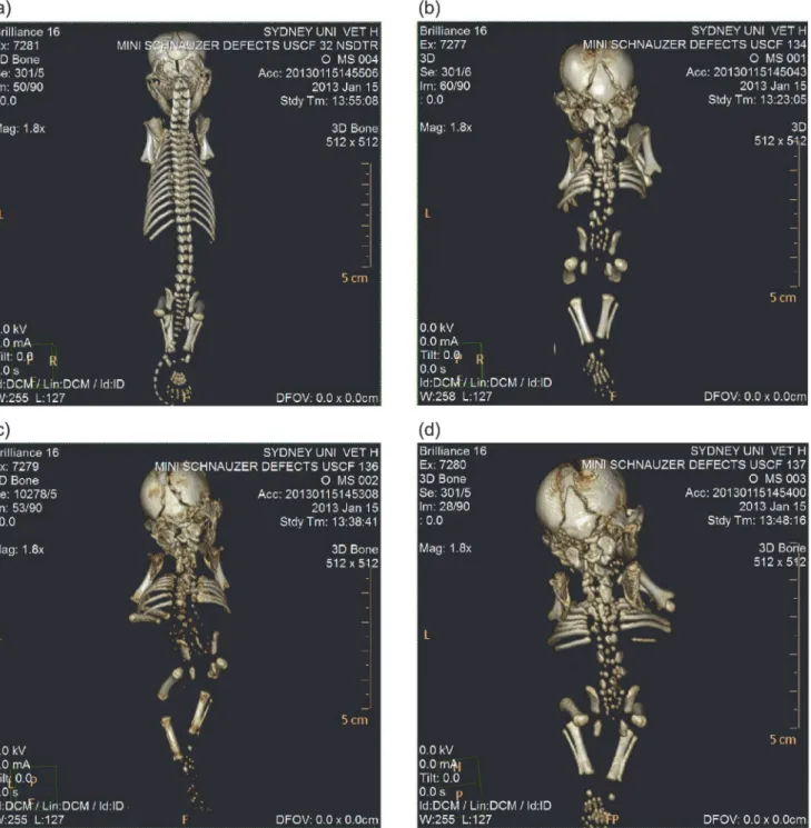
To characterize the underlying skeletal morphology of abnormal stillborn pups, computed to-mography (CT) scanning was carried out at the University of Sydney Veterinary Teaching Hospital using the Brilliance CT 16-Slice V3 (Philips, Amsterdam). In the absence of a normal age-matched counterpart of the same breed, an aged-matched Nova Scotia duck tolling re-triever sampled for unrelated reasons was used as control (USCF32).
Pedigree analysis
Australian National Kennel Club registered names and official pedigree transcripts were pro-vided by the breeder, and these were used to construct a three-generation pedigree of the affect-ed family. The paffect-edigree was visually assessaffect-ed for inheritance pattern.
Whole genome sequencing
Two samples (USCF134; USCF136) were selected for whole genome sequencing on the Illu-mina HiSeq 2000 (IlluIllu-mina, San Diego, CA). Tissue samples were taken from muscle of the af-fected pups and digested overnight with Proteinase K. Genomic DNA was extracted with the Nucleon BACC 2 Genomic DNA Extraction Kit (GE Healthcare). Paired-end libraries were prepared with the Illumina TruSeq preparation kits and sequenced according to vendor in-structions by the Ramaciotti Centre at the University of New South Wales, Kensington. Librar-ies were barcoded and both samples sequenced in a single lane of the sequencing machine.
Sequence alignment and variant calling
Reads were aligned as pairs to canFam2 [23] using Burrows-Wheeler Alignment (BWA) version
0.6.2 [24] with default parameters for paired-end data. Genome-wide single nucleotide
polymor-phisms (SNP) and insertion-deletion polymorpolymor-phisms (indels) were identified using SAMtools
mpileup version 1.18 [25] and filtered using custom Perl scripts. Only reads with mapping
Genome-wide association mapping
Ten miniature schnauzer samples including the three affected pups, sire, dam and five additional family members were genotyped on the Canine HD BeadChip (Illumina, San Diego, CA). For the seven unaffected miniature schnauzers, genomic DNA was extracted from EDTA anti-coagu-lated blood samples with the Nucleon BACC 2 Genomic DNA Extraction Kit (GE Healthcare). Array genotyping was performed by GeneSeek (Lincoln, NE). Association analysis was
con-ducted using PLINK [26], filtering out SNPs with minor allele frequency less than 0.1 and
geno-typing frequency less than 0.2. After frequency and genotype pruning, 73,921 SNPs remained in
the analysis. Unadjusted -log10 transformedPvalues were visualised in Haploview [27]. The top
100 SNPs were examined for evidence of regional clustering. The chromosomal region harbour-ing the largest proportion of the top 100 SNPs was taken forward as the candidate region.
Candidate gene and mutation identification
The candidate region was searched for potential candidate genes by first identifying the corre-sponding syntenic region/regions in the mouse genome. Since CT scans of the pups indicated
skeletal malformation, we used the Mouse Genome Browser (http://gbrowse.informatics.jax.
org/cgi-bin/gb2/gbrowse/mousebuild38/) to restrict to genes within these regions known to af-fect a skeletal phenotype. SNPs and indels homozygous for the non-reference allele within po-tential candidate genes were identified, focusing on those within exons according to the
refGene and xenoRefGene tracks on the UCSC Genome Browser (http://genome.ucsc.edu/),
canFam2 May 2005 build.
PCR-RFLP genotyping
An RFLP test from PCR amplicons (PCR-RFLP) was designed to genotype samples for the candidate mutation. Primers (forward CGGAGTTGGCGATGACCA; reverse
CGCTCTTCCTTTCCTGCTG) were selected using Primer3 [28] to amplify a 578 bp product
flanking the mutation. PCR was carried out in a total volume of 20μl using AmpliTag Gold
360Master Mix (Applied Biosystems) according to the manufacturer’s protocol and the
prod-uct size evaluated on 1% agarose gel. PCR conditions were: heat activation for 15 minutes (mins) at 95 degrees Celsius (°C) followed by 35 cycles of 30 seconds (s) at 95°C, 30 s at 58°C
and 30 s at 68°C, and terminated with a final elongation step at 72°C for 10 mins. 15μl of PCR
product was incubated with 0.3μl of the enzyme BsrBI for 7 hours at 37°C, followed by heat
in-activation. Two fragments of 472 bp and 107 bp were produced in homozygous wild type sam-ples. The mutation removed the restriction enzyme recognition sequence (TCCGCTCC) resulting in a single uncut 578 bp fragment for homozygous affected dogs.
A total of 133 samples were genotyped using this method. 127 were miniature schnauzers, including the three affected pups and seven additional family members, with the remaining six samples from the standard schnauzer breed. For these additional dogs, blood had been collect-ed for the routine investigation of a variety of disease conditions in two large veterinary diagnostic laboratories.
Results
Samples
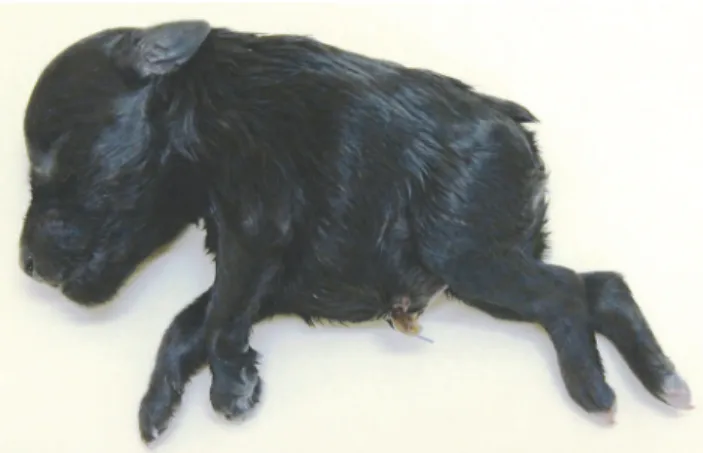
size compared to the forequarters, giving an overall comma-like morphology to the body (Fig. 1). Individual USCF134 displayed an umbilical hernia, while USCF136 had a cleft hard palate.
Computed tomography
All three affected miniature schnauzer pups displayed severe morphological defects of the axial
skeleton (Fig. 2). Sacrococcygeal agenesis was observed in addition to fully segmented
hemiver-tebrae in the cervical, thoracic and lumbar regions. The rib cage was poorly developed with a reduced number of ribs displaying malalignment and sporadic lateral fusions. Since the pro-bands were frozen after death, soft tissue imaging was unable to clearly depict the physical state of organs and cartilage.
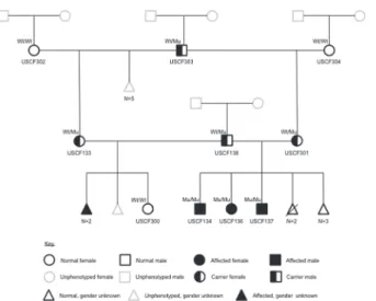
Pedigree analysis
The pattern of inheritance within the three-generation pedigree was consistent with a highly
penetrant autosomal recessive monogenic trait (Fig. 3). The reported relationships were
sup-ported by identity by descent (IBD) estimation using PLINK (S1 Table).
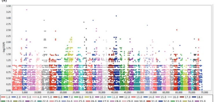
Genome-wide association mapping
PLINK genome-wide association analysis using ten individuals was hindered by limited statistical power, and this was evident when the results visualised with Haploview showed no strong signal
of association (Fig. 4A). We therefore approached the PLINK data using an alternate method,
se-lecting the 100 most significant SNPs and identifying which chromosomes were overrepresented in this group. Of the 100 most significant SNPs, 60% resided on chromosome 5 (CFA5), 13% on CFA18 and 8% on CFA12. Haploview plotting of all SNPs within chromosome demonstrated a
dispersed but contiguous signal of around 25 megabases (Mb) on CFA5 (Fig. 4B). Markers with
unadjustedPvalues less than 0.005 refined the peak association to CFA5:29.84 Mb–45.26 Mb.
Candidate gene and mutation analysis
Genes that have been linked to phenotypic groups in mouse models are a useful way of reduc-ing large gene lists to a handful of candidates. The associated region was found to be syntenic
with two mouse chromosomes on build GRCM38. CFA5:29.84 Mb–33.1 Mb was broadly
syntenic with mouse chromosome 9:5.29 Mb–8.54 Mb, and CFA5:33.1 Mb–45.26 Mb with
mouse chromosome 11:59.6 Mb–72.8 Mb. Searching these two regions for genes with a known
Figure 1. Photograph of miniature schnauzer pup USCF137 affected with Comma defect.
abnormal skeletal phenotype in the Mouse Genome Browser restricted the number of candi-date genes to 19. From a total of 27,724 SNPs and 5,808 indels identified from the sequence alignment of the affected samples within the candidate region, 274 SNPs and 12 indels resided within exons. Filtering these putatively coding variants to those occurring within one of the 19 genes with reported involvement in skeletal malformations in mice, four SNPs and one indel Figure 2. CT scans of miniature schnauzer pups affected with Comma defect and an age-matched unrelated control.(a) Unaffected Nova Scotia duck tolling retriever pup sampled for unrelated reasons. (b) Affected miniature schnauzer pup USCF134. (c) Affected miniature schnauzer pup USCF136. (d) Affected miniature schnauzer pup USCF137.
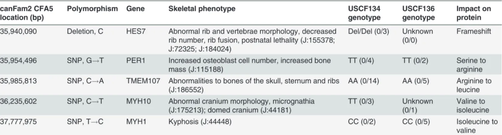
were observed (Table 1). Of these genes, mutations inHes7andTmem107have been linked to
phenotypes in mice with similarities to the affected pups in this study. A non-conservative
sub-stitution withinTMEM107was excluded as a candidate due to presence in the homozygous
state in whole genome sequence datasets of other breeds sequenced by our laboratory. A
gua-nine deletion at CFA5:35,940,090 (CFA5:32,945,846 in canFam3.1) within exon 2 ofHES7
(c.126delG) was taken forward for further analysis.
The predicted canineHES7mRNA sequence was obtained from NCBI (XM_844962.3). An
unknown nucleotide (N) present within the predicted mRNA sequence along with the three
preceding bases was corrected using the sequence data of USCF134 (S1 Dataset). The wild-type
canine HES7 protein is predicted to be 224 amino acids in length. The mutation was
intro-duced into this sequence and translated with the ExPASy translate tool (http://web.expasy.org/
translate/) (S1 Dataset). The deletion introduces a frameshift mutation, causing alteration from
the 43rdamino acid onwards and resulting in a premature termination codon in place of the
66thamino acid (p.(Thr43ProfsTer24)). The Conserved Domain Database (http://www.ncbi.
nlm.nih.gov/Structure/cdd/cdd.shtml) was used to identify important features in the predicted
wild-type protein. Significant hits were detected at residues 14–68 (Helix-loop-helix
DNA-binding domain; pfam00010) and at residues 91–128 (Hairy Orange domain; pfam07527).
Both of these functional sites are affected by the candidate deletion; the terminal half of the helix-loop-helix (HLH) domain being altered and the Hairy Orange domain being completely absent from the predicted mutant protein sequence.
PCR-RFLP genotyping
The PCR-RFLP test was designed to detect carriers by the presence of three bands, as opposed to two bands (472 bp and 107 bp) for homozygous wild type and a single uncut 578 bp
frag-ment for homozygous deleted (Fig. 5). Of 133 samples tested, only the three proband pups
test-ed homozygous for the deletion. Both parents of this litter testtest-ed heterozygous, as did the maternal grandsire and another of his female offspring. These within-family genotyping results
support an autosomal recessive mode of inheritance (Fig. 3). All remaining 126 samples tested
Figure 3. Three-generation pedigree for the miniature schnauzer family producing litters affected with Comma defect.Individuals with USCF identifiers (n= 10) are those that have been phenotyped and for
which DNA samples are available. Genotypes as determined by PCR-RFLP are indicated (Wt = wild type; Mu = mutant). Unsampled animals are depicted by grey outlines.
homozygous for the wild type allele. The Chi-squared value for association at this locus, X2(2,
N = 133) = 157, gives a genome-wide significant raw probability of 4.759e-36[25].
Discussion
Careful research into the expression of basic HLH genes and their influence on somitogenesis
by Besshoet al. [29,30] discovered Hes7 in the presomitic mesoderm of mouse embryos.
Ex-pression was found to be specific to the presomitic mesoderm, controlled by Notch signalling Figure 4. Genome-wide association analysis for Comma defect.The analysis included three cases and seven controls genotyped on the Canine HD BeadChip. Coordinates shown are from the canFam2 assembly. (a) Haploview plot of -log10transformedPvalues for 73,921 SNPs tested for association using PLINK. (b) Haploview plot of -log10transformedPvalues for SNPs on CFA5.
and oscillating in a two-hour cycle. The cyclic expression of HES7 is achieved through autore-gulation: the basic domain of HES7 protein binds DNA via an N-box, while the HLH region can heterodimerize with E47 transcription factor preventing interaction with E-boxes
[7,29,31]. Knockout experiments demonstrated thatHes7-deficient mice had severe
segmenta-tion defects of the axial skeleton, confirming the essential role of Hes7 in somitogenesis [30].
Later work established the critical importance of Hes7 autoregulation in the segmentation clock [32].
Since then,HES7mutations have been identified in a handful of human SCD cases, giving
rise to the disease subtype SCD4. A SNP in exon 2 (c.73C>T) created a substitution (p.
Arg25Trp) at a conserved site essential for DNA binding [7]. Functional analysisin vitro
dem-onstrated impaired transcriptional repression activity through both diminished affinity of the mutant HES7 protein to bind promoter N-box sequence and to heterodimerize with E47.
Com-pound heterozygosity for two non-synonymous SNPs in exon 3 (c.172A>G, p.Ile58Val) and
exon 4 (c.556G>T, p.Asp186Tyr) was identified in two siblings with SCD4 from a
non-consan-guineous family [33]. While only the substitution in exon 4 impaired transcriptional repression
ability of the mutated HES7 protein, both mutations were required to cause segmentation
de-fectsin vivo. In contrast to these cases where the patients survived, a 10-base duplication in
exon 4 (c.400_409dupAAACCGCCCC, p.Arg137GlnfsTer42) resulted in infant mortality in
two of seven affected individuals [34].
Table 1. List of candidate functional mutations observed in genome sequence of pups affected with Comma defect.
canFam2 CFA5 location (bp)
Polymorphism Gene Skeletal phenotype USCF134
genotype
USCF136 genotype
Impact on protein
35,940,090 Deletion, C HES7 Abnormal rib and vertebrae morphology, decreased rib number, rib fusion, postnatal lethality (J:155378; J:72325; J:184024)
Del/Del (0/3) Unknown (0/0)
Frameshift
35,954,496 SNP, G!T PER1 Increased osteoblast cell number, increased bone mass (J:115188)
TT (0/4) TT (0/2) Serine to arginine 35,985,813 SNP, C!A TMEM107 Abnormalities to bones of the skull, sternum and ribs
(J:186552)
AA (0/14) AA (0/5) Arginine to leucine 36,235,602 SNP, C!T MYH10 Abnormal cranium morphology, micrognathia
(J:175213); domed cranium (J:44181)
TT (0/3) Unknown (0/1)
Valine to isoleucine
37,777,975 SNP, T!C MYH1 Kyphosis (J:44448) CC (0/2) CC (0/5) Isoleucine to
valine
JBrowse references are provided for each mutant mouse skeletal phenotype. The number of quality sequence reads supporting the reference and alternate allele respectively are shown in brackets for each genotype.
doi:10.1371/journal.pone.0117055.t001
Figure 5. Comma defect PCR-RFLP test results for 11 miniature schnauzers.Samples in lanes 1–9 are homozygous for the wild-type genotype; lane 10 shows a carrier for the deletion; lane 11 is homozygous for the mutant allele. L = ladder (100 bp).
The frameshift mutation reported in the present study affects the same exon as that
de-scribed in [7], yet the severity of the phenotype differs. The human patient presented with a
number of syndromes but only minor motor and growth retardation. In contrast, the pups in this study were born stillborn or died within hours of birth. Similar to the deceased cases in
[34], mortality was likely due to impaired respiratory function resulting from truncal
shorten-ing and rib fusion. This differential prognosis is hardly surprisshorten-ing, given the more extreme changes at the protein level induced by frameshift mutations compared to SNPs. Where the SNP affected a single residue of the HLH domain, the indel affected multiple residues and eventually caused premature termination of the protein. The transcriptional repression ability of the mutant protein in our study would thus be attenuated or completely lost, preventing es-sential downregulation of HES7 required for somite boundary formation. While the indel
de-scribed in [34] occurs later in the mRNA sequence, the increased disruptiveness of a frameshift
mutation is reflected by the mortality rate of 29%. In both the present study and [34], the
posi-tion of the premature terminaposi-tion codon within the mRNA transcript would not enable
non-sense-mediated decay to prevent translation of the mutant mRNA into protein [35],
supporting that the deleterious effects of these mutations result from loss-of-function rather than gain-of-function. While previous work did not find HES7 stability or expression changes
due to additional missense amino acids [34], the impact of the 23 missense amino acids in the
mutated HES7 in the present study could alter protein stability or expression levels. Such changes if present would be unlikely to alter the outcome for the affected pups in this case, but may potentially influence phenotype in heterozygous individuals.
Haploinsufficiency has been reported in the literature forHES7mutations. Kinked tails
were observed in 43% of heterozygousHes7-knockout mice [30], and later investigation of the
developing axial skeleton ofHes7+/- mouse embryos found 53% with vertebral defects ranging
from mild to severe [36]. In this study, a human patient with scoliosis was found to be
hetero-zygous for a non-synonymous SNP inHES7that impaired the autorepressive activity of the
protein. The proband’s mother, also heterozygous for the SNP, did not display external signs of
vertebral malformation, nor did carrier family members of SCD4-affected individuals in other
studies, results which were further confirmed by radiograph [7,34]. These data suggest that the
penetrance ofHES7haploinsufficiency is incomplete. In the present study, three carriers were
identified. These are registered show dogs with no external signs of vertebral malformation, al-though one carrier has a docked tail which would prevent the detection of tail kinking if pres-ent. Due to ethical constraints, we were unable to obtain radiographs of these dogs for vertebral analysis. The potential for aberrant skeletal phenotypes to occur in dogs heterozygous for
HES7and other Notch-related gene mutations should be considered in the veterinary clinical
setting as well as in further canine SCD genetic investigations.
It is evident from human SCD studies that while penetrance of the vertebral defects is com-plete, the severity of the condition and presence of concomitant syndromes varies even between
patients with the same mutation. For example, three of seven patients with the sameHES7
mu-tation displayed dextrocardia withsitus inversus[34], while this syndrome was absent among
other SCD4 patients [7,33]. Although it could not be determined whether the probands in this
study presented with dextrocardia or any of the other syndromes described in [7,33,34] due to
the frozen state of the pups hindering soft tissue image clarity, umbilical hernia and cleft palate were detected. Umbilical hernia is observed in both SCD and STD, but cleft palate is associated
with STD but not SCD [37]. Study of the physiological difference between SCD4 phenotype in
dogs compared to humans may shed further light on the complex interaction of multiple genes required for normal somitogenesis and embryonic development. Dogs are an ideal model
spe-cies for human disease [38,39]. Hundreds of spontaneously occurring congenital disorders
within-breed homogeneity following centuries of selective breeding for specific traits. Further, dogs generally share the same home environment and levels of physical activity as their owners and receive roughly comparable levels of medical care. Whilst the importance of mouse models to gene and disease research is indisputable, the biological and epidemiological similarities be-tween human and canine disease cases makes dog a superior model system in which to study aspects such as genomic instability, gene-environment interaction and long term treatment outcomes. As dogs are larger than mice, certain procedures such as CT and ultrasonography
are possiblein utero. Further, since these disorders are occurring in a natural population, there
is no need to create and maintain a study colony, and the efforts which are invested to study the disorder for the benefit of human health simultaneously benefits canine health and welfare through increased understanding, diagnosis and treatment of the disease.
The predisposition towards inherited diseases in pedigree dogs is an area of strong public opinion. The PCR-RFLP test developed in this study can be used to accurately detect carriers for SCD4 in potential breeding animals, enabling breeders to reduce the incidence of neonatal mortality. The modest number of miniature schnauzer and standard schnauzer dogs sampled in this study suggests that SCD4 is rare, with carriers only detected within the immediate fami-ly. However, the pedigree is outbred, with imported animals from America and Europe con-tributing the recessive alleles on the sire and dam sides of the pedigree, respectively. The dams of the two affected litters (USCF301; USCF133) are half-siblings through a Swedish imported sire (USCF303), while the common sire of the affected litters (USCF138) was imported from Argentina. The respective imported sires were unrelated for more than four generations. This information suggests that the mutant recessive allele is globally dispersed. Among multiparous mammals, some percentage of neonatal loss or abnormality is considered normal. It is likely that this litter is not the first to be affected with SCD4, and that breeders were unable to appre-ciate the nature of the problem since the disorder was previously uncharacterized in dogs. The fact that we were able to successfully map the region using markers in the canine array, which were selected to be polymorphic in a number of dog breeds, supports that this mutation is not evolutionarily new and may have been present in the miniature schnauzer breed, and potential-ly other breeds, for many generations.
By publicizing the first case of SCD4 in dogs, we equip breeders and their veterinarians with the knowledge required to identify potential cases and verify the diagnosis by PCR-RFLP test. Wider testing of miniature schnauzers on an international scale is recommended to obtain more comprehensive prevalence parameters and increase breeder awareness of canine SCD4. It would also be useful to apply this test to breeds in which pathogenic hemivertebrae are ob-served. We refer to the disorder as Comma defect, terminology chosen to aid breeders in asso-ciating this characteristic abnormal body shape in stillborn pups with a disorder for which a reliable genetic test is available.
Supporting Information
S1 Dataset. Predicted wild-type and mutant canineHES7mRNA and protein sequences.
(DOCX)
S1 Table. Relationship estimation from genotypes.
(DOCX)
Acknowledgments
Author Contributions
Conceived and designed the experiments: CEW BH CMW. Performed the experiments: CEW BH CMW. Analyzed the data: CEW BH CMW. Contributed reagents/materials/analysis tools: CEW MM GR GT RM BH CMW. Wrote the paper: CEW.
References
1. Dequéant ML, Pourquié O (2008) Segmental patterning of the vertebrate embryonic axis. Nat Rev Genet 9: 370–382. doi:10.1038/nrg2320PMID:18414404
2. Turnpenny PD, Alman B, Cornier AS, Giampietro PF, Offiah A, et al. (2007) Abnormal vertebral seg-mentation and the notch signaling pathway in man. Dev Dyn 236: 1456–1474. PMID:17497699 3. Lewis J, Hanisch A, Holder M (2009) Notch signaling, the segmentation clock, and the patterning of
ver-tebrate somites. J Biol 8: e44.
4. Bulman MP, Kusumi K, Frayling TM, McKeown C, Garrett C, et al. (2000) Mutations in the human Delta homologue,DLL3, cause axial skeletal defects in spondylocostal dysostosis. Nat Genet 24: 438–441.
PMID:10742114
5. Whittock NV, Sparrow DB, Wouters MA, Sillence D, Ellard S, et al. (2004) MutatedMESP2causes
spondylocostal dysostosis in humans. Am J Hum Genet 74: 1249–1254. PMID:15122512
6. Sparrow DB, Chapman G, Wouters MA, Whittock NV, Ellard S, et al. (2006) Mutation of the LUNATIC FRINGE gene in humans causes spondylocostal dysostosis with a severe vertebral phenotype. Am J Hum Genet 78: 28–37. PMID:16385447
7. Sparrow DB, Guillén-Navarro E, Fatkin D, Dunwoodie SL (2008) Mutation of HAIRY-AND-ENHANC-ER-OF-SPLIT-7 in humans causes spondylocostal dysostosis. Hum Mol Genet 17: 3761–3766. doi: 10.1093/hmg/ddn272PMID:18775957
8. Warman ML, Cormier-Daire V, Hall C, Krakow D, Lachman R, et al. (2011) Nosology and classification of genetic skeletal disorders: 2010 Revision. Am J Med Genet A 155A: 943–968. doi:10.1002/ajmg.a. 33909PMID:21438135
9. Sparrow DB, McInerney-Leo A, Gucev ZS, Gardiner B, Marshall M, et al. (2013) Autosomal dominant spondylocostal dysostosis is caused by mutation inTBX6. Hum Mol Genet 22: 1625–1631. doi:10.
1093/hmg/ddt012PMID:23335591
10. Cornier AS, Staehling-Hampton K, Delventhal KM, Saga Y, Caubet JF, et al. (2008) Mutations in the
MESP2gene cause spondylothoracic dysostosis/Jarcho-Levin syndrome. Am J Hum Genet 82: 1334–
1341. doi:10.1016/j.ajhg.2008.04.014PMID:18485326
11. Cornier AS, Ramirez N, Arroyo S, Acevedo J, Garcia L, et al. (2004) Phenotype characterization and natural history of spondylothoracic dysplasia syndrome: A series of 27 new cases. Am J Med Genet A 128A: 120–126. PMID:15214000
12. Ramirez N, Cornier AS, Campbell RM Jr, Carlo S, Arroyo S, et al. (2007) Natural history of thoracic insufficiency syndrome: A spondylothoracic dysplasia perspective. J Bone Joint Surg Am 89A: 2663–2675.
13. Martínez-Frías ML, Bermejo E, Paisán L, Martín M, Egüés J, et al. (1994) Severe spondylocostal dys-ostosis associated with other congenital anomalies: a clinical/epidemiologic analysis and description of 10 cases from the Spanish registry. Am J Med Genet 51: 203–212. PMID:8074145
14. Thomsen B, Horn P, Panitz F, Bendixen E, Petersen AH, et al. (2006) A missense mutation in the bo-vineSLC35A3gene, encoding a UDP-N-acetylglucosamine transporter, causes complex vertebral
malformation. Genome Res 16: 97–105. PMID:16344554
15. Ghebranious N, Burmester JK, Glurich I, McPherson E, Ivacic L, et al. (2006) Evaluation ofSLC35A3
as a candidate gene for human vertebral malformations. Am J Med Genet A 140A: 1346–1348. 16. Schlensker E, Distl O (2013) Prevalence, grading and genetics of hemivertebrae in dogs. Eur J Comp
Anim Pract 23: 119–123. Available:http://dkfb.de/images/docs/THIO_Hannover_2.Paper_englisch. pdf. Accessed 12 August 2014.
17. Gutierrez-Quintana R, Guevar J, Stalin C, Faller K, Yeamans C, et al. (2014) A proposed radiographic classification scheme for congenital thoracic vertebral malformations in brachycephalic“screw-tailed” dog breeds. Vet Radiol Ultrasound doi:10.1111/vru.12172.
18. Kramer JW, Sande RD, Rantanen NW, Whitener EK (1982) Characterization of heritable thoracic hemi-vertebra of the German shorthaired pointer. J Am Vet Med Assoc 181: 814–815. PMID:7141980 19. Besalti O, Ozak A, Pekcan Z, Eminaga S (2005) Nasca classification of hemivertebra in five dogs. Ir
20. Australian National Kennel Council Ltd (2012) National registration statistics. Available:http://www. ankc.org.au/National-Registration-Statistics.aspx. Accessed 2013 Aug 21.
21. Gracis M, Keith D, Vite CH (2000) Dental and craniofacial findings in eight miniature schnauzer dogs af-fected by myotonia congenita: Preliminary results. J Vet Dent 17: 119–127. PMID:11968937
22. Bhalerao DR, Rajpurohit Y, Vite CH, Giger U (2002) Detection of a genetic mutation for myotonia con-genital among Miniature Schnauzers and identification of a common carrier ancestor. Am J Vet Res 63: 1443–1447. PMID:12371774
23. Lindblad-Toh K, Wade CM, Mikkelsen TS, Karlsson EK, Jaffe DB, et al. (2005) Genome sequence, comparative analysis and haplotype structure of the domestic dog. Nature 438: 803–819 PMID: 16341006
24. Li H, Durbin R (2009) Fast and accurate short read alignment with Burrows-Wheeler transform. Bioin-formatics 25: 1754–1760. doi:10.1093/bioinformatics/btp324PMID:19451168
25. Li H, Handsaker B, Wysoker A, Fennell T, Ruan J, et al. (2009) The Sequence Alignment/Map format and SAMtools. Bioinformatics 25: 2078–2079. doi:10.1093/bioinformatics/btp352PMID:19505943 26. Purcell S, Neale B, Todd-Brown K, Thomas L, Ferreira MAR, et al. (2007) PLINK: A tool set for
whole-genome association and population-based linkage analyses. Am J Hum Genet 81: 559–575. PMID: 17701901
27. Barrett JC, Fry B, Maller J, Daly MJ (2005) Haploview: analysis and visualization of LD and haplotype maps. Bioinformatics 21: 263–265. PMID:15297300
28. Rozen S, Skaletsky H (2000) Primer3 on the WWW for general users and for biologist programmers. In: Krawetz S, Misener S, editors. Bioinformatics methods and protocols: Methods in molecular biology. Totowa: Humana Press. pp 365–386.
29. Bessho Y, Miyoshi G, Sakata R, Kageyama R (2001)Hes7: a bHLH-type repressor gene regulated by Notch and expressed in the presomitic mesoderm. Genes Cells 6: 175–185. PMID:11260262 30. Bessho Y, Sakata R, Komatsu S, Shiota K, Yamada S, et al. (2001) Dynamic expression and essential
functions of Hes7 in somite segmentation. Genes Dev 15: 2642–2647. PMID:11641270
31. Bessho Y, Hirata H, Masamizu Y, Kageyama R (2003) Periodic repression by the bHLH factor Hes7 is an essential mechanism for the somite segmentation clock. Genes Dev 17: 1451–1456. PMID: 12783854
32. Hirata H, Bessho Y, Kokubu H, Masamizu Y, Yamada S, et al. (2004) Instability of Hes7 protein is cru-cial for the somite segmentation clock. Nat Genet 36: 750–754. PMID:15170214
33. Sparrow DB, Sillence D, Wouters MA, Turnpenny PD, Dunwoodie SL (2010) Two novel missense mu-tations in HAIRY-AND-ENHANCER-OF-SPLIT-7 in a family with spondylocostal dysostosis. Eur J Hum Genet 18: 674–679. doi:10.1038/ejhg.2009.241PMID:20087400
34. Sparrow DB, Faqeih EA, Sallout B, Alswaid A, Ababneh F, et al. (2013) Mutation ofHES7in a large
ex-tended family with spondylocostal dysostosis and dextrocardia withsitus inversus. Am J Med Genet A
161: 2244–2249. doi:10.1002/ajmg.a.36073PMID:23897666
35. Popp MWL, Maquat LE (2013) Organizing principles of mammalian nonsense-mediated mRNA decay. Annu Rev Genet 47: 139–165. doi:10.1146/annurev-genet-111212-133424PMID:24274751 36. Sparrow DB, Chapman G, Smith AJ, Mattar MZ, Major JA, et al. (2012) A mechanism for
gene-environ-ment interaction in the etiology of congenital scoliosis. Cell 149: 295–306. doi:10.1016/j.cell.2012.02. 054PMID:22484060
37. Cornier AS, Ramirez N, Carlo S, Reiss A (2003) Controversies surrounding Jarcho-Levin syndrome. Curr Opin Pediatr 15: 614–620. PMID:14631208
38. Tsai KL, Clark LA, Murphy KE (2007) Understanding hereditary diseases using the dog and human as companion model systems. Mamm Genome 18: 444–451. PMID:17653794