https://doi.org/10.1590/0004-282X20170044
ARTICLE
Autosomal recessive spastic ataxia of
Charlevoix-Saguenay: a family report from
South Brazil
Ataxia espástica autossômica recessiva de Charlevoix-Saguenay: relato de uma família do
sul do Brasil
Daniela Burguêz1,2, Camila Maria de Oliveira1, Marcio Aloísio Bezerra Cavalcanti Rockenbach3, Helena Fussiger1, Leonardo Modesti Vedolin3,4, Pablo Brea Winckler5, Marcelo Krieger Maestri6,
Alessandro Finkelsztejn5, Filippo Maria Santorelli7, Laura Bannach Jardim1,2,8,4, Jonas Alex Morales Saute1,2,8
Autosomal recessive spastic ataxia of Charlevoix-Saguenay (ARSACS) was initially reported in the Charlevoix-Saguenay region of northeastern Quebec, Canada, in 1978, as an early-onset,
neurodegenerative disorder1,2. he ARSACS is caused by bi-allelic
mutations in SACS (chromosome 13q12.12) that encode sacsin;
a protein highly expressed in large neurons, including cerebellar
1Hospital de Clínicas de Porto Alegre, Serviço de Genética Médica, Porto Alegre RS, Brasil;
2Hospital de Clínicas de Porto Alegre, Laboratório de Identificação Genética, Porto Alegre RS, Brasil;
3Hospital de Clínicas de Porto Alegre, Serviço de Radiologia, Porto Alegre RS, Brasil;
4Universidade Federal do Rio Grande do Sul, Departamento de Medicina Interna, Porto Alegre RS, Brasil.
5Hospital de Clínicas de Porto Alegre, Serviço de Neurologia, Porto Alegre RS, Brasil;
6Hospital de Clínicas de Porto Alegre, Serviço de Oftalmologia, Porto Alegre RS, Brasil;
7Molecular Medicine-Neurogenetics, IRCCS-Fondazione Stella Maris, Pisa, Italy;
8Universidade Federal do Rio Grande do Sul, Programa de Pós-Graduação em Medicina: Ciências Médicas, Porto Alegre RS, Brasil.
Correspondence: Jonas Alex Morales Saute; Hospital de Clínicas de Porto Alegre; Rua Ramiro Barcelos, 2350; 90035-903 Porto Alegre RS, Brasil; E-mail: [email protected]
Conflict of interest: There is no conflict of interest to declare. Received 08 November 2016; Accepted 14 February 2017.
ABSTRACT
Autosomal recessive spastic ataxia of Charlevoix-Saguenay (ARSACS) is an early-onset, neurodegenerative disorder caused by mutations in SACS, firstly reported in Quebec, Canada. The disorder is typically characterized by childhood onset ataxia, spasticity, neuropathy and retinal hypermyelination. The clinical picture of patients born outside Quebec, however, is often atypical. In the present article, the authors describe clinical and neuroradiological findings that raised the suspicion of an ARSACS diagnosis in two female cousins with Germanic background from Rio Grande do Sul, Brazil. We present a review on the neuroimaging, ophthalmologic and neurophysiologic clues for ARSACS diagnosis. The early-onset, slowly progressive, spastic-ataxia phenotype of reported patients was similar to ARSACS patients from Quebec. The SACS sequencing revealed the novel homozygous c.5150_5151insA frameshift mutation confirming the ARSACS diagnosis. ARSACS is a frequent cause of early onset ataxia/spastic-ataxia worldwide, with unknown frequency in Brazil.
Keywords: cerebellar ataxia; spastic paraparesis; genetics; magnetic resonance imaging; ophthalmology.
RESUMO
A ataxia espástica autossômica recessiva de Charlevoix-Saguenay (ARSACS) é uma doença neurodegenerativa de início precoce causada por mutações no gene SACS que foi inicialmente descrita na região de Quebec, Canadá. A apresentação típica de ARSACS é caracterizada por ataxia, espasticidade, polineuropatia e hipermielinização das fibras nervosas da retina de início infantil. No presente artigo, descrevemos os achados clínicos e neurorradiológicos que levaram à suspeita de ARSACS em duas primas descendentes de alemães naturais do Rio Grande do Sul, Brasil e revisamos os achados de neuroimagem, oftalmológicos e neurofisiológicos de ARSACS. O fenótipo de ataxia-espástica de início infantil precoce apresentado pelas pacientes era similar ao classicamente descrito em Quebec. O sequenciamento do SACS revelou a mutação nova c.5150_5151insA (mudança na matriz de leitura), em homozigose, confirmando o diagnóstico de ARSACS. A ARSACS é uma causa frequente de ataxia/ataxia-espástica de início precoce mundialmente, entretanto sua frequência é desconhecida no Brasil.
Purkinje cells. Sacsin contains a DNAJ motif, two domains similar to the N-terminal of the HSP90 class of heat-shock pro-tein and a ubiquitin-like domain, that is probably involved in the ubiquitin-proteasome system and chaperone-mediated protein folding3,4,5,6.
In Quebec, ARSACS was described as a homogenous syn-drome with childhood onset, between 12 and 18 months of age, typically characterized by slowly progressive signs such as spastic-ataxia of upper and lower limbs, dysarthria, slurred and dysrhythmic speech, distal amyotrophy and absence of ankle
relexes after 25 years of age. Furthermore, early non-progressive
signs were also reported, including generalized
hyperre-lexia, Babinski sign, broken-up ocular smooth pursuit and
retinal hypermyelination2. Now, ARSACS has been described
worldwide; however, the clinical picture of patients born
out-side Quebec is often diferent, including atypical indings such
as later onset, cognitive impairment, ophthalmoplegia and absence of spasticity and retinal hypermyelination7,8,9,10,11,12,13,14,15. Nerve conduction studies generally demonstrate signs of pro-gressive axonal-demyelinating peripheral neuropathy and mag-netic resonance imaging (MRI) shows predominant superior
cerebellar vermis atrophy2 and T2-luid-attenuated inversion
recovery (FLAIR) pontine linear hypointensities16,17.
We aimed to report clinical, imaging and molecular ind
-ings of a Brazilian family in which two cousins presented
with a similar phenotype to ARSACS families from Quebec.
METHODS
Case report study
Two cousins were referred to a neurogenetics outpatient
ser-vice to investigate a clinical picture of ataxia. hey were included in an ataxia screening panel from the Brazilian institution, after a written informed consent form was obtained. he present
work was approved by the Ethics Committee from the insti-tution at which the work was performed – Comissão de Ética em Pesquisa do Hospital de Clínicas de Porto Alegre, which follows the Code of Ethics of the World Medical Association (Declaration of Helsinki) and the standards established by the
author’s Institutional Review Board and granting agency.
After excluding other causes of hereditary ataxia, the cod-ing region and exon–intron boundaries of the nine codcod-ing
exons of SACS (NM_014363.5) were sequenced (Molecular
Medicine-Neurogenetics, Pisa, Italy), ampliied by poly -merase chain reaction. Amplicons were sequenced in both directions using a standard Sanger sequencing approach on
an ABI3130xl automatic sequencer.
All patients and their parents gave written informed
con-sent to publish this report and its media iles.
Patient description
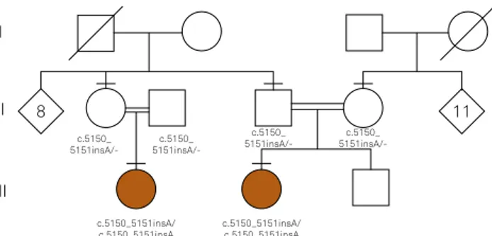
Both cousins were born to consanguineous parents (Figure 1) and reported multiple other consanguineous
marriages in the family. The surname related to consan-guinity was of Germanic background. The family lives in the countryside of Camaquã, a city with 63,000 inhab-itants, located in Rio Grande do Sul, the southernmost
state of Brazil, with mainly Azorean, Germanic and
Polish colonization. No other similar cases were reported in their family.
Case 1
Patient 1 was a 28-year-old woman presenting with an early-onset slowly progressive spastic-ataxic disorder.
Disease onset was probably in the irst or second year of life,
since she achieved independent walking at 20 months of age and never acquired a steady gait. Slurred speech started at the age of 10 and dysphagia was also reported in the fol-lowing years. On current neurological examination, she pre-sented with a spastic-ataxic gait; abnormal balance; moderate
lower limb spasticity; general hyperrelexia, except for absent Achilles relexes; bilateral Babinski sign; and mild limb ataxia. Other indings were broken-up smooth pursuit and
gaze-evoked nystagmus, dysarthria, pes cavus and hammertoes.
No cognitive deicit was observed.
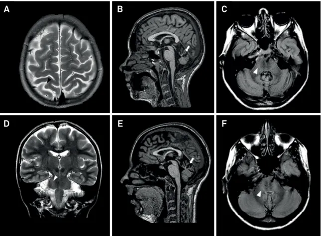
An MRI showed cerebellar vermis atrophy and T2-FLAIR central pontine linear hypointensities (Figure 2), raising the suspicion of an ARSACS diagnosis. Fundoscopy and optical coherence tomography showed thickening of the nerve iber retinal layer, compatible with hypermyelination (Figure 3). he electroretinogram was normal and pattern reversal visual
evoked potentials were prolonged bilaterally. Nerve conduc-tion studies revealed a length-dependent, predominantly sensory, axonal-demyelinating sensorimotor polyneuropathy.
Both upper and lower limb cortical somatosensory evoked
potentials were absent.
Case 2
Patient 2 was a 30-year-old woman also presenting with an early-onset, slowly progressive, spastic-ataxic disorder. She started independent walking at 13 months of age; frequent falls and gait difficulties were noticed at two years of age with progressive worsening thereafter. Progressive distal weakness in her lower limbs started at 15 years of age, resulting in foot drop. Neither significant
Figure 1. Simplified pedigree and genotypes.
c.5150_5151insA/ c.5150_5151insA
c.5150_5151insA/ c.5150_5151insA c.5150_
5151insA/-c.5150_
5151insA/-c.5150_
5151insA/-c.5150_
5151insA/-III
11 8
Figure 2. Brain MRI of ARSACS patients. Brain MRI of patient 1 (A-D) and patient 2 (E-F). A) Axial T2-weighted image showing mild parietal lobe atrophy (black arrowhead); B) sagittal T1-weighted image showing superior cerebellar vermian atrophy (white arrow); C) axial T2-FLAIR weighted image showing linear hypointensities in the pons (black arrows) and hyperintensities of the lateral pons merging into the middle cerebellar peduncles (white arrowhead); D) coronal T2-weighted image showing hyperintensities of the lateral pons merging into the middle cerebellar peduncles (white arrowhead); E) sagittal T1-weighted image showing superior cerebellar vermian atrophy (white arrow); F) axial T2-FLAIR weighted image showing linear hypointensities in the pons (black arrows) and hyperintensities of the lateral pons merging into the middle cerebellar peduncles (white arrowhead). FLAIR, fluid-attenuated inversion recovery.
A
B
C
D
E
F
Figure 3. Fundoscopy and optical coherence tomography of ARSACS patients. Ophthalmological evaluation of patient 1 (A-B) and patient 2 (C-D). A) and C) striated whitish aspect of the peripapillar retina suggestive of nerve fiber retinal layer hypermyelination on fundoscopy (white arrow); B) and D) increased thickness and reflectance of the nerve fiber retinal layer (black arrow) on optical coherence tomography.
A
B
dysphagia nor slurred speech were reported. A clinical picture very similar to Patient 1 was seen on neurologi-cal examination, with the addition of foot drop. Cognitive function was preserved.
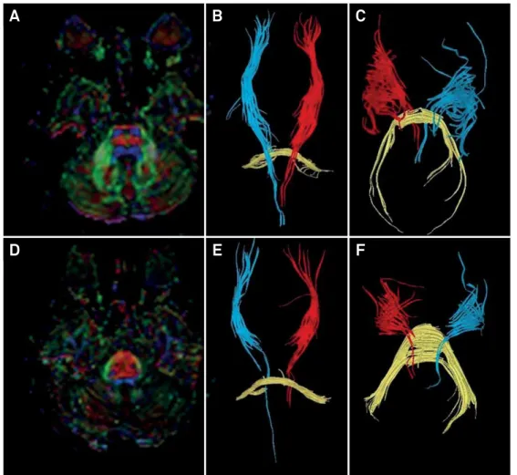
An MRI showed similar indings to Patient 1 (Figure 2). Additionally, tractography with difusion tensor imaging showed thick and displaced transverse pontine ibers with compressed corticospinal tracts in the pons (Figure 4).
Fundoscopy and optical coherence tomography showed thickening of the nerve iber retinal layer (Figure 3).
Electroretinogram and pattern-reversal visual evoked poten-tials were normal bilaterally. Nerve conduction studies revealed a length-dependent, predominantly sensory, axo-nal-demyelinating sensorimotor polyneuropathy. Cortical somatosensory evoked potentials were prolonged in upper, and absent in lower, limbs.
Diagnostic work-up
An extensive diagnostic work-up for autosomal reces-sive and dominant ataxic disorders was performed for
both cousins. Molecular analysis for Friedreich’s ataxia,
spinocerebellar ataxias type 1, 2, 3, 6, 7, 10, 12, 17, and dentatorubral-pallidoluysian atrophy were normal.
Normal blood levels of vitamin E, LDL and HDL choles -terols, triglycerides, alpha-fetoprotein, lactate, ceruloplas-min and immunoglobulins A, E, G and M; and normal leu-kocytes activities of arylsulfatase A, hexosaminidase A, galactocerebrosidase, beta-galactosidase and chitotriosi-dase, were found.
Their clinical pictures were highly suggestive of an autosomal recessive condition. Posterior fossa MRI and ophthalmological findings suggested an ARSACS
diag-nosis. The SACS sequencing revealed that Patients 1
and 2 were homozygous for the novel frameshift variant c.5151_5152insA, and that both pairs of parents were
het-erozygous for the variant (Figure 1). This DNA variant was absent from the Exome Aggregation Consortium and from
1000 genomes project databases, and it was classified as pathogenic according to the 2015 American College of
Medical Genetics and Genomics criteria18, confirming the
diagnosis of ARSACS.
Figure 4. Diffusion tensor imaging tractography in ARSACS. Fractional anisotropy images show the difference between a healthy individual (A-C) and patient 2 with ARSACS (D-F). While the healthy patient (A) has normal sized corticospinal (blue) and pontine transverse (red) tracts, patient 2 (D) has abnormally thickend pontine transverse tracts, leading to displaced and reduced corticospinal tract thickness. Tractography of the corticospinal tract (red and blue) and transverse pontine fibers (yellow) are shown in images B, C, E and F.
B
A
C
E
DISCUSSION
Autosomal recessive spastic ataxia of Charlevoix-Saguenay
was irst described in Quebec province, Canada. he inci -dence at birth of the disease in the Charlevoix-Saguenay region of Quebec is around 50/100,000 live born infants with
a carrier prevalence estimated to be 1/227,19, but it is largely
unknown in other countries. Since the identiication of sev -eral mutations in many populations, it is now recognized that ARSACS is not only limited to this region, but occurs
world-wide8-15. More than 100 SACS mutations have been described
to date. he majority of Quebecois harbor a homozygous sin
-gle nucleotide deletion (c.8844delT) due to a founder efect,
whereas about 4% of the total carrier frequency harbor the nonsense c.7504C > T, and additional mutations (including
four recurrent alleles) are less common20. In a few compound
heterozygous patients, this mutation was associated with
(5254C > T) nonsense mutation3.
he presence of certain brain MRI, retinal and nerve con -duction abnormalities in ataxic or spastic individuals might raise suspicion of an ARSACS diagnosis17,21,22,23,24,25.
Neuroimaging
At least three case series (64 patients in total) have
eval-uated qualitative brain MRI indings in ARSACS patients
born outside Quebec17,24,25. he MRI indings with greater
sensitivity for ARSACS diagnosis were predominant
supe-rior cerebellar vermis atrophy (range: 88–100%), T2-FLAIR
pontine linear hypointensities (range: 46–100%), enlarge-ment of middle cerebellar peduncles (range: 22–100%)
and T2-FLAIR hyperintensities of the lateral pons (range:
17–100%). As these studies only reported MRI abnor-malities in ARSACS patients, it was not possible to
esti-mate the speciicity of the indings. However, considering that T2-FLAIR pontine linear hypointensities were rarely
described for other diseases, this is probably the most
spe-ciic MRI sign of ARSACS.
Ophthalmological findings
Case series and reports have depicted low sensi-tivity (17-19%) of prominent myelinated retinal nerve fibers for an ARSACS diagnosis in patients born outside
Quebec21,24,25, except for a recent Italian study that reported
4/5 ARSACS patients with this finding22. Specificity of
ret-inal abnormalities for an ARSACS diagnosis, however, seems to be high, although not adequately addressed up to now. Importantly, some authors point out that ARSACS might be underdiagnosed due to erroneous fundoscopy interpretation as persistent myelination of the retina (present in 1–2% of normal population) and that optical coherence tomography might help to differentiate this pattern from thickening of peripapillar retinal fibers
typi-cal of ARSACS23.
Nerve conduction studies
At least two case series (46 patients evaluated with nerve conduction studies) evaluated peripheral nervous system
abnormalities in ARSACS patients born outside Quebec24,25.
Sensitivity of sensorimotor polyneuropathy ranged from 88-97%, with axonal involvement in 25%, demyelinating in
53% and both axonal and demyelinating in 14% of patients25.
herefore, peripheral neuropathy is another sensitive marker
for an ARSACS diagnosis. A demyelinating component of the sensorimotor peripheral neuropathy (present in 67–88% of cases) might help to distinguish ARSACS from other
com-mon recessive ataxias like Friedreich’s ataxia, POLG or ataxia
with oculomotor apraxia type 224.
ARSACS frequency worldwide, and in Brazil
Autosomal recessive spastic ataxia of Charlevoix-Saguenay has rarely been addressed in population stud-ies outside Quebec, in the context of autosomal recessive ataxias, spastic ataxias and hereditary spastic paraplegias.
he relative frequency of ARSACS varied from 0.3% of heredi -tary spastic paraplegia patients to 5% of early onset ataxia
patients from Germany24,26;and to 8% of spastic ataxia or
con-genital ataxia patients from multiple origins25. he ARSACS
frequency among ataxic or hereditary spastic paraplegia
families from Brazil is unknown.
he two individuals in the present report presented with
clinical, neuroimaging, electrophysiological and
ophthalmo-logical indings resembling the characteristics of the Quebec patients, which led to the suspicion of ARSACS. Before them, three Brazilian siblings presenting with a clinical diagnosis of typical ARSACS, without molecular conirmation, were
reported27. herefore, there is reason to expect other cases,
typical or atypical, in our population.
In conclusion, ARSACS is a spastic ataxic disorder, with worldwide distribution, being the second or third most com-mon cause of autosomal recessive ataxias in some series
out-side Quebec24. Brain MRI is a mandatory complementary
investigation for ataxic disorders of unknown diagnosis28
and ARSACS is one of the diseases where a speciic MRI pat
-tern may give rise to a correct diagnosis. Both ophthalmo -logical evaluation and nerve conduction studies should be routinely ordered for patients with ataxia for a better
pheno-typic characterization. he suspicion of persistent myelina
-tion of the retina in an ataxic patient should be conirmed
by optical coherence tomography; prominent myelinated
retinal nerve ibers on fundoscopy should lead to SACS
sequencing. A demyelinating or axonal-demyelinating sen-sorimotor polyneuropathy in an ataxic patient should also
raise ARSACS suspicion. Finally, atypical ARSACS cases
with Charcot-Marie-Tooth-like, or other complex pheno-types, might lack both MRI and fundoscopy abnormalities.
heir diagnosis may be made with a next-generation
References
1. Barbeau A, Bouchard R, Bouchard RW. Autosomal recessive spastic ataxia of Charlevoix-Saguenay. Can J Neurol Sci. 1978;5(1):61-9.
2. Bouchard JP, Richter A, Mathieu J, Brunet D, Hudson TJ, Morgan K et al. Autosomal recessive spastic ataxia of Charlevoix-Saguenay. Neuromuscul Disord. 1998;8(7):474-9. https://doi.org/10.1016/S0960-8966(98)00055-8
3. Richter A, Rioux JD, Bouchard JP, Mercier J, Mathieu J, Ge B et al. Location score and haplotype analyses of the locus for autosomal recessive spastic ataxia of Charlevoix-Saguenay, in chromosome region 13q11. Am J Hum Genet. 1999;64(3):768-75. https://doi.org/10.1086/302274
4. Engert JC, Doré C, Mercier J, Ge B, Bétard C, Rioux JD et al. Autosomal recessive spastic ataxia of Charlevoix-Saguenay (ARSACS): high-resolution physical and transcript map of the candidate region in chromosome region 13q11. Genomics. 1999;62(2):156-64. https://doi.org/10.1006/geno.1999.6003
5. Engert JC, Bérubé P, Mercier J, Doré C, Lepage P, Ge B et al. ARSACS, a spastic ataxia common in northeastern Québec, is caused by mutations in a new gene encoding an 11.5-kb ORF. Nat Genet. 2000;24(2):120-5. https://doi.org/10.1038/72769
6. Parfitt DA, Michael GJ, Vermeulen EG, Prodromou NV, Webb TR, Gallo JM et al. The ataxia protein sacsin is a functional co-chaperone that protects against polyglutamine-expanded ataxin-1. Hum Mol Genet. 2009;18(9):1556-65. https://doi.org/10.1093/hmg/ddp067
7. Bouhlal Y, Amouri R, El Euch-Fayeche G, Hentati F. Autosomal recessive spastic ataxia of Charlevoix-Saguenay: an overview. Parkinsonism Relat Disord. 2011;17(6):418-22. https://doi.org/10.1016/j.parkreldis.2011.03.005
8. Gerwig M, Krüger S, Kreuz FR, Kreis S, Gizewski ER,
Timmann D. Characteristic MRI and funduscopic findings help diagnose ARSACS outside Quebec. Neurology. 2010;75(23):2133. https://doi.org/10.1212/WNL.0b013e318200d7f8
9. Richter AM, Ozgul RK, Poisson VC, Topaloglu H. Private SACS mutations in autosomal recessive spastic ataxia of
Charlevoix-Saguenay (ARSACS) families from Turkey. Neurogenetics. 2004;5(3):165-70. https://doi.org/10.1007/s10048-004-0179-y
10. Grieco GS, Malandrini A, Comanducci G, Leuzzi V, Valoppi M, Tessa A et al. Novel SACS mutations in autosomal recessive spastic ataxia of Charlevoix-Saguenay type. Neurology. 2004;62(1):103-6. https://doi.org/10.1212/01.WNL.0000104491.66816.77
11. Criscuolo C, Saccà F, De Michele G, Mancini P,
Combarros O, Infante J et al. Novel mutation of SACS gene in a Spanish family with autosomal recessive spastic ataxia. Mov Disord. 2005;20(10):1358-61. https://doi.org/10.1002/mds.20579
12. Bouhlal Y, El Euch-Fayeche G, Hentati F, Amouri R. A novel SACS gene mutation in a Tunisian family. J Mol Neurosci. 2009;39(3):333-6. https://doi.org/10.1007/s12031-009-9212-9
13. Takiyama Y. Autosomal recessive spastic ataxia of Charlevoix-Saguenay. Neuropathology. 2006;26(4):368-35. https://doi.org/10.1111/j.1440-1789.2006.00664.x
14. Vermeer S, Meijer RP, Pijl BJ, Timmermans J, Cruysberg JR, Bos MM et al. ARSACS in the Dutch population: a frequent cause of early-onset cerebellar ataxia. Neurogenetics. 2008;9(3):207-14. https://doi.org/10.1007/s10048-008-0131-7
15. Baets J, Deconinck T, Smets K, Goossens D,
Van den Bergh P, Dahan K et al. Mutations in SACS cause atypical and late-onset forms of ARSACS. Neurology. 2010;75(13):1181-8. https://doi.org/10.1212/WNL.0b013e3181f4d86c
16. Martin MH, Bouchard JP, Sylvain M, St-Onge O, Truchon S. Autosomal recessive spastic ataxia of Charlevoix-Saguenay: a report of MR imaging in 5 patients. AJNR Am J Neuroradiol. 2007;28(8):1606-8. https://doi.org/10.3174/ajnr.A0603
17. Shimazaki H, Takiyama Y, Honda J, Sakoe K, Namekawa M, Tsugawa J et al. Middle cerebellar peduncles and Pontine T2 hypointensities in ARSACS. J Neuroimaging. 2013;23(1):82-5. https://doi.org/10.1111/j.1552-6569.2011.00647.x
18. Richards S, Aziz N, Bale S, Bick D, Das S, Gastier-Foster J et al. Standards and guidelines for the interpretation of sequence variants: a joint consensus recommendation of the American College of Medical Genetics and Genomics and the Association for Molecular Pathology. Genet Med. 2015;17(5):405-24. https://doi.org/10.1038/gim.2015.30
19. De Braekeleer M, Giasson F, Mathieu J, Roy M, Bouchard JP, Morgan K. Genetic epidemiology of autosomal recessive spastic ataxia of Charlevoix-Saguenay in northeastern Quebec. Genet Epidemiol. 1993;10(1):17-25. https://doi.org/10.1002/gepi.1370100103
20. Thiffault I, Dicaire MJ, Tetreault M, Huang KN,
Demers-Lamarche J, Bernard G et al. Diversity of ARSACS mutations in French-Canadians. Can J Neurol Sci. 2013;40(1):61-6. https://doi.org/10.1017/S0317167100012968
21. Hara K, Onodera O, Endo M, Kondo H, Shiota H, Miki K et al. Sacsin-related autosomal recessive ataxia without prominent retinal myelinated fibers in Japan. Mov Disord. 2005;20(3):380-2. https://doi.org/10.1002/mds.20315
22. Vingolo EM, Di Fabio R, Salvatore S, Grieco G, Bertini E, Leuzzi V et al. Myelinated retinal fibers in autosomal recessive spastic ataxia of Charlevoix-Saguenay. Eur J Neurol. 2011;18(9):1187-90. https://doi.org/10.1111/j.1468-1331.2010.03335.x
23. Desserre J, Devos D, Sautière BG, Debruyne P, Santorelli FM, Vuillaume I et al. Thickening of peripapillar retinal fibers for the diagnosis of autosomal recessive spastic ataxia of Charlevoix-Saguenay. Cerebellum. 2011;10(4):758-62. https://doi.org/10.1007/s12311-011-0286-x
24. Synofzik M, Soehn AS, Gburek-Augustat J, Schicks J, Karle KN, Schüle R et al. Autosomal recessive spastic ataxia of Charlevoix Saguenay (ARSACS): expanding the genetic, clinical and imaging spectrum. Orphanet J Rare Dis. 2013;8(1):41-53. https://doi.org/10.1186/1750-1172-8-41
25. Pilliod J, Moutton S, Lavie J, Maurat E, Hubert C,
Bellance N et al. New practical definitions for the diagnosis of autosomal recessive spastic ataxia of Charlevoix-Saguenay. Ann Neurol. 2015;78(6):871-86. https://doi.org/10.1002/ana.24509
26. Schüle R, Wiethoff S, Martus P, Karle KN, Otto S, Klebe S et al. Hereditary spastic paraplegia: clinicogenetic lessons from 608 patients. Ann Neurol. 2016;79(4):646-58. https://doi.org/10.1002/ana.24611
27. Pedroso JL, Braga-Neto P, Abrahão A, Rivero RL, Abdalla C, Abdala N et al. Autosomal recessive spastic ataxia of Charlevoix-Saguenay (ARSACS): typical clinical and neuroimaging features in a Brazilian family. Arq Neuropsiquiatr. 2011;69(2B):288-91. https://doi.org/10.1590/S0004-282X2011000300004