R E S E A R C H A R T I C L E
Open Access
Alterations of protein expression in conditions of
copper-deprivation for
Paracoccidioides lutzii
in
the presence of extracellular matrix components
Haroldo Cesar de Oliveira
1, Julhiany de Fátima da Silva
1, Marcelo Teruyuki Matsumoto
1, Caroline Maria Marcos
1,
Roberta Peres da Silva
1, Rosângela Aparecida Moraes da Silva
1, Mônica Teresa Veneziano Labate
2,
Carlos Alberto Labate
2, Ana Marisa Fusco Almeida
1and Maria José Soares Mendes Giannini
1*Abstract
Background:Paracoccidioidesspp is a fungi genus and the agent of paracoccidioidomycosis. The strategies of infection used by these pathogens involve the expression of proteins related to adaptation to the host, particularly regarding the uptake of micronutrients. This study analyzed the adhesion ofParacoccidioides lutziiduring conditions of copper (Cu) and iron (Fe) deprivation, while also evaluating the proteins expressed in conditions of Cu depletion in the presence of four extracellular matrix (ECM) components (laminin, fibronectin and types I and IV collagen).
Results:We cultured theP. lutziiin a chemically defined media without Cu and Fe. The fungus was then placed in contact with different ECM components and adhesion was evaluated. A significant increase in binding to all ECM components was observed when the fungus was cultured without Cu; which might be related to some adhesins expression. A proteomic assay was developed and revealed 39 proteins expressed that are involved in processes such as virulence, protein synthesis, metabolism, energy, transcription, transport, stress response and the cell cycle when the fungus was interacting with the ECM components. The up-regulated expression of two important adhesins, enolase and 14-3-3, was observed at the fungal cell wall during the interaction with the ECM components, indicating the role of these proteins in theParacoccidioides–host interaction.
Conclusions:This study is important for determining prospective proteins that may be involved in the interaction ofParacoccidioideswith a host. Understanding the adaptive response to different growth conditions, elucidating the processes of adhesion and cell invasion, and identifying the proteins that are differentially expressed during the fungus-host interaction may help elucidate mechanisms used for survival and growth ofParacoccidioidesin various human tissues.
Keywords:Paracoccidioidesspp, Copper, Adhesion, Protein expression, Paracoccidioidomycosis
Background
Paracoccidioides lutzii, a specie from a complex genus [1-4], are dimorphic fungi and the etiologic agents of para-coccidioidomycosis (PCM), which is the most important systemic mycosis in Latin America [5].Paracoccidioidesis a notably versatile pathogen, with the ability of infecting numerous systems and organs of the human body,
because it has developed mechanisms that enable adher-ence and invasion of host tissues [6].
TheParacoccidioidesspecies initiates host infection by adhering to components of the extracellular matrix (ECM); this adherence is mediated by a variety of adhe-sins on the fungal surface, with component recognition of adhesive matrix molecules, which plays an important role in the regulation of cell adhesion, differentiation,
migration and proliferation [7]. Some Paracoccidioides
adhesins have been described and are included in micro-organism strategies of evading the immune system and ensuring survival in hosts. Adhesion is closely associated
* Correspondence:[email protected]
1Departamento de Análises Clínicas, Faculdade de Ciências Farmacêuticas,
UNESP - Univ Estadual Paulista, Laboratório de Micologia Clinica, Rodovia Araraquara-Jaú, Km 1, Araraquara, SP, Brazil
Full list of author information is available at the end of the article
with the transcriptional control of several regulatory pathways that control the synthesis of these molecules. These pathways are activated in response to various con-ditions, such as nutrient limitation [8], which is vital for any pathogen.
To successfully colonize a host, Paracoccidioides must initially adhere to host tissues and simultaneously obtain essential nutrients for growth and survival. Iron (Fe) and copper (Cu) are required for survival, primarily due to their roles as cofactors for many essential metabolic func-tions. Cu is an essential micronutrient for all biological systems, with multiple proteins requiring one or more atoms of Cu to achieve the appropriate structure and function. To prevent the consequences of Cu deficiency, living organisms have evolved molecular mechanisms that regulate the uptake, intracellular traffic, storage and efflux of Cu. Some of the cellular responses to variations in Cu levels are related to changes in the expression of genes en-coding the molecular components of Cu metabolism. Cu serves as a catalytic and structural cofactor for enzymes involved many processes, including energy generation, Fe acquisition, oxygen transport and cellular metabolism [9]. Both the host and fungi have developed sophisticated strategies for acquiring the metals, even under conditions of limited availability. Several homeostatic mechanisms have been demonstrated in fungi, guaranteeing the main-tenance of sufficient concentrations of Cu for cell growth without causing damage. Additionally, posttranslational mechanisms, such as the intracellular trafficking of Cu transporters, have been identified in mammals. In these organisms, Cu homeostasis is also mediated by the tran-scriptional regulation of genes involved in Cu acquisition, mobilization, and sequestration [10,11].
During the infection process, the levels of free Fe and Cu are significantly reduced; the acquisition of these nu-trients is related to a higher adaptive process that is im-portant for microorganism virulence, as demonstrated in
several organisms. In Candida albicans, increased gene
expression was observed during Fe privation related to virulence, such as secreted hydrolase genes [12]. Simi-larly, mutations in the genes involved in Fe capture, such
as the Fe oxidase gene in C. albicans [13] and the Fur
gene from Helicobacter pylori [14], rendered these
mi-croorganisms incapable of colonizing host tissues and
causing infection. In Cryptococcus neoformans, the
in-creased expression of Cu transporters is related to the dissemination of the species in the host meninges [15]. InParacoccidioides, low Fe conditions have been associ-ated with fungal susceptibility to the antimicrobial action of monocytes [16]. The administration of exogenous Fe results in an increased fungal load in mouse tissues in-fected withParacoccidioides[17].
Proteomic methods have been used to study the biology of Paracoccidioides.Historically, studies have focused on
dimorphism and the characterization of single or a few protein targets in Paracoccidioides species [18-20], with the obtained information constituting a useful resource
for studying the dimorphism of Paracoccidioides [21].
Parenteet al. [17] used 2-DE to identify proteins related toParacoccidioidessurvival in a Fe-deficient environment. They discovered that, during Fe starvation, fungi use the glycolytic pathway to obtain energy instead of the oxida-tive pathway, which is dependent on enzymes containing Fe-S groups. In addition, those authors identified activa-tion of the Fe uptake systems as an indispensable survival mechanism required by the fungus in this type of
environ-ment [17]. Vallejo et al. [22] studied the secretome of
Paracoccidioides with regards to the fungal extracellular vesicles that are able to cross the cell wall and transport molecules that facilitate nutrient acquisition, cell defense, and modulation of the host defense machinery. In another
study, those authors determined that Paracoccidioides
shared secreted proteins amongHistoplasma capsulatum,
C. neoformans and Saccharomyces cerevisiae [23]. The
mycelia and yeast cell secretomes ofParacoccidioideshave also revealed that many proteins do not use the classical secretory pathway, while many other proteins likely exerted other activities, once secreted. Paracoccidioides data has indicated that it uses non-classical targeting mechanisms to direct protein export and also that it has molecules that can function as virulence factors [24]. De Arruda Grossklaus et al. [25] also studied the oxidative
stress response of Paracoccidioides using proteomic
analysis.
Our aim was to analyze the adhesion of
Paracocci-dioides lutzii(Pb01 isolate) in Cu and Fe depleted condi-tions, while also determining the proteins that may be
involved in the interaction among Paracoccidioides and
ECM components that lead to an increase in fungal ad-hesion when the fungus was deprived of Cu. These pro-teins are involved in distinct biological processes, such as the cell cycle, stress response and Fe transportation; additionally, several proteins have been described as virulence factors and possible candidates for Paracocci-dioides virulence. The results of this study provide new knowledge that may facilitate strategies used by Paracoc-cidioides to successfully parasitize host tissues, leading to the development of paracoccidioidomycosis.
Results
Adhesion ofParacoccidioides lutziito ECM components
during Cu and Fe depletion
Following incubation in MVM without Cu (MVM-W-Cu medium), theP. lutzii yeast phase presented a significant increase (p < 0.05) in adhesion (Figure 1) in the presence of all tested ECM components (laminin, fibronectin, and types I and IV collagen), while in MVM without Fe
(p < 0.05) in adhesion in the presence of laminin, fibronec-tin and type IV collagen and an increase in the presence of type I collagen, but this increase was significantly lower (p < 0.05) than the increase observed under Cu depletion.
Adhesin expression profile during Cu and Fe depletion in the presence of ECM components
The expression of genes that encoded most of the
known Paracoccidioidesadhesins during the interaction
with the host ECM components (laminin, fibronectin and types I and IV collagen) were analyzed. We used real-time PCR to determine whether the metal depletion modulated adhesin expression. As shown in Figure 2, we observed that different micronutrient depletion condi-tions led to different levels of adhesin expression.
Cu depletion led to increased expression of the main adhesins, except for gp43 and malate synthase (MLT), whereas Fe depletion led to decreased expression in all
studied adhesins. These effects corroborate the results of the adhesion experiments and revealed the importance
of these proteins to the Paracoccidioides-host
inter-action, in which increased and decreased adhesion may be related to adhesin expression.
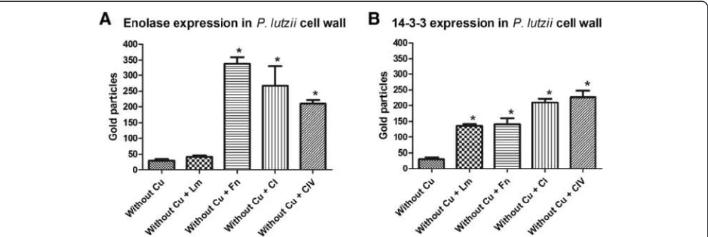
Immunogold expression analysis of enolase and 14-3-3 proteins inP. lutziiyeast cells during interaction with ECM
components after micronutrient depletion
Figure 2P. lutziiadhesins expression profile during interaction with laminin (A), fibronectin (B), collagen type I (C), and collagen type IV (D) after Cu and Fe depletion.
cells with and without Cu in contact with all the ECM components were processed by post embedding with gold particles. The immunocytochemistry assays re-vealed a ubiquitous distribution of the gold particles in all conditions, but we observed that, when the fungus was grown under simulated infection conditions, there was an increased expression of enolase and 14-3-3 (indi-cated with arrows in Figures 3 and 4) in the fungal cell wall. A quantitative analysis was developed by counting the number of expressed enolase and 14-3-3 proteins in the fungal cell wall and a real increase of protein expres-sion occurred in the cell wall during its interaction with all ECM components (Figure 5). These results showed that these proteins, when interacting with the host structures, are recruited to the cell wall and may be used during this interaction, as shown by da Silva et al. [26] and Marcos et al. [27].
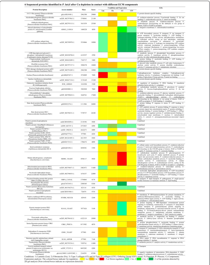
Paracoccidioides lutziiprotein expression identification by
mass spectrometry during Cu deprivation in the presence of different ECM components
The protein profile ofP. lutziigrown in an environment depleted of Cu was used as a master gel for comparison after contact with the ECM components (laminin, fibro-nectin, type I and type IV collagen). Based on these ana-lyses, differentially expressed proteins were selected for identification by mass spectrometry (MS), as well as the
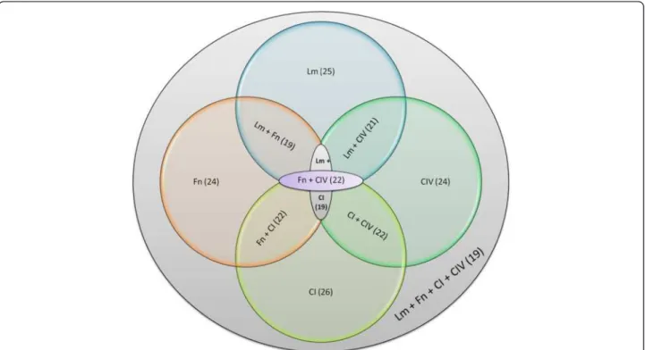
results of the expression analysis. The results indicate that the fungus uses an arsenal of proteins during its interaction with the host ECM components (Figure 6). This arsenal is composed of proteins that are related to different cell processes (Figure 7) and an extensive num-ber of proteins that may be involved with the interaction between the fungus and the host. Additionally, some of the identified proteins are involved in metabolism and the transport of micronutrients, such as Fe and Zinc (Zn), revealing the adaptation of the fungi to the envir-onment. We also observed proteins that are involved in DNA repair and heat shock proteins that are induced as a response to the stress that this fungus was submitted to during Cu starvation. The contact of the fungus with different ECM components led to the different re-sponses, forcing the pathogen to adapt to the environ-ment. The results found during this interaction with each of the different ECM components vary and these proteins can be shared to interact with the different components. We observed that 19 proteins are shared during the interaction with all ECM components, while
21 are common between the interaction ofP. lutzziwith
Lm and Fn, 19 with Lm and CI, 21 with Lm and CIV, 22 with Fn and CI, 22 with Fn and CIV and 22 with CI and CIV, as summarized in Figure 8. Comparative proteomic
analysis with P. lutzii maintained in a medium
supple-mented with Cu warrants future study.
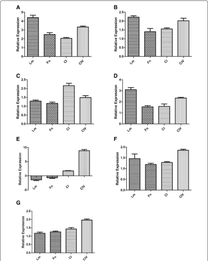
Real-time PCR to validate 2D-E assay results
Six of the proteins identified when using the proteomic approach were chosen to validate the regulation of gene expression by real-time PCR. The chosen genes for this evaluation were enolase, 14-3-3, aldolase, GAPDH, serine/ threonine-protein kinase CBK1, siderophore-Fe trans-porter ENB1 and vesicle-mediated transport-related pro-tein. These genes were selected based on their importance
in interactions between Paracoccidioides and the host.
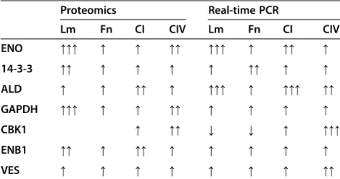
This analysis confirmed a regulation of the expression of the proteins identified using proteomics and showed that Cu depletion led to a regulation at the transcriptional levels of the proteins, since the observed gene expression levels were totally correspondent with the protein expres-sion levels (Figure 9). Table 1 summarizes these results.
In silicoanalysis of putative proteins identified in 2D gel
analysis
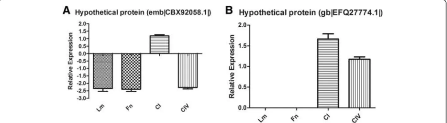
We observed 8 proteins that had unknown functions and made functional inferences using in silico analysis. It is known that surface proteins are important for Paracocci-dioides pathogenesis, so we performed in silico analysis to identify adhesin-like proteins using FaaPred software, which predicts fungal adhesins and adhesin-like proteins. This ana-lysis predicted 2 proteins as adhesin-like proteins (Table 2), revealing interesting candidates for further studies of the protein relationships withParacoccidioidesvirulence.
The expression of these adhesin-like proteins was eval-uated using real-time PCR during the interaction of
P. lutzii with the host ECM components (Figure 10).
These results demonstrated that proteomic assays could identify unknown molecules that may be related to the adhesion ofParacoccidioides.These results allow for the identification of some adhesin-like proteins that may be important for fungal adhesion to different ECM compo-nents in different sites of the host organism and deter-mining the contributions of these proteins to fungal establishment.
Discussion
All organisms most likely depend on efficient metal transport systems for survival. Fe and Cu are particularly important metals because they participate in vital reac-tions and are both cofactors of many metabolic enzymes and are essential nutrients for the maintenance of life [28]. In excess, these metals can be toxic; however, dis-turbances to the Fe and Cu levels can have serious ef-fects on cellular metabolism, growth and development [29]. Cu exists as a trace element, and is found in low concentrations in living organisms. However, Cu is an important catalytic cofactor that guides several import-ant biological processes that are essential for life. Cu reg-ulates changes in protein structure, catalytic activity and protein-protein interactions. In this manner, it controls a varied series of biochemical events; Cu may modulate critical fungal virulence factors [9].
The importance of our work lies precisely in the fact that Paracoccidioides is a facultative intracellular patho-gen. Because of this characteristic, any external changes in the intracellular environment can directly influence the pathogenicity of this organism. Additionally, the period evaluated and ECM components used in this work (lam-inin, fibronectin and collagen) are essential in the initial interaction of the pathogen with the host, thus any change of homeostasis (such as deprivation of micronutrients) would have a direct impact on this initial interaction and, consequently, virulence.
In our study, the depletion of Cu altered the adhesion
pattern of Paracoccidioides to ECM components. There
are important components in the Paracocccidioides-host interaction. When the fungus was grown in Cu-depleted conditions, overexpression of genes encoding adhesins was observed, whereas down-regulation of the same genes was observed during Fe-depletion. Complementing these results by using immunogold analysis, we clearly observed an increased expression of two important adhesins, eno-lase and 14-3-3 protein, when the fungus was in an
environment depleted of Cu and in contact with different ECM components. In addition, we observed that this in-crease occurred in the fungus cell wall, suggesting that the intensification of adhesion may be attributable to the great quantity of these adhesins in the fungal cell wall.
The identification of proteins involved in the initial phase of host-fungus interactions is very important and can result in the identification of potential drug targets for future therapeutic research to prevent the early in-fectious process. The 2-DE technique was used in this study to determine proteins that may be involved in the
interaction among Paracoccidioides and ECM
compo-nents when the fungus was deprived of Cu, leading to an increase in the adhesion of the fungi.
Our goal with the proteome was to understand what makes the fungus increase its capacity to adhere to all ECM components after Cu deprivation, which is why our main control was only the fungi maintained without Cu. All of the proteins identified in these results are expressed during Cu depletion, but they suffer modifica-tions in expression because of the contact of the fungi with the host.
Our proteomic assays identified 39 proteins expressed by the fungi after Cu depletion and during its interaction with the ECM components. These results contribute to
our knowledge of the molecular arsenal used by
Para-coccidioides to adhere, invade and cause systemic
mycosis.
Figure 8Proteins identified by Venn diagram, which interact with the different ECM components: overview of the number of the proteins differentially expressed byP. lutziiduring its interaction with the host components.
Among the identified proteins, some of them were already identified as being important to the interaction
of Paracoccidioides spp. with the host and could be
associated with dissemination of the infection. These proteins are 14-3-3, enolase, aldolase and glyceraldehy3-phosphate dehydrogenase; all of which are currently de-scribed as adhesins [26,27,30-32]. These adhesins have their expression up-regulated while interacting with the host with all ECM components. These results, once again, reveal the importance of these molecules in the inter-action ofP. lutziiwith the host. The experiment indicates that, during Cu starvation, these molecules are maintained at basal levels of expression, but contact with the host sig-naled the fungus to increase the expression of these pro-teins to adhere to the host and, from the host, obtain the necessary Cu for maintenance. With these results, we can understand how the fungus uses its protein arsenal to adapt to the host and have success during the infection process.
Increased enolase expression was verified; this protein was recently described as a fibronectin ligand related to Paracoccidioides adhesion and is considered a virulence factor [31]. Recent studies have shown that enolase has different distributions and biological functions [33] and is expressed at different sites in eukaryotes and prokaryotes
with distinct biological functions, such as the laminin lig-and inStaphylococcus aureus[34] and enolase binding of
human plasminogen in Trichomonas vaginalis [35],
Streptococcus pneumoniae [36], Leishmania mexicana
[37] andFasciola hepatica[38]. In our study, enolase was differentially expressed when the fungus was in contact with all of the ECM components, not only fibronectin, as previously described by Donofrioet al.[30]. However, it is worth noting that the strain used in the latter work was a Paracoccidioides brasiliensis strain named Pb18, which could be an important factor for further investigation [31]. Nevertheless, data from Nogueiraet al.[20] also demon-strated binding of enolase to laminin and type I collagen, corroborating the findings of this study. Moreover, data from Nogueiraet al.[20] also indicates that enolase is an importantParacoccidioidesmolecule used during its inter-action with the host. The expression of this protein in this present study corroborates earlier data suggesting that Paracoccidioides enolase can be used to adhere to and, perhaps, invade host cells through an interaction with hu-man plasminogen [31,33,36,38]. The ability to bind plas-minogen has been associated with invasive properties in pathogenic microorganisms [37,39,40] and can facilitate their penetration to the basement membrane and permit associations with fibronectin and laminin. Although Para-coccidioidesis considered a facultative intracellular patho-gen, fibronectin adhesin ligands can also mediate cellular invasion. Our electron microscopy results demonstrated an increase of enolase during the interaction between the fungus cell and the ECM components, mainly at the cell wall. These current results corroborate those of Marcos et al.[27], who reported a substantial increase of enolase in the cell wall during infection of pneumocytes (A549 cells), suggesting the importance of this protein in fungal adhesion to the host [41].
We also observed the overexpression of the 14-3-3 pro-tein when the fungus was in contact with all of the ECM components studied. The 14-3-3 proteins are a family of highly conserved acidic dimeric proteins that have been implicated in a variety of cellular processes in eukaryotes
[41-44]. In Paracoccidioides, a 30-kDa protein has been
Table 1 Comparison on the expression levels of the identified proteins and its coding genes
Proteomics Real-time PCR
Lm Fn CI CIV Lm Fn CI CIV
ENO ↑↑↑ ↑ ↑ ↑↑ ↑↑↑ ↑ ↑↑ ↑
14-3-3 ↑↑ ↑ ↑ ↑ ↑ ↑↑ ↑ ↑
ALD ↑ ↑ ↑↑ ↑ ↑↑↑ ↑ ↑↑↑ ↑↑
GAPDH ↑↑↑ ↑ ↑ ↑↑ ↑ ↑ ↑ ↑
CBK1 ↑ ↑↑ ↓ ↓ ↑ ↑↑↑
ENB1 ↑↑ ↑ ↑↑ ↑ ↑ ↑ ↑ ↑
VES ↑ ↑ ↑ ↑ ↑ ↑ ↑ ↑↑
Upregulation is represented by↑and downregulation is represented by↓and the number of arrows indicates the identified levels of expression (1 arrow - 1 to 2, 2 arrows - 2 to 3 and 3 arrows - 3 or more or less folds expressed when compared with the control).
Table 2In silicoanalysis of sequenced putative proteins using the software FaaPred
Protein Access number Prediction SVM Score
identified and characterized as a 14-3-3 protein that causes structural modification of polymerized actin micro-filaments and cytokeratin, induces apoptosis when inter-acting with epithelial cells and is capable of binding to laminin [45,46]. In addition, da Silva et al. [26] demon-strated that, during whenParacoccidioidesinteracted with A549 cells, an apparent increase of the 14-3-3 protein on the cell wall of the fungus occurs, suggesting that this pro-tein may be involved in host-parasite interactions.
Glyceraldehyde-3-phosphate dehydrogenase (GAPDH) and fructose 1,6-biphosphate aldolase (FBA) (part of the glycolytic pathway) were observed to be overexpressed dur-ing the interaction ofParacoccidioideswith all of the ECM components studied. Barbosaet al.[31] demonstrated that
GAPDH fromParacoccidioides is able to bind to laminin,
fibronectin and type IV collagen and the inhibition of this protein causes decreased adhesion rates of the fungus to epithelial cells [31]. FBA appears to be important in host-parasite interactions [47,48]. A FBA homologue was
previously described as an immunogenic protein of
Para-coccidioides [18]. Additionally, this fungus contains two genes encoding two different Class II FBAs. Phylogenetic analysis supports the concept of gene duplication for FBA genes, constituting a two-member family whose function could differ in the fungal cells. In addition, expression ana-lysis performed using northern blot and RT-PCR indicated a differential expression for Pbfba1 and Pbfba2 in Paracoc-cidioides cells, suggesting distinct functions for both pro-teins [32]. An interesting and unsuspected feature observed during this present work is that bothParacoccidioidesFBAs appeared to play roles in the fungal interaction with the host. PbFBA1 has immunogenic properties, as indicated by the fact that the native protein was recognized by the sera of infected patients [18].
Under conditions of Cu depletion, we observed an in-crease in the expression of ENB1, the enterobactin Fe transporter, a high-affinity siderophore in microbial sys-tems. This increase may have occurred because most
Fe-acquisition systems are Cu dependent, and Cu deficiency may force the fungi to use the siderophore system to ac-quire Fe from the culture media [49]. The siderophores are generally used by microorganisms in situations of low Fe concentrations. However, Froissardet al.[49] reported that ENB1 appears to be constitutively expressed in the plasma membrane of S. cerevisiae, and its expression is not affected by the substrate concentration.
Another protein that exhibited increased expression was CBK1, a serine/threonine protein kinase of the RAM signaling network. Cbk1p is similar to the human myotonic dystrophy kinase and is essential for normal morphogenesis in S. cerevisiae. It is involved in regulat-ing cellular morphogenesis, polarized growth, and septum destruction. Cbk1p activity is regulated by both phosphorylation and specific localization; the protein re-locates to the cytoplasm upon DNA replication stress [50]. Cbk1 has been previously implicated in regulating polarized morphogenesis, gene expression and cell integ-rity inS. cerevisiaeand is also critical for heat shock and cell wall stress signaling. These results obtained usingS. cerevisiae constitute the first evidence that Cbk1 kinase regulates MAPK-dependent stress signaling and provides mechanistic insight into Sdp1 phosphatase regulation via Bck2, a protein associated with the Pkc1-Mpk1 cell in-tegrity pathway [51]. In C. albicans,Cbk1 is a daughter cell protein involved in cell wall degradation from the daughter side during cell separation [52]; recently, this
protein was observed to be involved inC. albicans
bio-film formation [53]. In this current work, we verified that Cu depletion promotes stress and cellular changes
in the cell wall composition of Paracoccidioides. In
addition, the increased expression of all previously
de-scribed Paracoccidioidesadhesins and the increased
ex-pression of kinases may suggest that remodeling of the Paracoccidioidescell wall could occur through activation of the RAM pathway. This mechanism should be investi-gated in future studies.
The identification of a vesicle-mediated transport-related protein overexpressed during the interaction of Paracocci-dioides with all of the ECM components is an interesting finding because Vallejo et al. [54] recently described the presence of these structures in Paracoccidioides, including antigenic molecules, that are recognized by total sera from PCM patients. Vallejo et al. [22] also described the prote-omic analysis of extracellular vesicles and vesicle-free re-leased proteins in Paracoccidioides and demonstrated that these vesicles carry different molecules, including virulence factors previously described for Paracoccidioides. It is im-portant to emphasize that we observed molecules such as GAPDH, enolase, aldolase, 14-3-3 and malate dehydrogen-ase in the current study [23].
In our proteomic results, we also observed several puta-tive proteins with currently unknown functions.
There-fore, an in silico analysis was performed to identify
sequences that may play important roles as adhesins dur-ing the Paracoccidioides-host interaction. For this pur-pose, the FaaPred software [55] was used to predict fungal adhesins and adhesin-like proteins. In our analysis, we predicted 2 proteins that might be adhesin-like proteins. Although these are preliminary results, this type of analysis demonstrates that a considerable portion of the arsenal of molecules used byParacoccidioideswhile inter-acting with the host might still be unknown. These find-ings promote the need for further studies characterizing new virulence factors of this fungus.
Conclusions
Understanding the adaptive response to different growth conditions, elucidating the processes of adhesion and cell invasion, and identifying the genes that are differen-tially expressed during the parasite-host interaction may help elucidate the mechanisms used for survival and
growth of Paracoccidioidesin various human tissues. In
our study, we observed that proteins related to virulence were expressed against all ECM components, which might explain the increase in fungal adhesion. The fungus-host interaction includes a complex network of signaling pathways that are interconnected with regards to protein expression in a given situation or set of condi-tions. One goal of this study was to identify the
differen-tially expressed proteins of Paracoccidioides when in
contact with extracellular matrix components. In this sense, the 2-DE technique was extremely useful and pro-vided an overview of the cellular components at the par-ticular moment of contact.
Methods
Ethics statement
The 14-3-3 and enolase rabbit preimmune serum used in this study was obtained from two previous studies, Silva et al. [26] and Donofrio et al. [31] with the approval
of the Ethics Committee on Animal Experiments of the
Faculty of Pharmaceutical Sciences of Araraquara –
UNESP, Processes Protocols 10/2011/CEUA/FCF and 08/2001/CEUA/FCF respectively. The experiments in these previous studies were performed in strict accord-ance with Brazilian Federal Law 11,794 that established procedures for the scientific use of animals and the state law establishing the Animal Protection Code of the State of São Paulo. All efforts were made to minimize suffer-ing by the animals used in the current study.
Strain and culture conditions
The Paracoccidioides lutzii 01 strain (ATCC MYA-826)
was used in all experiments. The yeast phase was main-tainedin vitroin Fava-Netto’s medium [56] for 7 days at 36°C. For the adhesion assay, the fungus was incubated under three conditions: (1) complete McVeigh-Morton
(MVM) medium [57,58] containing glucose 1%, KH2PO4
11 mM, MgSO4.7H20 2 mM, CaCl2.2H20 1 mM, (NH4)
2SO415 mM, L-Asparagine 0.02%, L-Cystine 0.002%,
vita-min supplements 1%, and trace element supplement 0.1%, (2) MVM without Cu reagent (MVM-W-Cu) and (3) MVM without Fe reagent. Moreover, bathocuproinedisul-fonic acid (BCS) (Sigma-Aldrich, St. Louis, MO, USA) and bathophenanthroline disulfate (BPS) (Sigma-Aldrich, St. Louis, MO, USA) were used to chelate the Cu and Fe ions,
respectively. The fungus maintained in Fava-Netto’s
medium was transferred to the three conditions described above and incubated for 3 h at 37°C.
Fungal adherence assays
ECM proteins, represented by laminin, fibronectin, and types I and IV collagen (Sigma-Aldrich, St. Louis, MO, USA), were immobilized on 24-well microtiter plates at 50μg/ml diluted in carbonate-bicarbonate buffer (pH 9.6). The plates were incubated for 1 h at 25°C, incubated over-night at 4°C and washed with PBS. The fungal suspensions were washed again and suspended in PBS. Suspensions
(500 μL) of yeast containing 106 cells/ml (0.5 OD at
Expression analysis of enolase and 14-3-3 proteins in
Paracoccidioides lutziiyeast cells when interacting with
ECM components after micronutrient depletion using immunogold labeling
To examine the expression of the enolase and 14-3-3 pro-teins ofP. lutzii, we performed immunocytochemistry at the ultra-structural level using immunogold labeling. For each experiment, a suspension ofP. lutziiyeast cells without Cu and P. lutziiwithout Cu in the presence of ECM compo-nents (108cells/ml) were fixed (2.5% v/v glutaraldehyde in 0.1 M sodium cacodylate buffer, pH 7.2) for 24 h at 4°C and sent to the electron microscopy service of the Institute of Biomedical Sciences (ICB-I) USP-SP for preparation of ul-trathin sections. After fixation, the cells were rinsed several times using the same buffer, and free aldehyde groups were quenched with 50 mM ammonium chloride for 1 h, followed by block staining in a solution containing 2% (w/v) uranyl acetate in 15% (v/v) acetone for 2 h at 4°C. The ma-terial was dehydrated in a series of ascending concentrations of acetone (30 to 100% v/v) and embedded in LR Gold resin (Electron Microscopy Sciences, Washington, Pa.). The ultra-thin sections were collected on nickel grids, preincubated in 10 mM PBS containing 1.5% (w/v) bovine serum albumin (BSA) and 0.05% (v/v) Tween 20 (PBS-BSA-T). The sections were subsequently incubated overnight with a polyclonal antibody against the enolase and 14-3-3 proteins (diluted 1:50). After washing with PBS-BSA-T, the grids were incu-bated overnight with the labeled secondary antibody (rabbit IgG, Au conjugated, 10 nm; diluted 1:10). Controls were in-cubated with rabbit pre-immune serum at 1:50, followed by incubation with the labeled secondary antibody. After incu-bation, the grids were washed with the buffer described above, followed by a wash with distilled water, and staining with 3% uranyl acetate (w/v) and 4% lead citrate (w/v). Fi-nally, the grids were observed using a Jeol 1010 transmission electron microscope (Jeol, Tokyo, Japan). Using the software, Image Tool v. 3.00 [59], 10 different cells of each tested situ-ation were selected and the proteins marked with gold parti-cles were counted, providing quantitative analyses of the expression of this protein when interacting with the ECM components after Cu depletion.
RNA isolation and cDNA synthesis
Total RNA was extracted using the following conditions: (1) P. lutziiyeast cells maintained in MVM without Cu; and (2) yeast cells cultivated in MVM without Cu, incubated for 3 h at 37°C followed by the addition of the different ECM com-ponents (laminin, fibronectin, and types I and IV collagen) at 50 μg/ml, and incubated for an additional 2 h at 37°C. These preps were used for the Real Time PCR assays.
Total RNA was extracted from all experimental condi-tions using Trizol reagent (Invitrogen Life Technologies, Carlsbad, CA). First-strand cDNA synthesis was performed using reverse transcriptase (RevertAid™ H Minus Reverse
Transcriptase, Fermentas Life Sciences, Canada) and 1μg of total RNA. First-strand cDNA was used as a template to synthesize the second-strand of cDNA using the SMART PCR cDNA synthesis kit (Clontech Laboratories, Palo Alto, CA, USA).
Proteomic analysis ofParacoccidioides lutziiprotein
expression during Cu deprivation in the presence of different ECM components
The total protein extracts of P. lutzii were obtained in the yeast phase under different conditions, as shown in Table 3, to isolate and characterize the proteins differen-tially expressed in the different conditions.
Cells fromP. lutziiin the yeast phase submitted to the dif-ferent conditions (Table 3) were centrifuged (5000gfor 10 min) and washed 3–5 times with cold water to remove the
culture medium. Then, 10 mM Tris–HCl and protease
in-hibitors (1 mM pepstatin, 1 mM leupeptin, 1 mM aprotin, 1 mM antipain, 1 mM chymostatin and 1 mM PMSF) were added to the pellet, which was subsequently homogenized with liquid nitrogen, macerated with glass beads and vor-texed for 30 min. This preparation was then centrifuged for 45 min at 13000g, and the supernatant collected. The pro-tein concentration of the extracts was quantified using the Bradford Assay [60] (Bio-Rad Laboratories, California, USA). The samples were then analyzed by SDS-PAGE. The protein components of the extracts were subjected to iso-electric focusing using EttanIPGphor 3 (GE Healthcare, Buckinghamshire, UK). The second dimension, performed to separate proteins according to molecular weight, was conducted in a 12.5% polyacrylamide gel according to Laemmli et al. [61]. The gels were stained with Coomassie Brilliant Blue G-350 [62]. The data analysis was performed using Image Master 2D Platinum software (GE Healthcare, Buckinghamshire, UK) to compare the protein profile of the P. lutziidepleted of Cu before and after the fungus came into contact with the different ECM components. The 2D Platinum software analyzes the volumes of the spots and determines how much the expression of the selected spots were increased or decreased when compared with the control (P. lutziiwithout Cu) and our tests (P. lutzii with-out Cu in contact with the different ECM components)”.
Table 3 Conditionsof P. lutziitested by proteomic assays
Condition
Protein identification by mass spectrometry (MS)
Protein spots were excised from 2-DE gels, cut, distained, reduced, alkylated and subjected to tryptic digestion using 10 ng/ml of Trypsin Gold (Promega Corporation, Wisconsin, USA), according to Celedon
et al. [63]. After digestion, the peptides were
ex-tracted twice with [50 μL of 60% (v/v) methanol, 1%
(v/v) formic acid (FA)], twice with [50 μL of 50% (v/
v) acetonitrile (ACN) and MS-grade-water, 1% (v/v)
FA] and once with 50 μL of ACN (100%). All
super-natants were combined and vacuum dried. Peptides
were then suspended in 13 μL of 0.1% (v/v) FA for
MS analysis. Peptide masses were measured using a nanoelectrospray ionization quadrupole time-of-flight hybrid mass spectrometer (Q-TOF Ultima; Waters) coupled to a nano-HPLC (Cap-LC, Waters), as
described by Fiorani Celedon et al. [63]. The resulting
spectra were processed using ProteinLynx v.4.0 soft-ware (Waters) and MASCOT MS/MS Ion Search (www.matrixscience.com). The sequences were
com-pared with those in the NCBI and SwissProt
databanks.
Real-time PCR
Real-time PCR analysis was used for three different pur-poses in this study:
1) to evaluate the adhesin gene expression during contact between P. lutzii and the different ECM in Cu-depleted conditions. For this, we evaluated the gene expression of 6 known adhesins: enolase (ENO), Glyceraldehyde-3-phosphate dehydrogenase (GAPDH), GP43, malate synthase (MLT),
triosephosphate isomerase (TPI), and 14-3-3; 2) to confirm the differential expression of the
proteins found in the proteomics assay. To do so, we compared the cDNA of P. lutziigrown in Cu-depleted conditions with the cDNA of P. lutzii
grown in Cu-depleted conditions but in contact with one of the four ECM components. We used 12 different primers: cell surface protein (CS pro-tein), cytokine-inducing glycoprotein (CIGI), ENO, glucose-6-phosphate dehydrogenase (G6PD), glu-tamine synthetase (GSA), RDS1 protein (RDS1), 14-3-3, aldolase (ALD), GAPDH, serine/threonine-protein kinase CBK1 (CBK1), siderophore Fe transporter (ENB1), and vesicle-mediated transport-related protein (VES);
3) to evaluate the expression of the adhesin-like pro-teins identified in the proteomics assay. We used 2 primers for this purpose: emb|CBX92058.1| and gb| EFQ27774.1|. The SIGLA used to name the primers corresponded to the accession number of the stud-ied gene.
The reaction mixtures contained 2μL of cDNA (40 ng),
12.5μL of Maxima® SYBR Green/ROX qPCR Master Mix
(2×) (Thermo Fisher Scientific, Massachusetts, USA), and
0.5μM of forward and reverse primers. The volume was
brought to 25 μL with nuclease-free water. The reaction program was as follows: 50°C for 2 min, 95°C for 10 min, 40 cycles of 95°C for 15 s, and a period of annealing and synthesis at 60°C for 1 min. Following the PCR, a melting-curve analysis was performed, which confirmed that the signal corresponded to a single PCR product. The reac-tions were performed in an Applied Biosystems 7500 cy-cler. The data were analyzed using the 2−ΔΔCT method. The cycle threshold values for the duplicate PCRs for each
RNA sample were averaged, and then the 2−ΔΔCTvalues
were calculated. The constitutive gene encoding the 60S ribosomal L34 was used as the endogenous control. A negative-control sample containing all reagents exceptP. lutzii cDNA was used. After 40 rounds of amplification, no PCR products were detected in this reaction. These ex-perimental results represent numbers of three independ-ent experimindepend-ents in triplicate.
All of the primer sequences used in the real-time PCR are listed in Additional file 1: Table S1.
In silicoanalysis of putative proteins identified in 2D-E
analysis for adhesin-like protein predictions
To predict adhesin-like proteins, we used the software, FaaPred (Fungal Adhesins and Adhesin-like proteins pre-dictions), developed by Ramana and Gupta [55]; this soft-ware uses an SVM-based method (Support Vector Machine) to identify fungal adhesins available on-line (http://bioinfo.icgeb.res.in/faap/). This software searches for N-terminal carbohydrates or peptide-binding domains, central Ser-and Thr-rich glycosylated domains and C-terminal regions that mediate covalent cross-linking to the fungal cell wall through modified glycosyl phosphati-dyl inositol (GPI).
Statistical analyses
All statistical analyses were performed using one-way ANOVA with Tukey’s coefficient. The results of the
stat-istical analyses were considered significant when the p
value was <0.05. These analyses and graphs were made using Prism 5 (GraphPad Software Inc.).
Additional file
Additional file 1: Table S1.Primers used for the real-time PCR assays.
Competing interests
The authors declare that they have no competing interests.
Authors’contributions
CMM, RPS, MTVL, CAL, AMFA and MJSMG analysed the data. HCO, JFS, CMM, CAL, MJSMG drafted the manuscript. All authors read and approved the final manuscript.
Acknowledgments
We are very grateful to Gaspar Ferreira de Lima and Edson Rocha de Oliveira for their technical assistance with electron microscopy. This investigation was financially supported by the Brazilian Organizations: FAPESP (Processes Numbers 2008/56476-5, 2009/52046-9 and 2011/18038-9), National Council for Scientific and Technological Development (Process number 473119/2010-2), PADCFCF-São Paulo State University.
Author details 1
Departamento de Análises Clínicas, Faculdade de Ciências Farmacêuticas, UNESP - Univ Estadual Paulista, Laboratório de Micologia Clinica, Rodovia Araraquara-Jaú, Km 1, Araraquara, SP, Brazil.2Departamento de Genética,
Escola Superior de Agricultura“Luiz de Queiroz, Universidade de São Paulo, Laboratório Multiusuários Centralizado de Genômica Funcional Aplicada à Agropecuária e Agroenergia, Piracicaba, São Paulo, Brazil.
Received: 7 July 2014 Accepted: 19 November 2014
References
1. Matute DR, McEwen JG, Puccia R, Montes BA, San-Blas G, Bagagli E, Rauscher JT, Restrepo A, Morais F, Niño-Vega G, Taylor JW:Cryptic speciation and recombination in the fungusParacoccidioides brasiliensis as revealed by gene genealogies.Mol Biol Evol2006,23(1):65–73. 2. Matute DR, Sepulveda VE, Quesada LM, Goldman GH, Taylor JW, Restrepo A,
McEwen JG:Microsatellite analysis of three phylogenetic species of Paracoccidioides brasiliensis.J Clin Microbiol2006,44(6):2153–2157. 3. Carrero LL, Niño-Vega G, Teixeira MM, Carvalho MJ, Soares CM, Pereira M,
Jesuino RS, McEwen JG, Mendoza L, Taylor JW, Felipe MS, San-Blas G:New Paracoccidioides brasiliensisisolate reveals unexpected genomic variability in this human pathogen.Fungal Genet Biol2008,45(5):605–612. 4. Teixeira MM, Theodoro RC, de Carvalho MJ, Fernandes L, Paes HC, Hahn RC, Mendoza L, Bagagli E, San-Blas G, Felipe MS:Phylogenetic analysis reveals a high level of speciation in theParacoccidioidesgenus.Mol Phylogenet Evol2009,52(2):273–283.
5. Franco M:Host-parasite relationships in paracoccidioidomycosis.J Med Vet Mycol1987,25(1):5–18.
6. Mendes-Giannini MJ, Taylor ML, Bouchara JB, Burger E, Calich VL, Escalante ED, Hanna SA, Lenzi HL, Machado MP, Miyaji M, Monteiro Da Silva JL, Mota EM, Restrepo A, Restrepo S, Tronchin G, Vincenzi LR, Xidieh CF, Zenteno E: Pathogenesis II: fungal responses to host responses: interaction of host cells with fungi.Med Mycol2000,38(Suppl 1):113–123.
7. Mendes-Giannini MJ, Monteiro da Silva JL, de Fátima da Silva J, Donofrio FC, Miranda ET, Andreotti PF, Soares CP:Interactions ofParacoccidioides brasiliensiswith host cells: recent advances.Mycopathologia2008, 165(4–5):237–248.
8. Sampermans S, Mortier J, Soares EV:Flocculation onset inSaccharomyces cerevisiae: the role of nutrients.J Appl Microbiol2005,98(2):525–531. 9. Kim BE, Nevitt T, Thiele DJ:Mechanisms for copper acquisition,
distribution and regulation.Nat Chem Biol2008,4(3):176–185.
10. Rutherford JC, Bird AJ:Metal-responsive transcription factors that regulate iron, Zn, and copper homeostasis in eukaryotic cells.Eukaryot Cell2004, 3(1):1–13.
11. Silva MG, Schrank A, Bailão EF, Bailão AM, Borges CL, Staats CC, Parente JA, Pereira M, Salem-Izacc SM, Mendes-Giannini MJ, Oliveira RM, Silva LK, Nosanchuk JD, Vainstein MH, de Almeida Soares CM:The homeostasis of iron, copper, and Zn inparacoccidioides brasiliensis, cryptococcus neoformans var. Grubii, and cryptococcus gattii: a comparative analysis. Front Microbiol2011,2:49.
12. Lan CY, Rodarte G, Murillo LA, Jones T, Davis RW, Dungan J, Newport G, Agabian N:Regulatory networks affected by iron availability inCandida albicans.Mol Microbiol2004,53(5):1451–1469.
13. Ramanan N, Wang Y:A high-affinity iron permease essential forCandida albicansvirulence.Science2000,288(5468):1062–1064.
14. Gancz H, Censini S, Merrell DS:Iron and pH homeostasis intersect at the level of Fur regulation in the gastric pathogenHelicobacter pylori. Infect Immun2006,74(1):602–614.
15. Waterman SR, Hacham M, Hu G, Zhu X, Park YD, Shin S, Panepinto J, Valyi-Nagy T, Beam C, Husain S, Singh N, Williamson PR:Role of a CUF1/ CTR4 copper regulatory axis in the virulence ofCryptococcus neoformans. J Clin Invest2007,117(3):794–802.
16. Dias-Melicio LA, Calvi SA, Peraçoli MT, Soares AM:Inhibitory effect of deferoxamine onParacoccidioides brasiliensissurvival in human monocytes: reversal by holotransferrin not by apotransferrin.Rev Inst Med Trop Sao Paulo2005,47(5):263–266.
17. Parente AF, Bailão AM, Borges CL, Parente JA, Magalhães AD, Ricart CA, Soares CM:Proteomic analysis reveals that iron availability alters the metabolic status of the pathogenic fungusParacoccidioides brasiliensis. PLoS ONE2011,6(7):e22810.
18. da Fonseca CA, Jesuino RS, Felipe MS, Cunha DA, Brito WA, Soares CM: Two-dimensional electrophoresis and characterization of antigens from Paracoccidioides brasiliensis.Microbes Infect2001,3(7):535–542.
19. Borges CL, Bailão AM, Báo SN, Pereira M, Parente JA, de Almeida Soares CM: Genes potentially relevant in the parasitic phase of the fungal pathogen Paracoccidioides brasiliensis.Mycopathologia2011,171(1):1–9.
20. Nogueira SV, Fonseca FL, Rodrigues ML, Mundodi V, Abi-Chacra EA, Winters MS, Alderete JF, de Almeida Soares CM:Paracoccidioides brasiliensis eno-lase is a surface protein that binds plasminogen and mediates inter-action of yeast forms with host cells.Infect Immun2010,78(9):4040–4050. 21. Rezende TC, Borges CL, Magalhães AD, de Sousa MV, Ricart CA, Bailão AM, Soares
CM:A quantitative view of the morphological phases ofParacoccidioides brasiliensisusing proteomics.J Proteomics2011,75(2):572–587.
22. Vallejo MC, Nakayasu ES, Longo LV, Ganiko L, Lopes FG, Matsuo AL, Almeida IC, Puccia R:Lipidomic analysis of extracellular vesicles from the pathogenic phase ofParacoccidioides brasiliensis.PLoS ONE2012, 7(6):e39463.
23. Vallejo MC, Nakayasu ES, Matsuo AL, Sobreira TJ, Longo LV, Ganiko L, Almeida IC, Puccia R:Vesicle and vesicle-free extracellular proteome of Paracoccidioides brasiliensis: comparative analysis with other pathogenic fungi.J Proteome Res2012,11(3):1676–1685.
24. Weber SS, Parente AF, Borges CL, Parente JA, Bailão AM, de Almeida Soares CM:Analysis of the secretomes ofParacoccidioidesmycelia and yeast cells.PLoS ONE2012,7(12):e52470.
25. de Arruda Grossklaus D, Bailão AM, Vieira Rezende TC, Borges CL, de Oliveira MA, Parente JA, de Almeida Soares CM:Response to oxidative stress in Paracoccidioidesyeast cells as determined by proteomic analysis. Microbes Infect2013,15(5):347–364.
26. da Silva JF, de Oliveira HC, Marcos CM, da Silva RA, da Costa TA, Calich VL, Almeida AM, Mendes-Giannini MJ:Paracoccidoides brasiliensis30 kDa Adhesin: identification as a 14-3-3 protein, cloning and subcellular localization in infection models.PLoS ONE2013,8(4):e62533.
27. Marcos CM, de Fátima da Silva J, de Oliveira HC, da Silva RA M, Mendes-Giannini MJ, Fusco-Almeida AM:Surface-expressed enolase contributes to the adhesion ofParacoccidioides brasiliensisto host cells.FEMS Yeast Res2012,12(5):557–570. 28. Eide DJ:The molecular biology of metal ion transport inSaccharomyces
cerevisiae.Annu Rev Nutr1998,18:441–469.
29. De Freitas J, Wintz H, Kim JH, Poynton H, Fox T, Vulpe C:Yeast, a model organism for iron and copper metabolism studies.Biometals2003, 16(1):185–197.
30. Donofrio FC, Calil AC, Miranda ET, Almeida AM, Benard G, Soares CP, Veloso SN, Soares CM, Mendes Giannini MJ:Enolase fromParacoccidioides brasiliensis: isolation and identification as a fibronectin-binding protein. J Med Microbiol2009,58(Pt 6):706–713.
31. Barbosa MS, Báo SN, Andreotti PF, de Faria FP, Felipe MS, dos Santos FL, Mendes-Giannini MJ, Soares CM:Glyceraldehyde-3-phosphate dehydro-genase ofParacoccidioides brasiliensisis a cell surface protein involved in fungal adhesion to extracellular matrix proteins and interaction with cells.Infect Immun2006,74(1):382–389.
32. Carneiro LC, de Faria FP, Felipe MS, Pereira M, de Almeida Soares CM: Paracoccidioides brasiliensispresents two different cDNAs encoding homologues of the fructose 1,6-biphosphate aldolase: protein isolation, cloning of the cDNAs and genes, structural, phylogenetic, and expression analysis.Fungal Genet Biol2005,42(1):51–60.
33. Pancholi V:Multifunctional alpha-enolase: its role in diseases.Cell Mol Life Sci2001,58(7):902–920.
35. Mundodi V, Kucknoor AS, Alderete JF:Immunogenic and plasminogen-binding surface-associated alpha-enolase ofTrichomonas vaginalis. Infect Immun2008,76(2):523–531.
36. Bergmann S, Rohde M, Chhatwal GS, Hammerschmidt S:Alpha-Enolase of Streptococcus pneumoniaeis a plasmin(ogen)-binding protein displayed on the bacterial cell surface.Mol Microbiol2001,40(6):1273–1287. 37. Vanegas G, Quiñones W, Carrasco-López C, Concepción JL, Albericio F,
Avilán L:Enolase as a plasminogen binding protein inLeishmania mexicana.Parasitol Res2007,101(6):1511–1516.
38. Bernal D, de la Rubia JE, Carrasco-Abad AM, Toledo R, Mas-Coma S, Marcilla A:Identification of enolase as a plasminogen-binding protein in excretory-secretory products ofFasciola hepatica.FEBS Lett2004, 563(1–3):203–206.
39. Coleman JL, Gebbia JA, Piesman J, Degen JL, Bugge TH, Benach JL: Plasminogen is required for efficient dissemination ofB. burgdorferiin ticks and for enhancement of spirochetemia in mice.Cell1997, 89(7):1111–1119.
40. Lähteenmäki K, Edelman S, Korhonen TK:Bacterial metastasis: the host plasminogen system in bacterial invasion.Trends Microbiol2005, 13(2):79–85.
41. van Heusden GP:14-3-3 Proteins: insights from genome-wide studies in yeast.Genomics2009,94(5):287–293.
42. DeLille JM, Sehnke PC, Ferl RJ:The arabidopsis 14-3-3 family of signaling regulators.Plant Physiol2001,126(1):35–38.
43. Darling DL, Yingling J, Wynshaw-Boris A:Role of 14-3-3 proteins in eukaryotic signaling and development.Curr Top Dev Biol2005, 68:281–315.
44. Obsil T, Obsilova V:Structural basis of 14-3-3 protein functions.Semin Cell Dev Biol2011,22(7):663–672.
45. Mendes-Giannini MJ, Andreotti PF, Vincenzi LR, da Silva JL, Lenzi HL, Benard G, Zancopé-Oliveira R, de Matos Guedes HL, Soares CP:Binding of extracellular matrix proteins toParacoccidioides brasiliensis.Microbes Infect 2006,8(6):1550–1559.
46. Andreotti PF, Monteiro da Silva JL, Bailão AM, Soares CM, Benard G, Soares CP, Mendes-Giannini MJ:Isolation and partial characterization of a 30 kDa adhesin fromParacoccidioides brasiliensis.Microbes Infect2005,
7(5–6):875–881.
47. McCarthy JS, Wieseman M, Tropea J, Kaslow D, Abraham D, Lustigman S, Tuan R, Guderian RH, Nutman TB:Onchocerca volvulus glycolytic enzyme fructose-1,6-bisphosphate aldolase as a target for a protective immune response in humans.Infect Immun2002,70(2):851–858.
48. Pitarch A, Sánchez M, Nombela C, Gil C:Sequential fractionation and two-dimensional gel analysis unravels the complexity of the dimorphic fungusCandida albicanscell wall proteome.Mol Cell Proteomics2002, 1(12):967–982.
49. Froissard M, Belgareh-Touzé N, Dias M, Buisson N, Camadro JM,
Haguenauer-Tsapis R, Lesuisse E:Trafficking of siderophore transporters in Saccharomyces cerevisiaeand intracellular fate of ferrioxamine B conjugates.Traffic2007,8(11):1601–1616.
50. Racki WJ, Bécam AM, Nasr F, Herbert CJ:Cbk1p, a protein similar to the human myotonic dystrophy kinase, is essential for normal
morphogenesis inSaccharomyces cerevisiae.EMBO J2000, 19(17):4524–4532.
51. Kuravi VK, Kurischko C, Puri M, Luca FC:Cbk1 kinase and Bck2 control MAP kinase activation and inactivation during heat shock.Mol Biol Cell2011, 22(24):4892–4907.
52. Colman-Lerner A, Chin TE, Brent R:Yeast Cbk1 and Mob2 activate daughter-specific genetic programs to induce asymmetric cell fates. Cell2001,107(6):739–750.
53. Gutiérrez-Escribano P, Zeidler U, Suárez MB, Bachellier-Bassi S, Clemente-Blanco A, Bonhomme J, Vázquez de Aldana CR, d’Enfert C, Correa-Bordes J: The NDR/LATS kinase Cbk1 controls the activity of the transcriptional regulator Bcr1 during biofilm formation inCandida albicans.PLoS Pathog 2012,8(5):e1002683.
54. Vallejo MC, Matsuo AL, Ganiko L, Medeiros LC, Miranda K, Silva LS, Freymüller-Haapalainen E, Sinigaglia-Coimbra R, Almeida IC, Puccia R:The pathogenic fungusParacoccidioides brasiliensisexports extracellular vesicles containing highly immunogenicα-Galactosyl epitopes. Eukaryot Cell2011,10(3):343–351.
55. Ramana J, Gupta D:FaaPred: a SVM-based prediction method for fungal adhesins and adhesin-like proteins.PLoS ONE2010,5(3):e9695.
56. FAVA NETTO C:Contribuição para o estudo imunológico da blastomicose de Lutz (Blastomicosesulamericana). In., vol. 21.São Paulo: Rev. Inst. A. Lutz; 1961:99–194.
57. McVeigh I, Morton K:Nutritional studies ofHistoplasma capsulatum. Mycopathol Mycol Appl1965,25(3):294–308.
58. Restrepo A, Jiménez BE:Growth ofParacoccidioides brasiliensisyeast phase in a chemically defined culture medium.J Clin Microbiol1980, 12(2):279–281.
59. Wilcox D, Dove B, McDavid D, Greer D:UTHSCSA Image Tool 3.0.San Antonio, TX: UTHSCSA; 2002.
60. Bradford MM:A rapid and sensitive method for the quantitation of microgram quantities of protein utilizing the principle of protein-dye binding.Anal Biochem1976,7(72):248–254.
61. Laemmli UK:Cleavage of structural proteins during the assembly of the head of bacteriophage T4.Nature1970,227(5259):680–685.
62. Neuhoff V, Arold N, Taube D, Ehrhardt W:Improved staining of proteins in polyacrylamide gels including isoelectric focusing gels with clear background at nanogram sensitivity using Coomassie Brilliant Blue G-250 and R-G-250.Electrophoresis1988,9(6):255–262.
63. Fiorani Celedon PA, de Andrade A, Meireles KG, Gallo de Carvalho MC, Caldas DG, Moon DH, Carneiro RT, Franceschini LM, Oda S, Labate CA: Proteomic analysis of the cambial region in juvenileEucalyptus grandis at three ages.Proteomics2007,7(13):2258–2274.
Submit your next manuscript to BioMed Central and take full advantage of:
• Convenient online submission
• Thorough peer review
• No space constraints or color figure charges
• Immediate publication on acceptance
• Inclusion in PubMed, CAS, Scopus and Google Scholar
• Research which is freely available for redistribution