Priscilla Freitas Gerber
MOLECULAR AND SEROLOGICAL
DIAGNOSIS OF HEPATITIS E VIRUS
IN SWINE AND CHICKEN
A thesis submitted to the Escola de Veterinaria of Universidade Federal de Minas Gerais in partial fulfillment of the requirement of Doctor in Veterinary Science
Major: Preventive Veterinary Medicine Major Professor: Zélia Inês Portela Lobato
Belo Horizonte
G362h Gerber, Priscilla Freitas, 1984-
Molecular and serological diagnosis of hepatitis E virus in swine and chicken / Priscilla Freitas Gerber. – 2014
53 p. :il.
Orientador: Zélia Inês Portela Lobato
Tese (Doutorado) – Universidade Federal de Minas Gerais, Escola de Veterinária Inclui bibliografia
1. Suínos – Doenças – Teses. 2. Galinhas – Doenças – Teses. 3. Hepatite – Teses. I. Lobato, Zélia Inês Portela. II. Universidade Federal de Minas Gerais. Escola de Veterinária. III. Título.
DEDICATION
This thesis is dedicated to Sidney Gerber, vô Tote, vó Maria, and Lalinha (in memorian).
“Think of him still as the same. I say, He is not dead—he is just away.”
ACKNOWLEDGEMENTS
It is extremely difficult to succeed without the support and guidance of others. Fortunately I had the chance to meet extremely generous mentors throughtout this journey. My most sincere gratitude and appreciation are dedicated to my supervisor, Prof. Zélia Lobato, for supporting and guiding me through the process of learning the scientific method and encouraging me to make my own discoveries. These past eight years of working under her guidance were a truly invaluable and rewarding experience. I also thank my committee members, Profs. Nelson Martins and Roberto Guedes, for the critical reading of this manuscript.
I am very thankful to Prof. Tanja Opriessnig for receiving me at Iowa State University and for providing me the opportunity to work with her, for her expert guidance and mentorship, for her encouragement and critical perspective that enriched my research. I am truly appreciative of the opportunity to have worked with her.
There are many others who have helped me accomplish this task. I thank Prof. Xiang-Jin Meng from Virginia Polytechnic Institute and State University, USA for providing samples and proteins used in this thesis. I thank Drs. Darrell Trampel, Chao-Ting Xiao and Luis Giménez-Lirola for their collaboration. I also acknowledge Kevin O’Neill, Cody Branstad, and Megan Ropella who contributed in numerous activities and helped me to manage the lab at ISU. It was an immense pleasure to learn and interact with all of them.
I am thankful to the Vaccinia research team and LPVA friends and colleagues for creating a wonderful and rewarding atmosphere. I am indebted to Dr. Maria Isabel Guedes for all her support and lab management at LPVA. I also thank Drs. Amanda Soriano, Alessandra Dias, Tércia Oliveira, Ana Carolina Matos, Izabelle Rehfeld and Aristóteles Gomes. Their scholarship and friendship has influenced me professionally and personally. I specially thank Grazielle Galinari for providing not only a great technical assistance but also for her supportive friendship. I also thank Prof. Marcos Bryan and Dr. Alessandra Castro (USP) for their feedback, advice and for giving opportunity to work together and for believing me and Profs. Leonaro Lara and Nelson Martins for facilitating the sample collection in chicken farms in Minas Gerais State.
In addition, I thank all my friends from Minas Gerais, Iowa, and beyond for their support. Special thanks to Vinícius and Orli Castro for their kind friendship and support over the years and Saleh Shahinfar for all caring and understanding throughout this process.
Finally, thank to my family, mom and Keilah, for always being supportive of my academic endeavors and for providing all love, understanding and comfort that I can always count on.
TABLE OF CONTENTS
Abreviations ... 10
Resumo ... 10
Abstract ... 110
1. Introduction ... 12
2. Literature review ... 12
3. Objectives ... 22
Chapter 1.Comparison of real-time reverse transcriptase (RT)-PCR assays for detection of swine hepatitis E virus in fecal samples ... 23
Abstract ... 23
1. Introduction ... 23
2. Material and methods ... 24
3. Results ... 27
4. Discussion ... 30
Chapter 2.Detection and characterization of hepatitis E virus in domestic pigs in southeast Brazil... 32
Abstract ... 32
1. Introduction ... 32
2. Material and Methods ... 32
2.1 HEV biology and classification
... 12
2.2 HEV pathogenesis
... 15
2.3 HEV environmental contamination and waterborne transmission
... 17
2.4 HEV foodborne transmission
... 17
2.5 HEV diagnosis
... 18
2.6 Avian HEV infection and disease
... 19
2.7 HEV in other animals
... 21
2.8 HEV in Brazil and South America America
... 21
2.1 Experimental samples
... 24
2.2 Field samples
... 24
2.3 Sample processing and RNA extraction
... 24
2.4 Primers and probes
... 25
2.5 Construction of plasmid DNA standards for the real-time PCR reactions
... 26
2.6 Real-time RT-PCR assays
... 26
2.7 Efficiency, limit of detection, intra-assay and inter-assay precision of the PCR assays
... 26
2.8 Conventional nested RT-PCR
... 26
2.9 Sequencing and phylogenetic analysis
... 27
2.10 Statistical analysis
... 27
3.1 Evaluation of real-time RT-PCR assays
... 27
3.2 Limit of detection and inter- and intra-assay precision of the four real-time PCR assays
.
…
27
3.3 Detection of HEV RNA in experimental samples with known mHEV exposure by the four real-time RT-PCR assays... 29
3. Results ... 33
4. Discussion ... 34
Chapter 3. Development of a fluorescent microbead-based immunoassay for the detection of antibodies against avian hepatitis E virus ... 36
Abstract ... 36
1. Introduction ... 36
2. Materials and Methods ... 37
3. Results ... 39
4. Discussion ... 40
Chapter 4. Evidence of avian hepatitis E virus infection in Brazilian chicken flocks ... 41
Abstract ... 41
1. Introduction ... 41
2. Material and Methods ... 42
3. Results ... 43
4. Discussion ... 43
General conclusions ... 45
References ... 45
LIST OF TABLES
Literature review. Table 1. Nomenclature of the hepatitis E virus (HEV) as proposed by Meng et al. (2013). ... 14Chapter 1. Table 1.Primers and probes used in this study ………..25
Chapter 1. Table 2. Efficiency, regression coefficient, slope and intercept for real-time RT-PCR assays A, B, C and D obtained by quantification of serially diluted plasmid DNA containing HEV genotypes 3 or 4 ORF2 and ORF3 overlapping region from 1 x 10⁸ to 1 x 101 copies... 28
2.1 Samples
... 32
2.2 RNA extraction
... 32
2.3 RT-PCR
... 33
2.4 Sequencing and phylogenetic analysis
... 33
3.1 HEV detection
... 33
3.2 Phylogenetic analysis
... 33
2.1 Experimental samples
... 37
2.2 Field serum samples
... 37
2.3 Fluorescent microbead immunoassay (FMIA)
... 37
2.4 Reproducibility
... 38
2.5 Cut-off determination
... 38
3.1 Evaluation of the avian HEV FMIA
... 39
3.2 Detection of anti-aHEV antibodies in chickens experimentally infected with aHEV
... 39
3.3 Detection of anti-aHEV antibodies in chickens of unknown exposure status
... 39
2.1 Samples
... 42
2.2 Fluorescent microbead-based immunoassay (FMIA) to detect anti-HEV antibodies in chicken sera
... 42
2.3 RNA extraction and avian HEV RNA detection
... 42
3.1 Detection of anti-avian HEV IgG antibodies in chickens
... 43
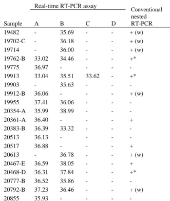
Chapter 1. Table 3. Limit of detection of four real-time RT-PCR assays and intra-assay precision results of 10-fold dilutions of HEV genotype 3 or HEV genotype 4 DNA plasmid controls tested in triplicate. ... 28 Chapter 1. Table 4. Detection rates for HEV RNA in fecal samples collected from pigs of unknown HEV
status by real-time RT-PCR assays A, B, C and D. ... 30 Chapter 1. Table 5. Detection of HEV RNA by conventional nested RT-PCR and real-time RT-PCR
assays in 20 swine field samples. ... 30
LIST OF FIGURES
Literature review. Figure 1. HEV genome organization ... 13 Literature review. Figure 2. Neighbor-joining phylogeny of the complete genomes of members of the Hepeviridae family ... 14 Literature review. Figure 3. Geographic distribution of HEV genotypes in viral isolates obtained from humans and animals ... 14 Chapter 1. Figure 1.Comparison of four real-time RT-PCR assays (A, B, C and D) in detecting and
quantifying HEV RNA on fecal samples after experimental inoculation …………...………29 Chapter 2. Figure 1. Percentage of HEV RNA positive fecal pools in each of 10 farms by real-time
RT-PCR……….……… 33
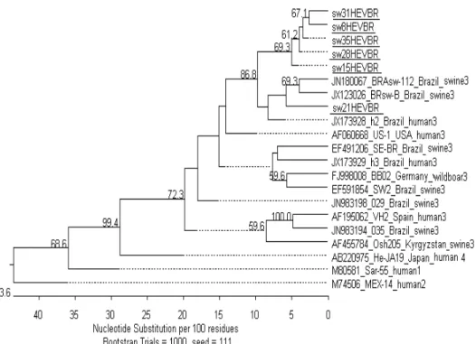
Chapter 2. Figure 2. Phylogenetic tree based on 304 nt region of the 5’ end of HEV isolates..………… 34 Chapter 3. Figure 1. Schematic representation of a FMIA workflow……… 38 Chapter 3. Figure. 2. Mean FMIA index value (±SEM) at different days post inoculation for chickens
infected experimentally with aHEV………39
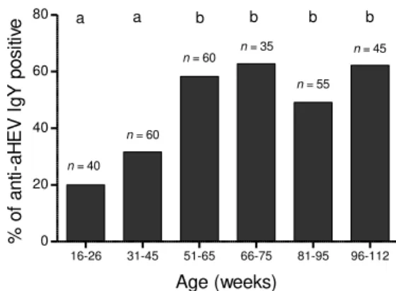
Chapter 3. Figure 3. Detection rates of IgY anti-aHEV antibodies in serum samples from chickens of
different age-groups……… 39
Chapter 4. Figure 1. Percentage of positive IgY anti-avian HEV antibodies in chickens of different
ABREVIATIONS
BLSD – big liver and spleen disease BLSV – big liver and spleen virus CTV – cutthroat trout virus DNA – deoxyribonucleic acid
ELISA – enzyme-linked immunosorbent assay FMIA – fluorescent microbead immunoassay HEV – hepatitis E virus
mHEV – mammalian hepatitis E virus aHEV – avian hepatitis E virus pHEV – piscine hepatitis E virus HSS – hepatitis splenomegaly syndrome
ICTV – International Committee on the Taxonomy of Viruses IgG – immunoglobulin G
IgY – immunoglobulin Y kb – kilo bases
nm – nanometers nt – nucleotide
ORF – open reading frame RNA – ribonucleic acid
RESUMO
O vírus da hepatite E (HEV) tem sido identificado em várias espécies animais. Baseado no hospedeiro, os variantes genéticos podem ser divididos em HEV mamífero (mHEV), HEV aviário (aHEV) e HEV piscino. O objetivo do primeiro estudo foi comparar a performance de dois testes de PCR de transcriptase reversa (RT-PCR) para detecção dos quatro genótipos de mHEV em um único teste (testes A e B) e dois testes RT-PCR duplex para detecção e diferenciação do mHEV-3 e -4 (testes C e D). RNA extraído de 28 amostras de fezes de suínos experimentalmente inoculados com HEV-3 e 186 amostras de suínos a campo com exposição desconhecida ao HEV foram testados. Para os testes A, B, C e D, o RNA do HEV foi detectado respectivamente em 96,4%, 39,2%, 14,2% e 0% das amostras experimentais e em 67,2%, 36,4%, 1,1%, and 0,5% das amostras de campo. Os testes tiveram baixa concordância. Testes A e B tiveram maior taxa de detecção do que os testes C e D (p < 0.05). No segundo estudo, 40 amostras fecais foram coletadas de suínos de 7, 10, 13 ou 17 semanas em 10 granjas. Vinte e nove (72,5%) amostras foram positivas para o RNA do HEV através de RT-PCR. As seis sequências obtidas eram do genótipo 3. No terceiro estudo, 160 amostras de soro de galinhas entre 6 a 118 semanas foram coletadas em 3 granjas e testadas para anticorpos anti-aHEV através de um teste the micro-esferas fluorescentes. Anti-aHEV IgY form detectados em todas as granjas estudadas e em 17% das galinhas. Quarenta amostras de fezes de 8 granjas foram testadas para o RNA do aHEV e 3 (8%) amostras foram positivas para o gene da helicase. Este estudo mostra evidência da circulação do aHEV em galinhas no Brasil.
Palavras-chave: HEV, suíno, galinha, diagnóstico
ABSTRACT
Hepatitis E virus (HEV) has been identified in several animal species. Based on the host tropism the strains can be clustered into mammalian HEV (mHEV), avian HEV (aHEV), and in piscine HEV strains. The aim of the first study was to compare the performance of two single-plex reverse transcriptase (RT)-PCR assays for broad detection of all four mHEV genotypes (assays A and B) and two duplex RT-(RT)-PCR assays for detection and differentiation of mHEV-3 and -4 (assay C and D). RNA extracted from 28 fecal samples from pigs experimentally inoculated with HEV-3 and 186 fecal samples from commercial pigs with unknown HEV exposure were tested. For assays A, B, C and D HEV RNA was detected respectively in 96.4%, 39.2%, 14.2%, and 0% of the experimental samples, and in 67.2%, 36.4%, 1.1%, and 0.5% of the field samples. Assays showed an overall poor agreement. Assays A and B had higher detection rates for HEV RNA than assays C and D (p < 0.05). In the second study, 40 fecal samples were collected from pigs at 7, 10, 13 or 17 weeks of age in 10 farms. Twenty nine (72.5%) samples tested positive for HEV RNA by RT-PCR. All 10 farms had at least one positive sample. All six yield sequences clustered in genotype 3. In the third study, 160 serum samples from chicken ranging from six to 118-weeks of age were collected on three farms and tested for aHEV antibodies by a fluorescent microbead-based assay. Anti-aHEV IgY were detected in 17% of the chickens. Forty pooled fecal samples from eight farms were tested for aHEV RNA by RT-PCR and three (8%) were positive for the helicase gene. This work provides evidence of circulation of aHEV in the Brazilian chicken population.
1. INTRODUCTION “Since its discovery 30 years ago, hepatitis E has been a neglected disease in terms of research funding. In fact, it has been so neglected that it does not even make the WHO [World Health Organization] list of
‘neglected tropical diseases’” (Scobie et al., 2013).
Hepatitis E was first recognized in 1980s by retrospective studies of water-borne epidemics of hepatitis in India (Khuroo, 1980). This agent was initially known as the enterically transmitted non-A, non-B hepatitis virus and was subsequently named the hepatitis E virus (HEV) based on its enteric transmission and association with hepatitis epidemics (Reyes et al., 1991). Currently, HEV is recognized as an pathogen worldwide (Kamar et al., 2012). HEV infection in pregnant women may cause particularly severe illness with a mortality rate of 10-20% and there is evidence of HEV persistent infection in immunocompromised patients (Kamar et al., 2012).Thus far, at least four recognized and two putative genotypes of mammalian HEV (mHEV) have been identified worldwide. The genotypes 1 and 2 of HEV infect only humans, while genotypes 3 and 4 have an expanded host range and are zoonotic (Meng, 2013). Confirmed zoonotic human infections arise primarily from consumption of contaminated pork products and deer meats in industrialized countries (Meng, 2013). The infection by mHEV is widespread in pigs and it appears to be asymptomatic (Meng et al., 2012).
Epidemiology of mHEV has been investigated in Brazil, and swine HEV RNA has been identified in pigs from different regions (dos Santos et al., 2011; de Souza et al., 2012; Gardinali et al., 2012). Consumption of pork meat has been suggested as the most probable cause of an autochthonous case of acute hepatitis E in the country (Lopes dos Santos et al., 2010). Because of the great genomic heterogeneity between human and swine strains from different geographic origin (Lopes dos Santos et al., 2010; dos Santos et al., 2011; Gardinali et al., 2012; Passos et al., 2013), more studies to better understand HEV molecular epidemiology and its impacts on Brazilian pigs herds and the HEV zoonotic transmission are needed.
Avian HEV (aHEV) was identified in chickens displaying decreased egg production and increased mortality and it was considered the most economically significant pathogen affecting broiler breeder flocks in Australia in the 1990s (Payne et al., 1999). aHEV is genetically and antigenically related to human and swine HEVs (Payne et al., 1999; Haqshenas et al., 2001). Phylogenetic analysis revealed that aHEV forms a separate genus, consisting of at least three different genotypes and it is not considered a zoonosis (Meng et al., 2012). It has been shown that a considerable proportion of chicken flocks in North America, Europe, and Asia were seropositive to aHEV infection although seropositive flocks did not necessarily suffer from disease (Huang et al., 2002b; Peralta et al., 2009a; Kwon et al., 2012). Although there is serological evidence of aHEV circulation in Brazilian chickens based on testing of a limited number of samples (n = 25) (Vitral et al., 2005), the status of avian HEV infection in South American chickens is largely unknown. Therefore, it is important to investigate the HEV circulation in Brazilian chicken farms.
In this study, HEV infections were investigated in pigs and chickens from commercial farms in Minas Gerais state, southeast Brazil.
2. LITERATURE REVIEW
2.1.1 Morphology, genome organization and replication
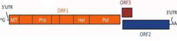
HEV is a spherical, non-enveloped virus of about 30 nm in size and with an icosahedral symmetry (Balayan et al., 1983; Xing et al., 1999). The viral genome is a single-stranded positive sense RNA strand of approximately 7.2 kb in length that consists of a short 5’ non -coding region (NCR) and a 3’ NCR, and three partially overlapping open reading frames (ORFs) (Tam et al., 1991). ORF1 encodes non-structural proteins. Putative functional motifs and domains such as methyltransferase, papain-like cysteine protease, helicase, and RNA-dependent RNA polymerase have been identified in ORF1 (Koonin et al., 1992) (Fig. 1). ORF2 encodes a capsid protein, and ORF3 encodes a small phosphorated protein that is associated with cytoskeleton (Zafrullah et al., 1997; Graff et al., 2005) (Fig. 1). aHEV is
genetically related to mHEV with conserved genomic organization and function despite a 600 nt deletion (Haqshenas et al., 2001; Huang et al., 2004). The capsid protein of aHEV contains both unique and conserved antigenic epitopes in comparison to the human and swine HEV capsid proteins (Dong et al., 2011). The HEV replication cycle is currently not well understood due to the limited success in generating an efficient cell culture system (Berto et al., 2013; Okamoto, 2013; Rogee et al., 2013). HEV attaches to the host cell via a specific high-affinity receptor and enters the cytoplasm by clathrin-mediated endocytosis (Kapur et al., 2012). However, virtually nothing is known about the mechanism by which HEV enters susceptible cells, or about processing of proteins, and mechanisms of virus assembly and release (Okamoto, 2013). The new in vitro culture systems that support HEV replication and release of encapsidated RNA will facilitate the understanding of the biology of this virus.
Fig. 1. HEV genome organization. The genomic RNA carries three open reading frames (orfs) that encode the nonstructural ORF1 (orange), capsid ORF2 (blue), and phosphorated ORF3 (brown) proteins. Reproduced from Aggarwal and Jameel, 2011.
2.1.2 HEV genetic diversity and
nomenclature
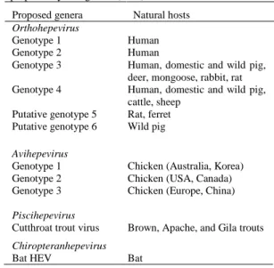
The classification of HEV is currently in transition. According to the most recent report from the International Committee on the Taxonomy of Viruses (ICTV) (Meng et al., 2012), HEV belongs to the familyHepeviridae, which posses a single genus,Hepevirus, that includes four recognized genotypes and at least two putative new genotypes, along with a floating species of aHEV. All the viruses within the genus Hepevirusinfect mammals and have been genetically identified from humans, pig, mongoose, deer, rat, rabbit, and ferret (Meng, 2011).
HEV genotypes 1 and 2 are restricted to humans and related to waterborne large outbreaks of hepatitis E in developing countries (Kamar et
al., 2012). HEV genotype 1 occurs mainly in Asia (Lu et al., 2006); however, it has also been reported in autochthonous cases in Cuba (Villalba et al., 2008) and Venezuela (Garcia et al., 2012). HEV genotype 2 consists of a single Mexican strain and some African strains (Lu et al., 2006). HEV genotype 3 circulates worldwide and contains strains from sporadic and cluster cases, and from chronic cases of hepatitis E in humans (Kamar et al., 2012) and from several animal species including pig, deer, rat, mongoose, and rabbit (Meng, 2013). HEV genotype 4 circulates mainly in Asia; however, it has been reported recently in Europe (Hakze-van der Honing et al., 2011; Colson et al., 2012; Tesse et al., 2012) and includes strains from sporadic and cluster cases of hepatitis E in humans and animal HEV strains from pigs. The two putative new genotypes of HEV include strains from rat and ferret (Johne et al., 2010; Purcell et al., 2011; Raj et al., 2012), and a novel strain of HEV from wild boars in Japan (Sato et al., 2011; Takahashi et al., 2011). A tentative genusOrthohepevirushas been proposed to contemplate the above mentioned mammalian strains of HEV (Meng, 2013) (Table 1, Fig. 2). The geographic distribuition of the tentative genusOrthohepevirusgenotypes 1 to 4 is shown on Fig. 3.
Avian HEV has only been reported in chickens and it is genetically and phylogenetically distinct from the mHEV, sharing only ∼50% nucleotide sequence identity (Bilic et al., 2009). The tentative genus Avihepevirushas been proposed to include the three genotypes of avian HEV thus far recognized: genotype 1 in Australia and Korea, genotype 2 in the United States, and genotype 3 in Europe and China (Bilic et al., 2009; Marek et al., 2010; Kwon et al., 2012) (Table 1, Fig. 2).
Fig. 2. Neighbor-joining phylogeny of the complete genomes of members of the Hepeviridae using the nucleotide percentage distance substitution matrix and complete deletion option in MEGA5. Adapted from Drexler et al. 2012.
Table 1. Nomenclature of the hepatitis E virus (HEV) as proposed by Meng et al. (2013).
Proposed genera Natural hosts
Orthohepevirus
Genotype 1 Human
Genotype 2 Human
Genotype 3 Human, domestic and wild pig,
deer, mongoose, rabbit, rat
Genotype 4 Human, domestic and wild pig,
cattle, sheep
Putative genotype 5 Rat, ferret
Putative genotype 6 Wild pig
Avihepevirus
Genotype 1 Chicken (Australia, Korea)
Genotype 2 Chicken (USA, Canada)
Genotype 3 Chicken (Europe, China)
Piscihepevirus
Cutthroat trout virus Brown, Apache, and Gila trouts
Chiropteranhepevirus
Bat HEV Bat
Fig.3. Distribution of HEV genotypes in viral isolates obtained from humans and animals (predominantly pigs). The colors used for a country and the circle associated with it represent the predominant HEV genotypes of human and animal isolates, respectively, from that country. The figure is based on data from Okamoto, 2007. Reproduced from Aggarwal and Jameel, 2011.
2.1.3 HEV classification: genotypes and subgenotypes
The ICTV does not include any consideration below the species level and this has been left to the initiative of specialty groups (Fauquet et al., 2005). The most current ICTV report recognizes four genotypes of HEV (Meng et al., 2012) and
(Arankalle et al., 1999; Worm et al., 2002; Lu et al., 2006; Zhai et al., 2006; Okamoto, 2007; Bouquet et al., 2012) contributing to differences in the classification among groups. Recently, there has been a controversy about whether isolates from rabbits and wild boar should be considered new genotypes or subtypes (Zhao et al., 2009; et al.; Cossaboom et al., 2011; Takahashi et al., 2011; Bouquet et al., 2012). The rapid increase in sequencing of different genomic regions and different fragment sizes in addition to the lack of commonly agreed criteria to define different taxonomic categories have resulted in multiple genotype/subgenotype definitions applied to HEV (Arankalle et al., 1999; Worm et al., 2002; Lu et al., 2006; Zhai et al., 2006; Okamoto, 2007; Bouquet et al., 2012; Smith et al., 2013), complicating the comparison of molecular epidemiology data worldwide. There is no agreement in the assignment of isolates to particular virus subtypes that would result, for example, in recognition of 4 genotypes mHEV subdivided into 24 (Lu et al., 2006) or 12 (Zhai et al., 2006) subtypes. Lu et al. (2006) in a comprehensive analysis of the bootstrap support for phylogenetic groupings and the nucleotide distances between these groupings, suggested that genotypes 1, 2, 3 and 4 could be classified into five (1a-1e), two (2a-2b), ten (3a-3j) and seven (4a-4g) subtypes, respectively. To make this taxonomic assignment, Lu et al. (2006) examined not only HEV complete genomes but sequences from the 5’ and 3’ end of ORF1 regions and three separate ORF2 regions. The 5’ end of the ORF2 was pointed as the region that better reflected the complete genomic sequence for HEV classification (Lu et al., 2006). More recently, Smith et al. (2013) suggested a classification system based on the amino acid sequences of concatenated ORF1/ORF2 coding regions excluding the hypervariable region. Using this criterion, the Hepevirus genus would include four species, including HEV genotypes 1 to 4, and the putative genotype 6 as proposed by the ICTV (Meng et al., 2012), avian HEV, bat HEV and rat/ferret HEV. Cutthroat trout virus would represent a second genus.
aHEV nomenclature also lacks a consensus criteria and the designations given in different studies still give risen to confusion. Phylogenetic analysis of near-complete aHEV
genomes from Australia, United States and Europe revealed three large clusters, corresponding to their geographical origin (Bilic et al., 2009). Subsequent analysis of a 130 nt fragment of the helicase gene using more isolates revealed greater diversity, and putative genotypes 4 and 5 were proposed (Marek et al., 2010). However, putative genotypes 4 and 5 strains clustered with aHEV genotype 3 when a 124 nt fragment of the capsid gene was used (Marek et al., 2010). More recently, a putative novel genotype was proposed based on a near-complete genome sequence from a Hungary isolate (Banyai et al., 2012); however, this sequence was not taken into consideration in the new Avihepevirus classification proposal (Meng, 2013). New genotypes have often been defined based on the presence of phylogenetic branches that have substantial bootstrap support. However, in many instances, there is a hierarchy of such branches so that it becomes difficult to decide which branches are taxonomically informative (Smith et al., 2013), leading to differences in the classification among groups.
The nomenclature and classification criteria of HEV need to be standardized to ensure that epidemiological, evolutionary and clinical observations among strains can be accurate and informative molecular epidemiological tools.
2.2.1 HEV infection in humans
Until recently, it was thought that HEV was only an acute self-limiting disease in the majority of patients (Aggarwal et al., 2011). However, awareness of HEV infection has expanded in the last few years and it is now known that the occurrence of HEV infection is underestimated, especially in developed countries (Kamar et al., 2013a). Clinical presentation of hepatitis E include acute icteric hepatitis, anicteric illness with nonspecific flu-like symptoms, and asymptomatic transaminase elevation (Aggarwal et al., 2011). The case fatality rate during epidemics was found to be between 0.2% and 4% (Kumar et al., 2013). However, fulminant hepatitis and high mortality are described, reaching 20% in cases of pregnant women infected with genotype 1 in developing countries and 70% in of patients infected with genotype 3 with underlying liver
disease (Kamar et al., 2012). HEV infection can become chronic in immunocompromised patients such as solid organ transplant recipients, patients receiving chemotherapy, and patients with HIV infection, and the course of disease progress to a chronic state and persistence of viral shedding in 65-80% of the cases and may rapdly progress to cirrhosis (Moal et al., 2013; Riveiro-Barciela et al., 2013; Zhou et al., 2013). Furthermore, HEV infection has been associated to extrahepatic manifestations, including neurologic symptoms, kidney injuries, pancreatitis, and hematological disorders (Kamar et al., 2013b).
To date, four major documented routes of HEV transmission have been reported: waterborne; foodborne; bloodborne, and vertical transmission (Kaba et al., 2013). Research among non-human primates has shown a direct association between infective dose and disease severity, but an inverse relationship to the incubation period (Tsarev et al., 1992; Tsarev et al., 1994). Incubation time ranges from 2 weeks to 2 months, viremia is transient and occurs mainly during the prodromic phase, and disappears at the onset of clinical symptoms, when viral shedding begins and regresses at the onset of jaundice within 2 to 3 weeks (Aggarwal et al., 2000). Vertical transmission from mother to fetus has been reported in 33 to 100% of the cases (Kaba et al., 2013). Human to human transmission of HEV is considered rare (Kaba et al., 2013). HEV infection is still an underdiagnosed disease and further studies are required to determine its prevalence, incidence, and control.
2.2.2 HEV infection in swine
Since its discovery in domestic swine in the United States in 1997, swine HEV strains have been identified worldwide in swine with widely variable prevalence (Meng, 2010). A retrospective study performed in Spain showed that HEV was endemic in the Spanish pig farms at least since 1985 (Casas et al., 2009). Studies of prevalence across Japan revealed that anti-HEV antibody is present in 93% of all domestic pig farms tested and that all swine HEV isolates belong to either genotype 3 or 4 (Takahashi et al., 2013). In Spain, the prevalence of anti-HEV antibodies on commercial swine farms reached 98%, while the anti-HEV prevalence in New Zealand and Brazil is 90% and 81%,
2.3.1 Waterborne and shellfish transmission
HEV is typically transmitted via fecal-oral route within an animal species, from animals to humans in infectious body fluids, and from contaminated water sources to human and other animals (Yugo et al., 2013). In developing countries, outbreaks usually occur during the rainy season, floods or the monsoon, conditions that facilitate the contamination of sources of drinking water by human excreta (Kaba et al., 2013). In both developing and developed countries, infectious HEV strains has been isolated from sewage, water treatment plants and rivers` water (Jothikumar et al., 1993; Vaidya et al., 2003; Borgen et al., 2008; Clemente-Casares et al., 2009; Rutjes et al., 2009). HEV contamination related to consumption of food items receiving sewage sludge, such as shellfish, that are most often eaten raw or slightly cooked have been reported in European and Asian countries (Cacopardo et al., 1997; ; Renou et al., 2008; Said et al., 2009; Song et al., 2010; Namsai et al., 2011; Diez-Valcarce et al., 2012). In Scotland, 92% of bivalve mussels collected were tested positive for HEV RNA with the viral sequences clustering with HEV genotype 3 identified in human and swine (Crossan et al., 2012). In addition, HEV genotype 3 RNA has been detected on irrigated, field-grown strawberries (Brassard et al., 2012). Drinking water from non-public supplies was identified as a risk factor for acquiring hepatitis E in Spain (Galiana et al., 2008) and France (Renou et al., 2008). Therefore, as in developing countries, the environment should be considered as a potential source of HEV transmission in developed countries.
2.3.2 Environmental and animal handling transmission
Swine is a major reservoir for HEV and occupational contact with infected swine is a risk factor for zoonotic HEV transmission in humans. Professionals working in close proximity to swine, swine manure, or sewage may become infected with HEV during occupational activities (Galiana et al., 2008). Run-offs from animal facilities such as hog
operations have been implicated in human HEV infections with the detection of infectious HEV genotype 3 in the animal manure and wastewater (Borgen et al., 2008; Rutjes et al., 2009). Swine veterinarians in the United States were shown to have 27% seropositivity to HEV in comparison to 16% of the normal blood donors (Meng et al., 2002). Swine workers in Valencia, Spain were found to be 5.4 times more likely to be positive for anti-HEV IgG than those not exposed to swine (Galiana et al., 2008). In the Netherlands, 11% of swine veterinarians were positive in comparison to 6% of non-swine veterinarians and 2% of the general population (Bouwknegt et al., 2008a). Similarly, swine farmers showed at least two-fold more chance to be seropositive than general population in Sweden and Maldova (Olsen et al., 2006; Bouwknegt et al., 2008a). In addition, an acute hepatitis E case was reported in a person who had been given a pet pig 2 months before the onset of symptoms. HEV sequences recovered from the patient were closed related to the sequence identified from the animal (Renou et al., 2007).
The multitude of novel strains of HEV in wildlife and other domestic animal species suggest additional mechanisms of transmission. For example, field workers at the Iowa Department of Natural Resources who work with a variety of wildlife species had a higher prevalence for HEV antibodies in comparison to normal blood donors (Karetnyi et al., 1999). In studies conducted in France and Japan, hunting has been found to be associated with the higher prevalence of anti-HEV antibodies in blood donors (Mansuy et al., 2008; Toyoda et al., 2008). Finally, contact with horses and pets have been associated with a higher prevalence of anti-HEV antibodies in multivariate analysis (Christensen et al., 2008; Kuniholm et al., 2009). While exposure to HEV, identified by the presence of anti-HEV antibodies in these populations, does not in itself indicate a disease, it does identify a potential route of transmission and exposure.
Many sporadic and cluster cases of hepatitis E have been linked to the consumption of contaminated raw or undercooked animal meat and meat products (Masuda et al., 2005;
2.3 HEV environmental contamination and
waterborne transmission
Matsubayashi et al., 2008). The strongest evidence of zoonotic transmission of HEV has come from studies conducted in Japan, and was associated with eating not thoroughly cooked deer or wild boar meat; HEV sequences recovered from leftover frozen meats and from patients infected with HEV were identical (Li et al., 2005; Pavio et al., 2010). Further evidence supporting the zoonotic transmission of HEV is through the consumption of contaminated raw or undercooked food products from pigs purchased in supermarkets or from wild boars (Borgen et al., 2008; Wichmann et al., 2008; Colson et al., 2010; Wilhelm et al., 2011; Miyashita et al., 2012). HEV sequences recovered from suspected commercial pig livers or pig liver sausages were closely related, or identical in some cases, with those recovered from patients infected with HEV (Matsubayashi et al., 2008; Colson et al., 2010; Wilhelm et al., 2011). In addition, based on case-control studies conducted in France and Germany, consumption of uncooked pig liver sausage, game meats or offal was identified as a risk factor for HEV infection (Wichmann et al., 2008; Colson et al., 2010; Legrand-Abravanel et al., 2010).
Approximately 2% of the pig livers sold in local grocery stores in Japan (Yazaki et al., 2003), 4% in Germany (Wenzel et al., 2011), 6.5% in the Netherlands (Bouwknegt et al., 2007), and 11% in the United States (Feagins et al., 2008a) tested positive for the HEV genotype 3 RNA. Investigations in pork product chains in Italy, the Czech Republic, Spain, and the United Kingdom revealed detectable, infectious HEV at both processing locations and point of sale (Di Bartolo et al., 2011; Di Bartolo et al., 2012). The potential widespread dissemination of HEV through pork production chains and the potential foodborne transmission are of significant concern (Purcell et al., 2010).
2.5.1 Human HEV diagnosis
Diagnosis of hepatitis E relies on laboratory abnormalities in liver enzyme and liver function tests. Since cases of hepatitis E are difficult to distinguish clinically from other types of acute viral hepatitis, the diagnosis is established by a combination of serological and molecular techniques (Kumar et al., 2013).
Several assay systems have been developed for the detection of IgM and IgG anti-HEV antibodies, although there is considerable variability in their sensitivities and specificities (Aggarwal, 2013). The most frequently used format for anti-HEV assays has been indirect enzymatic immunoassays, because of its high sensitivity, relative ease of performance and adaptability to high-throughput testing (Aggarwal, 2013). Drobeniuc et al. (2010), in a pan-genotypic validation of six commonly available IgM assays have found that only two of these assays had sensitivity and specificity above 95%. Sensitivity of the different assays ranged from 72% to 98%, and specificity from 78% to 96%. κ-coefficients for agreement between results of various pairs of tests varied from 0.42 to 0.80 (mean = 0.53). Analytical sensitivities of various tests were compared using serial dilutions of a particular serum specimen and varied up to 15-fold. This lack of consistency makes comparison of the diagnosis of HEV infection using different tests difficult. Analysis of HEV RNA by using nucleic acid amplification techniques is also used for diagnosis; this method can identify active infection and help confirm serologic results (Aggarwal, 2013). Several in-house conventional and real-time RT-PCR assays targeting either ORF2 or ORF3 genes have been developed. However, the sensitivity of these assays varies significantly, depending on the targeted region and viral genetic diversity (Abravanel et al., 2012). In a study published in 2011, a panel of 22 plasma specimens that were positive for HEV RNA and included serial 10-fold dilutions of four different strains of HEV genotypes 3 or 4 and two control specimens negative for HEV RNA were tested by 20 laboratories (using 12 assays) in 10 countries (Baylis et al., 2011). The majority (59%) of the 17 laboratories that provided method details were using the same assay (i.e., Jothikumar et al., 2006). The analysis revealed a 100– 1,000-fold difference in sensitivity between different assays, and the assay developed by Jothikumar et al., (2006) presented the highest sensitivity (limit of detect detection of approximately 4 HEV genome equivalents per reaction). The high sensitivity of this assay associated to its ability to detect all four mHEV that infect humans contributed to its widespread usage in diagnostic laboratories. However, false negative
RT-PCR results have been reported using it and a modification in the probe has been suggested to restore the sensitivity of this assay (Garson et al., 2012). Accurate RT-PCR assays are needed to avoid misdiagnosed infections, because serological testing is less sensitive in immunocompromised patients.
2.5.2 Swine HEV diagnosis
Since HEV infection is asymptomatic in swine, HEV diagnosis is not performed routinely in pigs. HEV serology or detection of HEV RNA is performed usually in research laboratories that have developed assays adapted to swine (Engle et al., 2002; de Deus et al., 2008a; Casas et al., 2009; Zhang et al., 2011). However, pig is now a recognized reservoir for zoonotic HEV infection (Meng, 2013) and it is important to measure level of viral contamination in pig herds. HEV has been cited recently as a pathogen that should be eliminated from any herd used for xenotransplantation (Fishman et al., 2012), and therefore, appropriate diagnosis are required for detection and elimination of HEV in pigs used for this purpose.
Conventional or real-time RT-PCR assays have been developed in independent laboratories targeting ORF1, ORF2 or ORF3 of HEV (Wang et al., 1999; Huang et al., 2002a; Jothikumar et al., 2006; Gyarmati et al., 2007; Zwettler et al., 2012). Broadly reactive RT-PCRs developed to detect human HEV have also been used to monitor HEV in pig herds (Cooper et al., 2005; Andraud et al., 2013). Since only one serotype of HEV has been described, detection of anti-HEV antibodies in swine can be performed using commercial kits for humans adapted to swine or in-house ELISA tests based on genotypes 1 and/or 3 antigens (Zhang et al., 2011). Recently, a fluorescent micro-beaded immunoassay (FMIA) has been developed for the detection of anti-HEV IgG antibodies in pigs and compared to an in-house ELISA based on the same antigen, a recombinant HEV ORF2 protein (Owolodun et al., 2013). Results indicate an almost perfect agreement between the two assays; however the FMIA has the advantage of utilizing considerably less amount of antigen compared to an ELISA and also allows screening of antibodies to multiple pathogens or antigens simultaneously in one reaction well (Owolodun et al., 2013). Therefore, it reduces the time and labor required
for assay performance and also requires a smaller amount of sample thereby further reducing cost.
Although various assays have been reported for the monitoring of HEV infection in pig herds, it is worth noting that these assays have not been validated due to the absence of appropriate gold standards (Pavio et al., 2010).
The first descriptions of an infectious disease in broiler breeders named big liver and spleen disease (BLSD) were reported in Australia in 1980s (Handlinger et al., 1988). Based on a 523-nt sequence, the causative agent, big liver and spleen virus (BLSV), was found to be genetically related to human HEV with about 62% nucleotide sequence identity (Payne et al., 1999). In this period, it was considered the most economically significant disease affecting commercial breeder flocks in Australia, causing reduced egg production and a slight increase in mortality (Payne et al., 1999). A similar disease, hepatitis-splenomegaly syndrome (HSS) was reported in Canada in 1991 (Ritchie et al., 1991) and a HEV-like virus was identified in the USA from chickens affected with HSS (Haqshenas et al., 2001). Based on the genetic relatedness of this novel chicken virus with HEV, it was designated avian HEV to distinguish from human HEV and swine HEV. aHEV shared about 80% nucleotide sequence identity with BLSV, and about 57-61% nucleotide sequence identity with mammalian HEVs (Haqshenas et al., 2001). These two previously identified syndromes (HSS and BLSD) are now recognized to be caused by variant strains of the aHEV (Bilic et al., 2009; Marek et al., 2010; Meng, 2013).
2.6.1 HEV infection in chickens
aHEV RNA can be detected in broiler breeders hens and laying hens with and without clinical signs (Huang et al., 2002b; Peralta et al., 2009a; Sun et al., 2004a). aHEV has been shown to cross species barriers and infect turkeys (Sun et al., 2004b), however, rhesus monkeys and mice are not susceptible to infection by aHEV under experimental conditions (Huang et al., 2004). Based on serological evidence, aHEV is widespread in chicken flocks with seropositive rates of approximately 71% in the USA, 90% in
Spain, and 57% in Korea (Huang et al., 2002b; Peralta et al., 2009a; Kwon et al., 2012). aHEV infection in chickens is age-dependent with 17% of seropositive chickens under 18 weeks of age and 36% of seropositive adult chickens (Huang et al., 2002b; Zhao et al., 2013). Under natural conditions, chickens become infected at approximately 3-4 months of age (Sun et al., 2004a). The relatively low doses of virus that can be transmitted among chickens via the fecal-oral route has been speculated as the reason of the subclinical infection in the majority of chickens (Sun et al., 2004a). Differences in virus strain, virus dose, diet and age have been implicated as potential co-factors for the manifestation of the full spectrum of clinical disease (Agunos et al., 2006; Meng et al., 2008). Nevertheless, aHEV strains recovered from healthy chickens in normal flocks and previously considered to be avirulent were only slightly attenuated in an experimental infection model (Billam et al., 2009). There was no clear clustering of aHEV sequences based on whether viruses were sampled from chickens displaying clinical signs or not (Marek et al., 2010).
Clinical signs usually affects 30 to 72 weeks-old chickens and may include egg drop in some flocks up to 20%, enlargement of the liver and spleen, and acute death of affected chickens (Meng et al., 2008). In affected flocks, mortality rates can be up to 0.3-1.0% a week (Meng et al., 2008; Morrow et al., 2008). Post-mortem evaluations show enlarged livers, enlarged spleens, and serosanguineous fluid in their coelomic cavities accompanied by a drop in egg production and high mortality rates (Billam et al., 2005; Meng et al., 2008). Typical histopathological changes include necrotizing, hemorrhagic, and non-specific hepatitis (Meng et al., 2008). In specific-pathogen-free (SPF) chickens experimentally inoculated either by oronasal route or by intravenous route with aHEV strains recovered from a chicken with HSS developed microscopic liver lesions characterized by lymphocytic and heterophilic periphlebitis, phlebitis and fibrinoid necrosis (Billam et al., 2005; Billam et al., 2009). Also, approximately 25% of the infected chickens developed sub capsular hemorrhages or enlargement of the right intermediate lobe of the liver (Billam et al., 2005). Similar gross and microscopic findings have been described in
chickens naturally infected with aHEV in field outbreaks of HSS (Agunos et al., 2006; Morrow et al., 2008).
2.6.2 aHEV diagnosis
aHEV diagnosis is primarily based on detection of avian HEV RNA by RT-PCR in feces, liver, or bile, and anti-aHEV IgY antibodies by ELISA (Meng, 2010). An indirect ELISA was developed by using a truncated ORF2 protein of aHEV genotype 2, and its cross-reactivity with human and swine HEVs was shown (Haqshenas et al., 2002). On the basis of this ELISA, several studies about the seroprevalence of avian HEV were carried out in North America, Europe and Asia (Huang et al., 2002b; Peralta et al., 2009a; Kwon et al., 2012). More recently, another in-house indirect ELISA using truncated ORF2 protein of aHEV genotype 3 has been developed and applied in a serological survey in Chinese chicken flocks (Zhao et al., 2013).The sensitivity and specificity of these assays are not known.
Most of the RT-PCR methods for detection of aHEV RNA are based on degenerate primers targeting ORF1 or ORF2 (Payne et al., 1999; Huang et al., 2002b; Sun et al., 2004a; Bilic et al., 2009; Troxler et al., 2011). Although the most used primers were developed for aHEV genotype 2 (Huang et al., 2002b; Sun et al., 2004a), it has been successfully used for detection aHEV genotypes 1 and 3 (Massi et al., 2005; Morrow et al., 2008; Peralta et al., 2009a; Marek et al., 2010; Kwon et al., 2012).However, the sensitivity of the RT-PCR assays for detection of different aHEV genotypes is not known since strains identified in different geographic regions are genetically heterogenic (Meng, 2010).
HSS livers do not contain excessive fat and HFLS livers do not have massive necrosis (Agunos et al., 2006). Other causes of liver lesions and splenomegaly include bacterial septicemia, lymphoid tumours (lymphoid leukosis, Marek´s disease), and avian adenoviruses infections which have characteristic histological lesions. A fractured liver resulting from external trauma to the body wall should also be considered as a differential diagnosis and is macroscopically characterized by hemorrhage in the coelomic cavity with coagulated blood adhering to the fracture site on the liver surface (Meng et al., 2008). Until now, avian HEV appeared to be regarded as a minor causative agent in several diagnostic cases and ignored when other poultry diseases have been diagnosed. However, avian HEV should be tested as one of the potential causative agents since chickens showing HSS with decreased egg production have been identified worldwide when the reasons of decreased egg production and increased mortality in chickens are uncertain (Kwon et al., 2012).
Other known animal species infected by different HEV strains genetically identified thus far include rat, mongoose, rabbits, ferrets, cutthroat trout, bats, and deer (Meng, 2013). Anti-HEV antibodies have been detected in a number of other animal species including cattle, sheep, and goats with the potential to carry novel strains of HEV (Meng, 2013). The zoonotic potentials of these novel animal strains of HEV are not altogether understood.
HEV genotype 3 strains have been genetically identified in different species of deer and wild boar in Asia and Europe and foodborne transmission to humans have been reported (Tei et al., 2003; Sonoda et al., 2004; Takahashi et al., 2004; Li et al., 2005; Masuda et al., 2005; Nishizawa et al., 2005; de Deus et al., 2008b; Kaci et al., 2008; Reuter et al., 2009; Schielke et al., 2009; Kaba et al., 2010). Rabbits may also serve as reservoir hosts for HEV transmission to humans given the genetic identification of HEV strains related to the zoonotic genotype 3 from rabbits in China, the United States, and France (Lhomme et al., 2013). The ability of rabbit HEV to cause cross-species infection in a pig
model has been demonstrated (Cossaboom et al., 2012).
2.8.1 HEV in humans
The first serological evidence of human HEV infection in South America was found in 1994 in Venezuela (reviewed by Echevarria et al., 2013). Most prevalence rates reported among either urban or rural populations ranged from 1 to 10% (Echevarria et al., 2013). In Brazil, the anti-HEV IgG positive detection rates in blood donors ranged from 2.0% to 3.0% and from 1.68% to 4.3% in the general population (Assis et al., 2002; Bortoliero et al., 2006; Carrilho et al., 2005; Kiesslich et al., 2002; Lyra et al., 2005; Parana et al., 2000; Silva et al., 2012; Trinta et al., 2001; Vitral et al., 2005). In a recent serological study conducted on individuals living in Mato Grosso state, contact with pig or pig carcasses was not associated with an increase in HEV exposure (Silva et al., 2012).
South American countries, including Argentina, Brazil, Chile, Peru, and Uruguay have diagnosed patients with acute hepatitis E by anti-HEV IgM and/or HEV RNA detection (Echevarria et al., 2013). Autochthonous hepatitis E cases due to HEV genotype 3 have been reported in Argentina, Brazil and Uruguay (Lopes Dos Santos et al., 2010; Mirazo et al., 2013; Munne et al., 2011; Passos et al., 2013). Anti-HEV IgM antibodies have been detected retrospectively from patients with acute non-A-C hepatitis in Brazil since the 1990s (Lyra et al., 2005). However, the first molecular identification and characterization of an autochthonous human acute HEV infection was only reported in 2010, and a foodborne zoonotic transmission has been pointed as the most probable cause of infection (Lopes Dos Santos et al., 2010). Recently, another retrospective study has identified HEV infection in renal transplant recipients with unexplained increase in alanine aminotransferase and aspartate aminotransferase levels in Brazil (Passos et al., 2013). Diagnosis for hepatitis E is still neglected in Brazil and public health strategies are needed to include HEV as a possible agent in cases of acute non-A–C hepatitis.
2.7 HEV in other animals
2.8.2 HEV in domestic animals
The first evidence of the circulation of HEV in commercial pig farms in South America were reported in Argentina in 2006, where HEV RNA detection rates ranged from 4% to 98% of pigs within farms (Munne et al., 2006b). The Argentinean strains were closely related to human HEV genotype 3 strains identified from sporadic cases in Argentina (Munne et al., 2006a), and from an acute case of hepatitis E in Brazil (Lopes Dos Santos et al., 2010). HEV genotype 3 has also been identified in Bolivia (Dell'Amico et al., 2011) and Chile (Ibarra et al., 2007).
In Brazil, HEV genome was detected in six of eight stool pools from piglets 40 to 60 days old in São Paulo state (Paiva et al., 2007). In Parana state, HEV RNA was detected in 62.5% pig farms and 15.3% fecal samples (Gardinali et al., 2012). More recently, two different HEV genotype 3 subtypes occurring in swine from Para state have been reported (de Souza et al., 2012). HEV strains isolated were always from genotype 3. HEV RNA has been detected in raw effluents from a slaughterhouse in Rio de Janeiro (dos Santos et al., 2011). Sequencing and phylogenetic analysis revealed that the sewage strains were closely related to human and swine HEV strains recorded in the state (dos Santos et al., 2009; Lopes Dos Santos et al., 2010).
Anti-HEV IgG has also been detected in cows (1.42%, 1/70), dogs (6.97%, 3/43), chickens (20%, 5/25), and rodents (50%, 2/4) in Brazil (Vitral et al., 2005). To the author’s knowledge, this is the only report of serological detection of aHEV in South America and there is no report of HSS or aHEV occurrence in chickens in Brazil.
3. OBJECTIVES
The present study was divided in four chapters, with the following objectives:
1. To develop a broadly reactive RT-PCR assay capable of detecting mHEV genotypes 1-4 and to develop a duplex RT-PCR for detection and identification of mHEV genotypes 3-4; 2. Genetically identify and characterize HEV virus that may be found in pig fecal samples in Brazil;
3. To develop a fluorescent micro-beaded immunoassay for the detection of chicken HEV IgY antibodies;
CHAPTER 1
COMPARISON OF REAL-TIME
REVERSE TRANSCRIPTASE
(RT)-PCR ASSAYS FOR DETECTION OF
SWINE HEPATITIS E VIRUS IN
FECAL SAMPLES
ABSTRACT
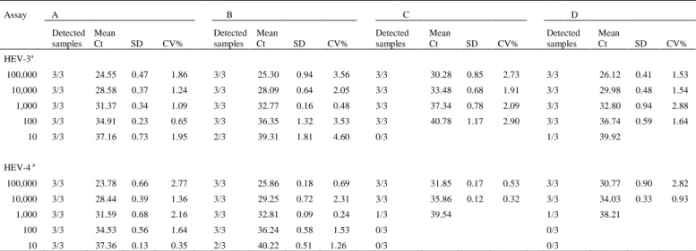
Hepatitis E virus (HEV) is a major cause of acute viral hepatitis in people in many developing countries and is also endemic in many industrialized countries. Mammalian HEV (mHEV) isolates can be divided into at least four recognized major genotypes. Several nucleic acid amplification techniques have been developed for mHEV detection with great differences in sensitivity. The aim of this study was to compare the performance of two single-plex real-time reverse transcriptase (RT)-PCR assays for broad detection of all four mHEV genotypes (assays A and B) and two duplex real-time RT-PCR assays for detection and differentiation of mHEV genotypes 3 and 4 (assay C and D). RNAs extracted from 28 fecal samples from pigs experimentally inoculated with HEV genotype 3 and 186 fecal samples from commercial pigs with unknown HEV exposure were tested by all four assays. In experimental samples, HEV RNA was detected in 96.4% (assay A), 39.2% (assay B), 14.2% (assay C), and 0% (assay D) of the samples. In field samples with unknown HEV exposure, HEV RNA was detected in 67.2% (assay A), 36.4% (assay B), 1.1% (assay C), and 0.5% (assay D) of the samples. Assays showed an overall poor agreement (κ = 0.19 to 0.03) with differences in detection rates between assays (p < 0.01). Assays A and B that broadly detect HEV genotypes 1-4 had significantly higher detection rates for HEV RNA than the duplex assays C and D that were both designed to detect and differentiate between HEV genotypes 3 and 4.
Keywords: Diagnosis, multiplex, hepatitis E virus, HEV RNA, genotypes, real-time RT-PCR
1. INTRODUCTION
Hepatitis E virus (HEV) is the causative agent of hepatitis E in humans (Purcell et al., 2008). HEV infection in pregnant women may cause particularly severe illness with a mortality rate
of 10-20%, and recently there are numerous reports of persistent and chronic HEV infection in immunocompromised patients such as organ transplant recipients (Kamar et al., 2012). Currently, HEV is classified in the genus Hepevirus in the Hepeviridae family (Meng et al., 2012). The virus is a non-enveloped, positive-sense, single-stranded RNA virus that encodes three open reading frames (ORFs). ORF1 encodes for non-structural proteins, ORF2 encodes the viral capsid, and ORF3, which overlaps with ORF2, encodes a multi-functional small protein (Meng et al., 2012). HEV has been identified in several animal species including domestic pigs, chickens, deer, wild boars, mongooses, rabbits, rats, ferrets, bats, and fish (Meng, 2013) and, based on the host tropism, the strains genetically identified thus far can be clustered into mammalian HEV (mHEV), avian HEV (aHEV), and in piscine HEV (pHEV) strains. Within mHEV, there are at least four recognized genotypes capable of infecting humans. Genotypes 1 and 2 are associated with epidemics and restricted to humans in developing countries, whereas genotypes 3 and 4 can infect a wide variety of species including humans and pigs, and are associated with sporadic and cluster cases of human hepatitis E in both developing and industrialized countries (Kamar et al., 2012).While mHEV genotype 3 has worldwide distribution (Lu et al., 2006), genotype 4 were reported in Asia (Lu et al., 2006), and more recently in Europe (Colson et al., 2012; Hakze-van der Honing RW et al., 2011). In humans, infections with genotypes 1 and 2 are mainly transmitted via fecally-contaminated water while infections with genotypes 3 and 4 appear to occur primarily by food-borne zoonotic transmission through the consumption of raw or undercooked meat from pigs, wild boars or deer (Meng, 2013).
et al., 2013), and only two of six commonly available IgM anti-HEV detection assays had sensitivities and specificities above 95% (Drobeniuc et al., 2010). Due to this overall low sensitivity, a combination of antibody detection and nucleic acid detection has been suggested for optimizing mHEV diagnosis (Huang et al., 2010; Baylis et al., 2011).
Considering the heterogeneity of mHEV strains circulating in humans and other animal species, several conventional reverse transcription (RT)-PCR and real-time RT-(RT)-PCR assays have been developed for the detection of HEV RNA in various types of samples including sera, feces and environmental samples (Meng et al., 2001; Ahn et al., 2006; Enouf et al., 2006; Jothikumar et al., 2006; Gyarmati et al., 2007; Legrand-Abravanel et al., 2009). Comparisons of RT-PCR assays have shown a 10 to 1,000-fold variation in sensitivity when samples were tested in parallel in the same laboratory (Ward et al., 2009; Mokhtari et al., 2013). In a blinded study to investigate the performance of conventional and real-time RT-PCR assays in 20 laboratories that performed HEV RNA detection on a regular basis, variations in sensitivity in the order of 100- to 1,000-fold were found using a standard panel of HEV genotype 3 and 4 strains (Baylis et al., 2011). Currently, a real-time RT-PCR designed in 2006 (Jothikumar et al., 2006) is the most widely used assay for detection of HEV infection in humans (Baylis et al., 2011; Baylis et al., 2013) primarily based on the reported high sensitivity (limit of detection of 4 genome equivalents of HEV genome) and its ability to detect all four recognized mHEV genotypes that are capable of infecting humans (Garson et al., 2012).
Although real-time PCR assays targeting conserved regions can provide accurate detection of the HEV genomes and yield results more rapidly compared to conventional RT-PCR, commonly a second molecular method such as sequencing or subtyping is required to further characterize strains. Recently, a real-time duplex RT-PCR assay for detection and identification of HEV genotype 3 and 4 in amounts as low as 50 genomic equivalents copies per reaction has been reported (Zhang et al., 2013). This assay, targeting the ORF2/ORF3 overlapping region, was designed to allow for a sensitive and rapid detection of the zoonotic
HEV genotypes to potentially facilitate epidemiological investigations and to better understand outbreak situations. The aim of this study was to compare the performance of two single-plex real-time RT-PCR assays for broadly detection of all 4 recognized mHEV genotypes (assays A and B) and two duplex real-time RT-PCR assays for detection and differentiation of mHEV genotypes 3 and 4 (assay C and D). Each single-plex and one duplex real-time RT PCR assays had been previously described while the other single-plex assay is an in-house assay.
2. MATERIAL AND METHODS
The experimental protocol was approved by the Virginia Polytechnic and State University Institutional Animal Care and Use Committee and by Virginia Polytechnic and State University Institutional Biosafety Committee. Twenty-eight serial fecal samples were collected daily from two pigs experimentally inoculated with human HEV genotype 3 strain US-2 (GenBank accession number AF060669) or swine HEV genotype 3 strain Meng (GenBank accession number AF082843) from day post-inoculation (dpi) 2 to 14. The fecal samples were suspended in saline (10% w/v), and the fecal suspensions were stored −80 °C until use.
A total of 186 fecal samples of pig origin were chosen arbitrarily from routine diagnostic cases submitted during May 2013 to the Iowa State University Veterinary Diagnostic Laboratory (ISU-VDL). These samples originated on 86 farms located in 12 US states, Iowa, Illinois, Indiana, Minnesota, Missouri, North Carolina, North Dakota, Nebraska, Ohio, South Dakota, Texas, and Wisconsin, with samples obtained from age group: suckling (1-2 weeks of age), nursery (3-7 weeks of age) and grow-finish pigs (8-25 weeks of age).
Fecal samples of ~1 g were resuspended in phosphate buffered saline (PBS) to obtain a final 10% suspension (w/v), vigorously vortexed and centrifuged at 1500 ×
g
for 102.1 Experimental samples
2.2 Field samples
2.3 Sample processing and RNA
min. Viral RNA extraction was carried out on 50 µl fecal supernatant using a MagMAX 96 Viral Isolation kit (Ambion, Foster City, CA, USA) according to the manufacturer’s instructions on an automated extraction platform (KingFisher Flex; Thermo Fisher Scientific). Negative controls, using water as a sample, and positive controls, using fecal suspensions from a pigs infected either with mHEV genotype 3 or 4, were added to each extraction plate. The extracted RNA was stored at −80 °C until use.
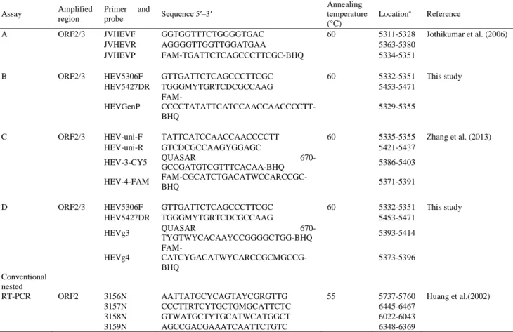
All primers and probes used in this study are listed in Table 1.
Primers and probes from assays B and D developed in this study were designed manually based on a multiple sequence alignment of mHEV genotypes 1-4 in GenBank. Sequences were aligned using CLUSTAL W within DNASTAR (Lasergene 8). A pair of primers (HEV5606F/HEV5427DR) and a probe (HEVGenP) located in the conserved ORF2/ORF3 overlapping region broadly reactive with mHEV genotypes 1-4 were designed (assay B). Additionally, probes specific for the detection of mHEV genotypes 3 or 4 (HEVg3 and HEVg4; assay D) were designed in this same region. Oligonucleotide primers/probes were analyzed for the absence of possible hairpins and dimers by Primer Express software (Version 3.0; Applied Biosystems).
Table 1. Primers and probes used in this study
a Nucleotide positions are in accordance with GenBank accession number AF60669, except for the HEV genotype 4 probes that are
in accordance with GenBank accession number HQ634346.
2.4 Primers and probes
Assay Amplified
region
Primer and
probe Sequence 5′–3′
Annealing temperature (°C)
Locationa Reference
A ORF2/3 JVHEVF GGTGGTTTCTGGGGTGAC 60 5311-5328 Jothikumar et al. (2006)
JVHEVR AGGGGTTGGTTGGATGAA 5363-5380
JVHEVP FAM-TGATTCTCAGCCCTTCGC-BHQ 5334-5351
B ORF2/3 HEV5306F GTTGATTCTCAGCCCTTCGC 60 5332-5351 This study
HEV5427DR TGGGMYTGRTCDCGCCAAG 5453-5471
HEVGenP
FAM- CCCCTATATTCATCCAACCAACCCCTT-BHQ
5329-5355
C ORF2/3 HEV-uni-F TATTCATCCAACCAACCCCTT 60 5335-5355 Zhang et al. (2013)
HEV-uni-R GTCDCGCCAAGYGGAGC 5421-5437
HEV-3-CY5 QUASAR GCCGATGTCGTTTCACAA-BHQ 670- 5386-5403
HEV-4-FAM
FAM-CGCATCTGACATWCCARCCGC-BHQ 5371-5391
D ORF2/3 HEV5306F GTTGATTCTCAGCCCTTCGC 60 5332-5351 This study
HEV5427DR TGGGMYTGRTCDCGCCAAG 5453-5471
HEVg3 QUASAR
670-TYGTWYCACAAYCCGGGGCTGG-BHQ 5393-5414
HEVg4
FAM- CATCYGACATWYCARCCGCMGCCG-BHQ
5373-5396
Conventional nested
RT-PCR ORF2 3156N AATTATGCYCAGTAYCGRGTTG 55 5737-5760 Huang et al.(2002)
3157N CCCTTRTCYTGCTGMGCATTCTC 6445-6467
3158N GTWATGCTYTGCATWCATGGCT 6022-6043