Ciência Rural, v.48, n.2, 2018.
Detection of
Campylobacter
spp. in chilled and frozen broiler carcasses comparing
immunoassay, PCR and real time PCR methods
Detecção de Campylobacter spp. em carcaças de frango de corte resfriadas e congeladas pelos métodos imunoenzimático, PCR e PCR em tempo real
Luciana Pimenta Reis1 Liliane Denize Miranda Menezes2 Graciela Kunrath Lima1
Ethiene Luiza de Souza Santos1 Elaine Maria Seles Dorneles3 Débora Cristina Sampaio de Assis1 Andrey Pereira Lage1 Silvana de Vasconcelos Cançado1 Tadeu Chaves de Figueiredo1*
ISSNe 1678-4596
Received 11.18.16 Approved 11.28.17 Returned by the author 01.05.18 CR-2016-1034.R3
INTRODUCTION
Campylobacteriosis is a foodborne disease associated with human infection by thermophilic Campylobacter spp., mainly C. jejuni and C. coli (BUTZLER, 2004). These bacteria are some of the most widespread causative agents of human gastroenteritis, with many animals, wild and domestic, serving as potential reservoirs (CDC, 2014). Birds, especially broiler chickens, are the main natural reservoir of Campylobacter spp., and there by consumption and handling of poultry and poultry products have been implicated as the main source of infection to humans (HUE et al., 2010, EFSA, 2011, Oliveira et al., 2013). The species that are pathogenic towards humans have
a narrow temperature range for growth (30-46°C) and cannot survive the heating processes used for food preparation (Humphrey et al., 2007).
The detection of Campylobacter spp. in food samples is generally performed using traditional culture-based methods, immunoenzymatic assays, or Polymerase Chain Reaction (PCR)-based methods. The traditional culture-based methods for the isolation,
identification, and differentiation of Campylobacter species are time-consuming and challenging because of the fastidious nature of the microorganisms, which have complex nutritional and environmental requirements (BARROS-VELAZQUEZ et al., 1999; FITZGERALD et al., 2008). The sandwich Enzyme-Linked Immunosorbent Assay (ELISA), which is based on the
1Escola de Veterinária, Universidade Federal de Minas Gerais (UFMG), 31270-901, Belo Horizonte, MG, Brasil. E-mail: [email protected]. *Corresponding author.
2Instituto Mineiro de Agropecuária (IMA), Contagem, MG, Brasil.
3Departamento de Medicina Veterinária, Universidade Federal de Lavras (UFLA), Lavras, MG, Brasil.
ABSTRACT: In order to detect and identify Campylobacter spp. in broiler chicken carcasses, and to compare detection methods, 43 chilled and 43 frozen carcasses were collected and analyzed. Three methodologies were evaluated: an automated Enzyme Linked Fluorescent Assay
(ELFA) VIDAS®30, Polymerase Chain Reaction (PCR) and real-time PCR. Only four chilled carcasses (4.6%) were considered positive for
Campylobacter spp. by VIDAS®30 and no sample was positive when the conventional PCR technique was used. However, real-time PCR
showed a higher incidence of contamination by Campylobacter spp. in broiler carcasses, with 45 (52.3%) positive samples. C. jejuni was the species most frequently reported in the samples (88.8%). No differences in the frequencies of Campylobacter spp. were observed between the chilled and frozen broiler carcasses. In conclusion, real-time PCR was the most sensitive method for the detection of Campylobacter spp. in chilled or frozen broiler carcasses, which were mainly contaminated by C. jejuni.
Key words: C. jejuni, C. coli,detection methods, VIDAS®30.
RESUMO: Com o objetivo de detectar e identificar Campylobacter spp. em carcaças de frango de corte utilizando três metodologias distintas - ensaio imunoenzimático VIDAS®30, Reação em Cadeia da Polimerase (PCR) e PCR em tempo real - foram coletadas e analisadas 43
carcaças de frango resfriadas e 43 congeladas. Quatro carcaças refrigeradas (4,6%) foram consideradas positivas para Campylobacter spp. pelo VIDAS®30 e nenhuma amostra positiva foi identificada quando utilizada a técnica de PCR. Porém, ao analisar as carcaças pela
metodologia da PCR em tempo real, foi observada uma maior incidência de Campylobacter spp., com 45 amostras (52,3%) positivas, sendo
que Campylobacter jejuni foi a espécie mais frequentemente encontrada nas amostras (88,8%). Não foi observada diferença na frequência do micro-organismo entre carcaças de frangos resfriadas e congeladas. Concluiu-se que a técnica de PCR em tempo real apresentou maior sensibilidade na detecção de Campylobacter spp. em carcaças de frangos de corte e que foi encontrada elevada presença de carcaças
contaminadas, especialmente por C. jejuni.
Palavras-chave: C. jejuni, C. coli,métodos de detecção, VIDAS®30.
detection of an antigenic reaction against the bacteria after their cultivation on selective media, is often used for the analysis of Campylobacter spp. in food.This
assay is faster, more sensitive, and more specific than the
traditional culture-based methods. The Enzyme Linked Fluorescent Assay (ELFA) employs a similar principle
featuring a final detection step by fluorescence and is
used in the VIDAS® automated immunoassay system
(bioMérieux, Marcy-l’Étoile, France) (AOAC, 2015). Methods based on PCR and real-time PCR have also been used for the detection of Campylobacter spp. in
food samples due to their high specificity and sensitivity.
In addition to their great accuracy, PCR-based methods are also faster and more easily performed than the other techniques for detecting Campylobacter spp. (SCHNIDER et al., 2010).
Among the control measures that have been recommended to prevent the growth of Campylobacter spp. in broiler carcasses, cold treatment is one of the most important. Several studies
have reported a significant reduction in the percentage
of carcasses contaminated by C.jejuni after freezing (EL-SHIBINY et al., 2009; SAMPERS et al., 2010). However, the effects of cooling and freezing on the survival of Campylobacter spp. in broiler carcasses have not been completely elucidated.
Therefore, the aims of the present study were to detect and identify Campylobacter spp. in chilled and frozen broiler carcasses, and to compare the performance of three detection methods, namely the VIDAS®30
immunoassay method, PCR, and real-time PCR.
MATERIAL AND METHODS
Samples and experimental conditions
Eighty-six broiler carcasses from 43 different production batches were collected in a slaughterhouse industry under the Federal Inspection Service located in Minas Gerais State, Brazil. From each of the 43 batches, one frozen (n = 43) and one chilled (n = 43) broiler carcass were collected and analyzed on the second day after processing. Hence, each carcass sample represented one batch or one repetition.
Microbiological analyses
VIDAS®30 immunoenzymatic assay
The detection of Campylobacter spp. in the broiler carcasses was performed using three distinct methodologies, namely an automated ELFA (VIDAS®30) (AOAC, 2015), PCR (HARMON et al.,
1997; VILARDO et al., 2006), and real-time PCR (SCHNIDER et al., 2010).
Each carcass was aseptically placed in a sterile bag containing 100mL of Bolton broth with a selective supplement (SR183E, Oxoid, Basingstoke, UK) and was subjected to a washing process by rinse friction for approximately 1min (bioMérieux, 2015). After rinsing, the solution was incubated at 37 ± 0.5°C for 4h and then at 42 ± 0.5°C for 24h under microaerophilic conditions (5% O2, 10% CO2, and 85% N2) [Combibag with microaerophilia generator GENbox (bioMérieux, Hazelwood, USA)]. After incubation, 2mL aliquots of the Bolton broth were transferred to sterile tubes to perform the immunoassay by VIDAS®30 (bioMérieux) and 1.5mL aliquots were
transferred to centrifuge microtubes for PCR assays. The immunoassay was performed with the VIDAS®30 system, using the Campylobacter (CAM)
kit (bioMérieux), according to the manufacturer’s instructions. From the aliquot after incubation in Bolton broth, 0.5mL was heated at 100°C for 15min and then transferred to a test well of a reagent strip and inserted into the VIDAS®30 system. For the confirmation of positive results, a loop from
the remaining Bolton broth, that was not heated,
was plated on modified charcoal cefoperazone
deoxycholate agar (mCCDA) (Oxoid) and incubated at 42 ± 0.5°C for 48h under microaerophilic conditions (bioMérieux, 2015). Subsequently, colonies with growth typical of Campylobacter spp. were selected and plated on blood agar and incubated at 42 ± 0.5°C for 24h. Then, they were transferred to a Neisseria-Haemophilus (NH) card for biochemical tests using the VITEK 2® system (bioMérieux) (VALENZA et
al., 2007).
PCR analyses
For Campylobacter spp. determination by PCR, the microtubes containing 1.5mL of inoculated Bolton broth were centrifuged at 20,000 ×g at room temperature and submitted to DNA extraction (PITCHER et al., 1989). DNA purity and concentration were determined by spectrophotometry (GeneQuant™ 100 Classic, GE Healthcare Life Sciences, Little Chalfont, UK)(SAMBROOK & RUSSELL, 2001).
In the multiplex PCR assays, the primers
pg3 (5′- GAACTTGAACCGATTTG-3′) and pg50 (5′-ATGGGATTTCGTATTAAC-3′) were used for the amplification of the flaA gene (460 bp), which is present in Campylobacter species, especially C. jejuni and C. coli. Additionally, the primers C1
Ciência Rural, v.48, n.2, 2018.
of C. jejuni (160 bp), according to HARMON et al. (1997) and VILARDO et al. (2006) (Table 1). The reactions were performed in a total volume of 25µL containing 20ng of DNA, 1× buffer (10mM Tris-HCl, 50mM KCl) (Phoneutria, Belo Horizonte, MG, Brazil), 5.5mM of MgCl2 (Phoneutria), 200µM of each deoxyribonucleotide (dATP, dCTP, dGTP, and
dTTP), 1.0µM of each primer (Eurofins Scientific,
Luxembourg), and 1.25 units of Taq DNA polymerase (Phoneutria). The thermocycling conditions (Veriti® Thermal Cycler, Thermo Fisher Scientific, Waltham,
MA, USA) were as follows: initial denaturation step at 94°C for 4min followed by 25 cycles of denaturation at 94°C for 1min, primer annealing at 45°C for 1min, and elongation at 72°C for 1min.
The final extension step was performed at 72°C for
7min. The amplified products were submitted to
electrophoresis in a 1.5% agarose gel in Tris-borate-EDTA (TBE) buffer (1.0M Tris, 0.01M boric acid, 0.01M EDTA, pH 8.2) and stained with 0.5mg mL-1
ethidium bromide. Products were visualized under ultraviolet light and photographed (L-Pix EX, Loccus Biotecnologia, Cotia, SP, Brazil). A 100-bp DNA size marker (Life Technologies, Carlsbad, CA, USA) was used to estimate amplicons length.
Real-time PCR analysis
Real-time PCR assays were performed using the primers Ccj FusA-L1
(5′-GCCTTGAAGAGATTAAAACAGGGATT-3′)
and Ccj FusA-R1
(5′-TTTAAACGCTGTACCGCAAAGCA-3′)
to amplify the fusA gene of C. jejuni (83 bp). In addition, the primers Ccc FusA-L2
(5′-GCCTTGAGGAAATTAAAACTGGTATT-3′)
and Ccc FusA-L2
(5′-TTTAAATGCAGTTCCACAAAGCA-3′)
were used to amplify the fusA gene of C. coli(83 bp).The probes used were Cj_fusA-probea (FAM- AAGTCTTTCTATCGTTCC -MGBNFQ) for C. jejuni and Cc_fusA-probea (NED- AAGTCTTTCTATTGTTCC -MGBNFQ) for C. coli (SCHNIDER et al., 2010) (Table 1). Real-time PCR assays were performed with the two primer pairs and probes simultaneously, in triplicate. Reactions were performed in a total volume of 25µL containing 1× TaqMan Universal PCR Master Mix (Applied Biosystems, Foster City, CA, USA), 0.3µM of each primer, 0.2µM of each probe, and 2µL of genomic DNA. Amplification was carried out at 50°C for 2min and 95°C for 10min, followed by 40 cycles of 95°C for 15s and 60°C for 1min using anABI 7500 Thermal Cycler Real-Time PCR System (Applied Biosystems). The result was considered positive when at least two of the triplicate samples were
defined as positive, with a threshold cycle (Ct) value
less than 40.
Limits of detection of the PCR and real-time PCR assays
To assess the limits of detection of the PCR and real-time PCR assays, two standard curves were generated. One standard curve was performed with serial dilutions of DNA from C. jejuni and C. coli in ultrapure water. To obtain the other standard curve, DNA from C. jejuni and C. coli were diluted in Bolton broth that had been used to rinse a broiler carcass. The DNA of C. jejuni and C. coli were subjected to serial
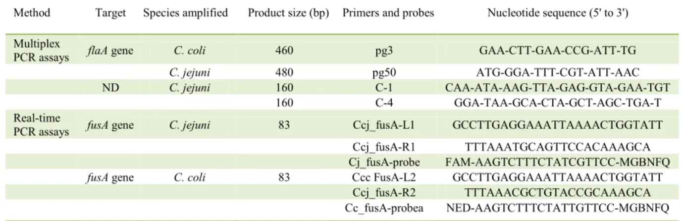
Table 1 - Primers, probes and sequences for C. jejuni and C. coli PCR and real-time PCR assays.
Method Target Species amplified Product size (bp) Primers and probes Nucleotide sequence (5' to 3')
Multiplex
PCR assays flaA gene C. coli 460 pg3 GAA-CTT-GAA-CCG-ATT-TG
C. jejuni 480 pg50 ATG-GGA-TTT-CGT-ATT-AAC
ND C. jejuni 160 C-1 CAA-ATA-AAG-TTA-GAG-GTA-GAA-TGT
160 C-4 GGA-TAA-GCA-CTA-GCT-AGC-TGA-T Real-time
PCR assays fusA gene C. jejuni 83 Ccj_fusA-L1 GCCTTGAGGAAATTAAAACTGGTATT Ccj_fusA-R1 TTTAAATGCAGTTCCACAAAGCA Cj_fusA-probe FAM-AAGTCTTTCTATCGTTCC-MGBNFQ
fusA gene C. coli 83 Ccc FusA-L2 GCCTTGAGGAAATTAAAACTGGTATT Ccj_fusA-R2 TTTAAACGCTGTACCGCAAAGCA
Cc_fusA-probea NED-AAGTCTTTCTATTGTTCC-MGBNFQ
Ciência Rural, v.48, n.2, 2018.
tenfold dilutions from 20ng to 2fg, corresponding to 107 to 100 genome copies of Campylobacter spp.,
respectively (GenBank accession numbers: C. jejuni, CP000814 and C. coli, CP006702) (DOLEZEL et al., 2003). Reference strains, which were used as controls in the PCR and real-time PCR assays, were C. jejuni (NCTC 11351) and C. coli (NCTC 11366). These reference strains were cultivated in blood agar at 42 ± 0.5°C for 48h under microaerophilic conditions.
Statistical analysis
The experiments were conducted using a completely randomized 2 × 3 factorial design, including two types of broiler carcasses (chilled and frozen) and three analytical methodologies (immunoenzymatic VIDAS®30, PCR, and real-time PCR) with 43 repetitions
each. Comparisons among the analytical methodologies were performed by the McNemar test and the analyses of chilled and frozen samples were performed by
the Fisher test at the 5% significance level. Statistical
analysis was performed using R Statistical Software version 3.1.2 (R Foundation for Statistical Computing, Vienna, Austria).
RESULTS AND DISCUSSION
Limits of detection of the PCR and real-time PCR assays
In the present study was observed that real-time PCR proved to be more sensitive than conventional PCR to detect Campylobacter spp. in broiler carcasses (Table 2). Conventional PCR results using the primers designed by HARMON et al. (1997) and VILARDO et al. (2006) showed that those primer pairs were unsuitable for use with food samples. They were designed for the detection of C. jejuniand C. colifrom cultured bacteria using a large amount of microorganisms per sample (HARMON et al. 1997, VILARDO et al., 2006). Several substances
present in food and clinical materials, such as broiler
carcasses, can inhibit or significantly decrease the efficiency of the PCR reactions (SCHRADER et
al., 2012). Moreover, it is important to consider that besides the presence of inhibitors, the amount of Campylobacterspp. usually present in contaminated food is relatively low, owing to their high sensitivity to atmospheric oxygen concentrations and the low temperatures typically used for food preservation (BARROS-VELAZQUEZ et al., 1999).
Detection of Campylobacter spp. using VIDAS®30,
PCR, and real-time PCR assays
The detection of Campylobacter spp. in chicken skin is important for assessing the incidence of contamination among the broiler carcasses that reach the market. Table 3 summarizes results of Campylobacter spp. detection in chilled and frozen broiler carcasses. Among the total chilled carcasses, only four (9.3%) samples were considered positive for Campylobacter spp. by the VIDAS®30 assay
and no sample was considered positive when the conventional PCR technique was used. However, results of the real-time PCR showed a higher incidence of Campylobacter spp. in broiler carcasses, with 45 (52.3%) samples being considered positive, being 24 (55.8%) from chilled and 21 (48.8%) from frozen carcasses. Hence, the results of the present study highlighted the potential public health risks linked to the consumption of chicken and the usefulness of the real-time PCR assay for the detection of Campylobacter spp. in broiler carcasses.
Detection of C. jejuniand C. coli by real-time PCR The real-time PCR assay allowed differentiation between C. jejuni and C. coli and showed that C. jejuni was the most frequently detected Campylobacter species reported in the tested samples (Table 4). C. jejuni was detected in
Table 2– Limit of detection of the PCR and real-time PCR assays used to detect Campylobacter spp. in broiler carcasses.
Primers Dilution Analytical sensitivity
Number of genome
copies Technique Specificity
pg3 / pg50 Water 20 pg 104 PCR Campylobacter spp.
pg3 / pg50 Broiler carcass rinsea 200 pg 105 PCR Campylobacter spp.
C1 / C4 Water 200 pg 105 PCR C. jejuni
C1 / C4 Broiler carcass rinse 2 ng 106 PCR C. jejuni
FusA-L1 / FusA-R1 Water 200 fg 102 Real-time PCR C. jejuni FusA-L1 / FusA-R1 Broiler carcass rinse 200 fg 102 Real-time PCR C. jejuni
FusA-L2 / FusA-R2 Water 200 fg 102 Real-time PCR C. coli
FusA-L2 / FusA-R2 Broiler carcass rinse 200 fg 102 Real-time PCR C. coli a
Ciência Rural, v.48, n.2, 2018.
40 (88.8%) of the 45 positive samples, while C. coli was found in 15 (33.3%) samples. C. jejuni and C. coli were simultaneously reported in ten (22.2%) of the 45 positive broiler carcasses. Among the chilled carcasses that were positive for Campylobacter spp., C. jejuni was detected in 20 carcasses (83.3%) and C. coli was detected in 9 carcasses (37.5%). C. jejuni and C. coli. were simultaneously detected in
five chilled carcasses (20.8%). Among the frozen
carcasses that were positive for Campylobacter spp.,
C. jejuniwas detected in 20 carcasses (95.2%) and
C. coliwas reported in six carcasses (28.5%). Both
C. jejuni and C. coli were simultaneously detected
in five frozen samples (23.8%). It is noteworthy that, usually, 95% of human cases of campylobacteriosis are related to infection by C. jejuni, while C. coli accounts for 4% of cases and C. lari accounts for about 1% (NACHAMKIN et al., 2008).
Comparison among the methods used for Campylobacter spp. detection
A comparison among the three methods used for Campylobacter spp. detection in
broiler carcasses showed a significant difference
(P<0.001) between real-time PCR and the other studied techniques (Table 3). The VIDAS®30
immunoenzymatic assay did not present superior results to those of conventional PCR for the detection of Campylobacter spp. in broiler carcasses. The low bacterial counts in the carcasses could have
influenced the performance of the VIDAS®30 assay
in this study, since only four chilled carcasses were found to be positive for contamination when using this technique. These results may suggest that the VIDAS®30 immunoenzymatic assay was only able to
detect contamination in carcasses with higher loads
of microorganisms. Thus, comparing results ofthe VIDAS®30 assay with those of real-time PCR, the
lower incidence of Campylobacter spp. detected by the former method may suggest that the bacterial counts in the tested broiler carcasses were below the limit of detection (> 103 CFU mL-1) (SAHIN et al., 2008) or
that viable but non culturable bacteria predominated in the samples. Low incidences of the contamination of broiler carcasses by Campylobacter spp. recorded by PCR and VIDAS®30 immunoenzymatic assay
was unexpected, since high rates of contamination has been widely demonstrated in the world and in different Brazilian states (DIAS et al., 1990; FREITAS & NORONHA, 2007; KAAKOUSH et al., 2015).
Findings from real-time PCR showed a completely different situation, i.e., that a high incidence (52.3%) of broiler carcasses were contaminated by Campylobacter spp.. These results
most probably reflect the high analytical sensitivity
of the method, which has a limit of detection of 102
cells mL-1. Lower detection limit of real-time PCR
as compared to those of conventional PCR (104–106
cells mL-1) and the VIDAS®30 assay (> 103 CFU
mL-1) (SAHIN et al., 2003) presumably enabled the
detection of Campylobacter spp. in samples in which the concentration of microorganisms was relatively low, which is the typical situation in food samples.
It should be noted that amplification by real-time
PCR was observed even for low concentrations of Campylobacter spp. DNA (20fg, equivalent to 10cells mL−1). Moreover, the Bolton broth showed no inhibitory effects during the assessment of the detection limit of the real-time PCR assays. However,
since the efficiency of the reaction with this lowest
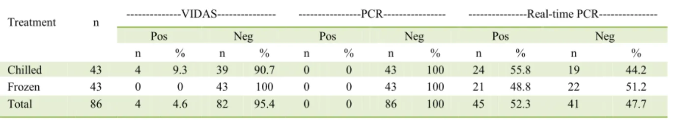
tested concentration of DNA was substantially lower Table 3– Detection of Campylobacter spp. in chilled and frozen broiler carcasses using the immunoassay method VIDA®30, PCR and
real-time PCR.
Treatment n ---VIDAS--- ---PCR--- ---Real-time PCR---
Pos Neg Pos Neg Pos Neg
n % n % n % n % n % n %
Chilled 43 4 9.3 39 90.7 0 0 43 100 24 55.8 19 44.2
Frozen 43 0 0 43 100 0 0 43 100 21 48.8 22 51.2
Total 86 4 4.6 82 95.4 0 0 86 100 45 52.3 41 47.7
McNemar χ2= 2.25; P = 0.1336; gl = 1; VIDAS®30 x PCR.
McNemar χ2 = 39.0244; P = 4.185 x 10⁻¹⁰; gl = 1;VIDAS®30 x real-time PCR. McNemar χ2 = 43.0222; P = 5.412 x 10⁻¹¹; gl = 1; PCR x real-time PCR.
Fisher test: VIDAS – chilled x frozen – P = 0.1162; real-time PCR – chilled x frozen P=0.0625. Pos = positive for Campylobacter spp.
than that observed for the other tested concentrations, these results were disregarded when interpreting the limit of detection. It is also important to consider that very low quantities of C. jejuni, around 8.0× 10² CFU
mL-1, are sufficient to cause infection and diarrhea in
humans (BLACK et al., 1988).
Regarding the temperature treatment of broiler carcass, it is interesting to observe that despite the higher frequency of carcasses contaminated by Campylobacterspp. among chilled samples compared to frozen ones (Table 3), this difference was not
significant (P>0.05). The fact that real-time PCR detects
the genetic material of the microorganism, even if it is no longer viable, could be the reason for the absence of a difference between these storage conditions, since Campylobacterspp. is usually more sensitive to freezing than refrigeration (EL-SHIBINY et al., 2009; SAMPERS et al., 2010). It is possible to speculate that if the immunoenzymatic technique used had shown a higher analytical sensitivity, or if we had incorporated conventional methods for the isolation of Campylobacter
spp., a significantly higher proportion of positive samples
among chilled carcasses would have been observed. Therefore, although the real-time PCR assay detected a high level of Campylobacter spp. contamination among broiler carcasses, it is not possible to make statements about the viability of this microorganism. Thus, it is not possible to assess the real risk to public health using real-time PCR alone. Alternative methods for the detection of viable Campylobacter spp. cells have been researched. Combination of real-time PCR with propidium monoazide was tested by Josefsen et al. (2010) and provided a fast and reliable methodology for
detection and quantification of viable Campylobacter bacteria in broiler carcasses.
CONCLUSION
Real-time PCR was more sensitive than conventional PCR and the VIDAS®30 immunoassay
for the detection of Campylobacter spp. in broiler carcasses. The level of Campylobacter spp.
contamination among chilled or frozen broiler carcasses was high, especially for C. jejuni.
ACKNOWLEDGMENTS
The authors acknowledge the assistance of the Colegiado de Pós-Graduação em Ciência Animal of the Escola de Veterinária at the Universidade Federal de Minas Gerais – UFMG, the Conselho Nacional de Desenvolvimento Científico e Tecnológico (CNPq), the Coordenação de Aperfeiçoamento de Pessoal de Nível Superior (Capes) and the Fundação de Amparo à Pesquisa do Estado de Minas Gerais (FAPEMIG). The authors also thank the Instituto Mineiro de Agropecuária (IMA) for assisting with the analyses.
REFERENCES
AOAC Performance Tested, Certificate No. 051201, 2015.
Vidas®Campylobacter (CAM). Acessado em 15 feb. 2016. Available from: <http://www.aoac.org/imis15_prod/AOAC_Docs/ RI/16PTM/16C_051201_BMC[gwutxn].pdf.>. Accessed: Jul. 25, 2016.
BARROS-VELAZQUEZ, J. et al. Isolation and typing methods for the epidemiologic investigation of thermotolerant campylobacters.
International Microbiology, v.2, p.217-226, 1999. Available from: <http://www.im.microbios.org/08december99/04%20 Barros.pdf>. Accessed: Jul. 25, 2016.
bioMERIÉUX. 2015. Vidas®Campylobacter (CAM). Ref.30.111, 07999 O - en - 2015/01. Available from: <https://techlib. biomerieux.com/wcm/techlib/techlib/documents/docLink/ Package_Insert/50443001-50444000/Package_Insert_-_07999_-_O_-_en_-_30111.pdf.>. Accessed: Fev. 05, 2015.
BLACK, R.E. et al. Experimental Campylobacter jejuni infection in humans. The Journal of Infectious Diseases, v.157, p.472-479, 1988.Available from: <https://academic.oup.com/jid/article-abstr act/157/3/472/859012?redirectedFrom=PDF>. Accessed: Jul. 08, 2016.doi:10.1093/infdis/157.3.472.
BUTZLER, J.P. Campylobacter, from obscurity to celebrity. Clinical Microbiology and Infectious, v.10, p. 868-876, 2004. Available from:<http://dx.doi.org/10.1111/j.1469-0691.2004.00983.x>. Accessed:Jul. 08, 2016. doi: 10.1111/j.1469-0691.2004.00983.x.
CDC. 2014. Campylobacter. Atlanta.Available from: <http:// www.cdc.gov/foodsafety/diseases/campylobacter/index.html>. Accessed: Jul. 08, 2016. Epub 03-Jun-2014.
DIAS, T.C. et al. Chicken carcasses as a source of Campylobacter
jejuni in Belo Horizonte, Brazil. RevistadoInstitutodeMedicina
Table 4 - Detection of C. jejuni and C. coli in chilled and frozen broiler carcasses.
Treatment ---C. jejuni / C. Coli--- ---C. jejuni--- ---C. coli--- Total
n % n % n % n
Chilled 5 20.8 20 83.3 9 37.5 24
Frozen 5 23.8 20 95.2 6 28.5 21
Ciência Rural, v.48, n.2, 2018. TropicaldeSão Paulo, v.32, p. 414-418, 1990. Available from:
<http://dx.doi.org/10.1590/S0036-46651990000600005>. Accessed: Jul. 10, 2016. doi: 10.1590/S0036-46651990000600005.
DOLEZEL J. et al. Nuclear DNA content and genome size of trout and human. Cytometry A, v. 51, p. 127-128, 2003. Available from: <http://onlinelibrary.wiley.com/doi/10.1002/cyto.a.10013/epdf>. Accessed:Jul. 10, 2016. doi: 10.1002/cyto.a.10013.
EFSA – European Food Safety Authority. Scientific opinion on
Campylobacter in broiler meat production: control options and performance objectives and/or targets at different stages of the food chain. EFSA Journal. v.9, p. 1-141. 2011. Available from: <https://www.efsa.europa.eu/en/efsajournal/pub/2105>. Accessed: May. 25, 2017. doi:10.2903/j.efsa.2011.2105.
EL-SHIBINY, A. et al. Survival at refrigeration and freezing temperatures of Campylobacter coli and Campylobacter jejuni
on chicken skin applied as axenic and mixed inoculums.
The International Journal of Food Microbiology, v.131, p.197-202, 2009. Available from: <http://dx.doi.org/10.1016/j. ijfoodmicro.2009.02.024>. Accessed:Jul. 08, 2016. doi: 10.1016/j. ijfoodmicro.2009.02.024.
FITZGERALD, C. et al. Diagnosis and antimicrobial susceptibility
of Campylobacter species. In: Nachamkin, I.; Szymanski, C.M.
Campylobacter. Washington, DC.: ASM Press, 2008.
p.227-243.Available from: <http://www.asmscience.org/content/ book/10.1128/9781555815554.ch12>. Accessed: Jul. 08, 2016.doi: 10.1128/9781555815554.
FREITAS, J.A.; NORONHA, G.N. Occurence
of Campylobacterspp. in chicken meat and giblets exposed
to consumption in Northern Brazil. Arquivo Brasileiro de Medicina Veterinária e Zootecnia, v.59, p.813-815, 2007. Available from:<dx.doi.org/10.1590/S0102-09352007000300038>. Accessed: Jun. 12, 2016. doi: 10.1590/S0102-09352007000300038.
HARMON, K.M. et al. Differentiation of Campylobacter
jejuni and Campylobacter coli by polymerase chain reaction.
MolecularandCellular Probes, v.11, p.195-200, 1997. Available from: <http://dx.doi.org/10.1006/mcpr.1997.0104>. Accessed:Jul. 05, 2016. doi: 10.1006/mcpr.1997.0104
HUE, O. et al. Prevalence of and risk factors for Campylobacter
spp. contamination of broiler chicken carcasses at the slaughterhouse. Food Microbiology. v.27, p.992-999, 2010. Available from:<http://dx.doi.org/10.1016/j.fm.2010.06.004>. Accessed:Jul. 08, 2016. doi: 10.1016/j.fm.2010.06.004.
HUMPHREY, T. et al. Campylobacters as zoonotic pathogens: a food production perspective. International Journal of Food Microbiology. v.117, p.237-257, 2007. Available from: <https:// doi.org/10.1016/j.ijfoodmicro.2007.01.006>. Accessed:Jul. 08, 2016. doi:10.1016/j.ijfoodmicro.2007.01.006.
KAAKOUSH, N.O. et al. Global Epidemiology of Campylobacter Infection. Clinical Microbiology Reviews. v.28, p.687-720, 2015. Available from: <http://cmr.asm.org/content/28/3/687.full>. Accessed:Jul. 08, 2016. doi: 10.1128/CMR.00006-15.
NACHAMKIN, I. et al. Campylobacter.Washington, DC: ASM
Press, 2008. 3th ed. 716p. Available from:<http://www.asmscience.
org/content/book/10.1128/9781555815554>. Accessed: Fev. 22,2016. doi: 10.1128/9781555815554.
OLIVEIRA, A.L., OLIVEIRA, R.B.P. Enumeration of Campylobacter spp. and presence ofCampylobacter jejuni in broiler carcasses in the state of Minas Gerais, Brazil.Ciência Rural. v.43, p.480-484, 2013. Available from: <http://dx.doi. org/10.1590/S0103-84782013005000007>. Accessed: Jan. 05, 2016. doi:10.1590/S0103-84782013005000007.
PITCHER, D.G. et al. Rapid extraction of bacterial genomic DNA with guanidium thiocyanate. Lettersin AppliedMicrobiology.
v.8. p.151-156, 1989. Available from:<http://dx.doi.org/10.1111/ j.1472-765X.1989.tb00262.x>. Accessed:Fev. 05, 2015. doi: 10.1111/j.1472-765X.1989.tb00262.x.
SAHIN, O. et al. Detection of Campylobacter. In: Torrence, M.E.; Isaacson, R.E. Microbial Food Safety in Animal Agriculture: Current Topics. New Jersey: Wiley Online Library. 2003. Cap.
20, p.183-194. Available from: <http://onlinelibrary.wiley.com/ book/10.1002/9780470752616>. Accessed:Jul. 08, 2016.doi: 10.1002/9780470752616.
SAMBROOK, J., RUSSELL. D.W. Molecular Cloning: A Laboratory Manual. New York : Cold Spring Harbor Laboratory Press, 2001.
SAMPERS, I. et al. Survival of Campylobacter spp. in poultry meat preparations subjected to freezing, refrigeration, minor salt concentration, and heat treatment. International Journal of Food Microbiology. v.137, p.147-153, 2010. Available from:<http:// dx.doi.org/10.1016/j.ijfoodmicro.2009.11.013>. Accessed:Fev. 05, 2015. doi: 10.1016/j.ijfoodmicro.2009.11.013.
SCHNIDER, A. et al. Comparison of real-time PCR assays for
detection, quantification, and differentiation of Campylobacter
jejuni and Campylo bactercoli in broiler neck skin samples.
Journal of Food Protection. v.73, p.1057-1063, 2010. Available from:<http://jfoodprotection.org/doi/pdf/10.4315/0362-028X-73.6.1057?code=fopr-site>. Accessed:Fev. 22,2016.
SCHRADER, C. et al. PCR inhibitors - occurrence, properties and removal. Journal of Applied Microbiology. v.113, p.1014-1026, 2012. Available from: <https://doi.org/10.111 1/j.1365-2672.2012.05384.x>. Accessed: Jul. 10, 2016. doi: 10.1111/j.1365-2672.2012.05384.x.
VALENZA, G. et al. Microbiological Evaluation of the New VITEK 2 Neisseria-Haemophilus Identification Card. Journal of Clinical Microbiology. v.45, p.3493-3497,2007. Available from: <https://doi.org/10.1128/JCM.00953-07>. Accessed: Fev. 05. 2015. doi: 10.1128/JCM.00953-07.
VILARDO, M.D.C.B. et al. Application of biochemical
and polymerase chain reaction assays for identification of
Campylobacter isolates from non-human primates. Memórias