INFLUÊNCIA DA LAPAROSCOPIA E DA LAPAROTOMIA NA
GASOMETRIA, LEUCOMETRIA DIFERENCIAL E CITOCINAS EM
MODELO DE SEPSE ABDOMINAL EM RATOS
Dissertação apresentada à Coordenação do Programa de Pós-graduação em Ciências da Saúde, do Centro de Ciências da Saúde da Universidade Federal do Rio Grande do Norte, como requisito para obtenção do título de Mestre em Ciências da Saúde.
Irami Araújo Filho
INFLUÊNCIA DA LAPAROSCOPIA E DA LAPAROTOMIA NA
GASOMETRIA, LEUCOMETRIA DIFERENCIAL E CITOCINAS EM MODELO DE SEPSE ABDOMINAL EM RATOS
Dissertação apresentada à Coordenação do Programa de Pós-graduação em Ciências da Saúde, do Centro de Ciências da Saúde da Universidade Federal do Rio Grande do Norte, como requisito para obtenção do título de Mestre em Ciências da Saúde.
Orientador: Prof. Dr. Aldo da Cunha Medeiros
NATAL - RN
Catalogação da Publicação na Fonte. UFRN / Biblioteca Setorial do CCS
Araújo Filho, Irami.
Influência da laparoscopia e da laparotomia na gasometria, leucometria diferencial e citocinas em modelo de sepse abdominal em ratos / Irami Araújo Filho. – Natal, RN, 2005.
50 f. : il.
Orientador : Aldo da Cunha Medeiros.
Dissertação (mestrado) – Universidade Federal do Rio Grande do Norte. Centro de Ciências da Saúde. Programa de Pós-Graduação em Ciências da Saúde.
1. Laparoscopia – Dissertação. 2. Pneumoperitôneo – Dissertação. 3. Sepse – Dissertação. 4. Acidose – Dissertação. 5. Leucócitos – Dissertação. I. Medeiros, Aldo da Cunha. II. Título.
UNIVERSIDADE FEDERAL DO RIO GRANDE DO NORTE CENTRO DE CIÊNCIAS DA SAÚDE
CURSO DE PÓS-GRADUAÇÃO EM CIÊNCIAS DA SAÚDE DEPARTAMENTO DE CIRURGIA
Chefe do Departamento: Prof. Antônio Medeiros Dantas Filho
Irami Araújo Filho
INFLUÊNCIA DA LAPAROSCOPIA E DA LAPAROTOMIA NA
GASOMETRIA, LEUCOMETRIA DIFERENCIAL E CITOCINAS EM MODELO DE SEPSE ABDOMINAL EM RATOS
Presidente da Banca: Prof. Dr. Aldo da Cunha Medeiros - UFRN
BANCA EXAMINADORA
Prof. Dr. Aldo da Cunha Medeiros – UFRN
Prof. Dr. José Lamartine Andrade de Aguiar - UFPE
Prof. Dr. Jeancarlo Fernandes Cavalcante - UFRN
Profª Dra. Maria José Pereira Vilar - UFRN (Suplente)
Dedico esta dissertação aos meus pacientes, que na dor de seus sofrimentos, depositaram nas minhas mãos a sua confiança e esperança de cura, sendo sempre o motivo maior do meu sacrifício, dedicação e estudo.
9Agradeço a Deus pela minha existência e por ter me dado o dom de ser Cirurgião;
9Aos meus pais, Irami e Terezinha pelos sentimentos de responsabilidade, lealdade e amizade a mim transmitidos;
9Ao Professor Aldo Medeiros, meu segundo pai, pela confiança em mim depositada, sem a qual não teria despertado meu gosto pela ciência e não teria concluído esta etapa tão importante de minha vida;
9A Amália Rêgo minha amada e fiel companheira nos momentos mais difíceis; a Irami Neto, meu filho, razão do meu viver, por compreender a ausência de seu pai e a privação do lazer;
9A Raquel e Ricardo, meus irmãos, pelo apoio fraterno e torcida alegre;
9A todos que contribuíram e contribuem para a minha formação como Médico Cirurgião e pessoa humana, em especial aos Professores Dr. Antônio Medeiros, Dr. Edílson Pinto, Dr. Tertuliano Aires, Dr. Isaú Gerino e ao Dr. Ernani Rosado.
9Aos alunos do Programa de Iniciação Científica, Amália Cínthia
Meneses do Rêgo, Ticiana Cabral da Costa, Daniele Fernandes
Pimentel, Ana Cláudia Medeiros de Amorim Garcia, Abraão Allen
Honorato Sobrinho e Thaís Medeiros Cruz, por terem colaborado
decisivamente na realização do presente trabalho.
Dedicatória... vi
Agradecimentos ... vii
Resumo... viii
1 INTRODUÇÃO... 01
2 REVISÃO DA LITERATURA... 04
2.1.Laparoscopia e função imune... 05
2.2.Resposta ao trauma e citocinas ... 05
2.3.Gasometria ... 07
2.4.Leucócitos periféricos e cirurgia laparoscópica ... 07
3 ANEXAÇÃO DOS ARTIGOS... 09
3.1. ARTIGO I Influence of laparoscopy and laparotomy on gasometry, leukocytes and cytokines in a rat abdominal sepsis model... 09
3.2. ARTIGO II Total gastrectomy with substitution of stomach by jejunal pouch with and without duodenal passage. Study in rats... 22
3.3. ARTIGO III Association of advanced gastric carcinoma with Helicobacter pylori infection... 31
4 COMENTÁRIOS, CRÍTICAS E CONCLUSÕES... 41
5 REFERÊNCIAS... 45
Abstract
A cirurgia laparoscópica está associada com trauma reduzido e baixa
resposta metabólica na fase aguda do trauma, quando comparada com a
cirurgia aberta. As citocinas e o balanço ácido-base são fatores importantes
da resposta biológica ao trauma cirúrgico-anestésico. O objetivo deste
estudo foi determinar se o pneumoperitônio com CO altera a expressão
das citocinas intraperitoneais, a gasometria do sangue arterial, dos
exsudatos intraperitoneal e subperitoneal, e a contagem diferencial de
leucócitos em ratos com sepse abdominal. Método: Ratos Wistar foram
aleatoriamente distribuídos em cinco grupos: controle (somente 2
anestesia),
laparotomia, pneumoperitônio com CO2, ligadura e punção do ceco por
laparotomia, ligadura e punção do ceco por laparoscopia. Após 30 minutos
dos procedimentos, sangue arterial foi colhido para leucometria em
hemocitômetro. FNTD, IL-1E e IL-6 foram dosadas no lavado intraperitoneal
(por ELISA). Os parâmetros gasosos foram medidos no sangue arterial e
nos exsudatos intraperitoneal e subperitoneal. Resultados: Os valores de
FNTĮ, IL-1ȕ e IL-6 foram significantemente menores nos ratos submetidos
ao pneumoperitônio do que em todos os outros grupos (p<0.05). Expressão
de FNTĮ, IL-1ȕ e IL-6 foi menor no grupo sepse induzida por laparoscopia
do que por laparotomia (p<0.05). Os ratos submetidos à ligadura e punção
do ceco por via laparoscópica desenvolveram acidose hipercárbica no
sangue arterial e exsudato subperitoneal, mais intensa do que no grupo
sepse laparotômica. Leucopenia e linfopenia foram mais acentuadas no
grupo sepse laparoscópica (p<0.01). Entretanto, os animais submetidos a
eosinófilos quando comparados com os controles (p<0.05). Conclusões:
Este estudo demonstrou que o pneumoperitônio com CO contribuiu para
reduzir a resposta inflamatória e imunológica em ratos submetidos a modelo
de sepse abdominal, no que diz respeito a citocinas intraperitoneais e
leucometria diferencial. O pneumoperitônio também contribuiu para
instalação de acidose hipercárbica nos ratos sépticos.
2
Palavras chave: Pneumoperitônio, Gás carbônico, Sepse, Acidose,
Leucócitos.
1 INTRODUÇÃO
A cirurgia videoendoscópica teve início em 1987, e tem como base provocar um mínimo trauma aos pacientes, reduzir a permanência hospitalar e a ausência às atividades no trabalho e na família. Na videocirurgia por via laparoscópica é indispensável a insuflação de CO2 na cavidade peritoneal, para o acesso visual aos órgãos abdominais com segurança1,2.
De um modo geral, a imunossupressão é uma conseqüência bem estabelecida do estresse cirúrgico e da lesão 4,6,7. Isto não só tem sido definido no que diz respeito aos níveis séricos de citocinas, mas também em relação às alterações celulares da resposta ao trauma5,12. Vários estudos têm examinado como a cirurgia laparoscópica afeta vários componentes da resposta inflamatória e do sistema imunológico3-6,8,9.
A cirurgia laparoscópica pode atenuar a imunossupressão celular induzida pelo trauma operatório em termos de contagem de leucócitos totais, populações de leucócitos específicos, e subpopulações de leucócitos3,4. Foi demonstrado em alguns estudos um aumento significante nos leucócitos periféricos nas operações pela técnica aberta, quando comparado com pacientes submetidos à colecistectomia laparoscópica 4-7. Kloosterman et al (1984)8 demonstraram um aumento passageiro no número de granulócitos após colecistectomia aberta, fato que não ocorreu após colecistectomia laparoscópica.
intracelular, criando um ambiente mais ácido. Sabe-se que a função dos macrófagos é prejudicada através da queda no pH intra e extracelular12,13. West et al(1995)12 especulam que a diminuição na produção de citocinas pelos macrófagos peritoneais pode contribuir para uma aparente depressão na resposta sistêmica inflamatória durante a cirurgia laparoscópica. Os autores especulam que um mecanismo molecular potencial, ainda não descrito, possa explicar a imunossupressão dos macrófagos peritoneais.
Uma hipótese alternativa foi sugerida por Watson et al(1995)14 em um estudo em ratos submetidos à laparoscopia com ar atmosférico e gás carbônico. Os animais do grupo de controle sofreram laparotomia aberta. Neste estudo, macrófagos do tecido peritoneal liberaram superóxido e FNT após laparotomia e laparoscopia com ar atmosférico em quantidades significativamente maiores, quando comparado com o procedimento de controle e laparoscopia com gás carbônico. Porém, a fagocitose dos macrófagos peritoneais foi significativamente menor na laparoscopia com ar e na laparotomia, quando comparados com insuflação de gás carbônico. Os autores especularam que algum fator no ar, talvez uma pequena quantidade de endotoxina contaminante, em lugar de gás carbônico, era responsável pelas alterações na função dos macrófagos. Tudo indica que o pneumoperitônio com gás carbônico, através de mecanismos ainda obscuros, parece atenuar a resposta imune dos macrófagos peritoneais.
Sem dúvida, a eficácia clínica da cirurgia laparoscópica está bem estabelecida. Está cada vez mais aparente que aquelas respostas sistêmicas imunes e metabólicas da cirurgia aberta podem não se aplicar totalmente à cirurgia laparoscópica. A linha de pesquisa nesta área tem procurado explicar esses fenômenos e, como esses esforços se multiplicam, serão em breve melhor entendidas as conseqüências sistêmicas, metabólicas e imunes da cirurgia laparoscópica e os pacientes serão os reais beneficiários. No que diz respeito à cirurgia laparoscópica em casos de peritonite, permanece a dúvida se a atenuação da resposta imune, provavelmente provocada pelo pneumoperitôneo com CO2, seria prejudicial ou benéfica para os operados.
2 REVISÃO DA LITERATURA
A partir de 1990 a cirurgia laparoscópica vem progressivamente substituindo com vantagens a cirurgia abdominal aberta, também denominada convencional. Tem resultado em menos dor pós-operatória, retorno mais rápido às atividades normais e melhor efeito cosmético. Outros fatores distinguem a cirurgia laparoscópica da cirurgia aberta: uso do pneumoperitôneo com atmosfera de CO2, menor dissecção de estruturas intra-abdominais, mínimas mudanças de temperatura e uma melhor preservação da resposta imune tanto sistêmica quanto intraperitoneal. A principal vantagem da cirurgia laparoscópica sobre a cirurgia aberta é que o estado da normalidade é recuperado mais rapidamente. Vários estudos têm mostrado que ocorre menor lesão tecidual e conseqüente menor repercussão na resposta inflamatória15-19 e preservação da imunidade10,20-22 . Além disso, a incidência de infecção cirúrgica é reduzida com a cirurgia laparoscópica23-25. Várias séries publicadas têm demonstrado que os procedimentos laparoscópicos prolongados, mesmo em casos de infecção intraabdominal, são bem tolerados e não aumentam a translocação bacteriana ou a disseminação intra-abdominal da sepse26,27.
Desde sua introdução em 1987, a cirurgia laparoscópica e, especialmente a colecistectomia laparoscópica foi rapidamente aceita para uso na clínica cirúrgica. Este procedimento minimamente invasivo requer o pneumoperitôneo para a visualização adequada da cavidade peritoneal e das estruturas a serem operadas28-31. O gás carbônico tem sido usado para insuflação por ser de baixo custo e não inflamável. Sua capacidade de difusão é alta, com subseqüente rápida absorção e excreção29,32. Alguns efeitos colaterais do pneumopetitôneo com CO2têm sido descritos, como hipercapnia, acidose e hipertensão pulmonar33-36. Além do CO2, outros gases como hélio, argônio e óxido nitroso (N2O), como também a laparoscopia sem gás têm sido investigados28,30,31,37.
imune4,39. Partindo do princípio de que este aspecto é importante, é preciso que seja esclarecido se a laparoscopia está ou não associada com significativas vantagens ou desvantagens imunológicas e se este fato tem implicações nos resultados das ressecções oncológicas e especialmente na presença de sepse abdominal.
2.1. Laparoscopia e função imune
Como a cirurgia laparoscópica causa menos trauma tecidual do que a cirurgia aberta, é de se esperar que esteja associada com uma melhor preservação da função imune sistêmica. A causa exata dos melhores resultados clínicos está sendo investigada, mas a melhor preservação da função imune sistêmica deve estar contribuindo para a melhora na recuperação dos doentes40-42. Após a cirurgia aberta convencional, a função imunológica em geral está deprimida, com alterações importantes nos níveis de citocinas e mudanças nas funções dos componentes celulares da resposta sistêmica imune40,43,44.
A resposta fisiológica ao trauma é um aumento imediato nos níveis de uma série de hormônios e conseqüentemente, diminuição na resposta imune celular. Tal resposta manifesta-se como uma redução na interação entre linfócitos e macrófagos, redução na atividade das células killer,
diminuição na quimiotaxia de linfócitos e neutrófilos, como também atenuação nas respostas de sensibilidade retardada45. Em geral, a resposta imune à cirurgia depende principalmente, de alterações nas funções e níveis das citocinas e do sistema imunológico exercido pelas células.
2.2. Resposta ao trauma e citocinas
inflamatórios, pode levar a efeitos deletérios nos tecidos. No pós-operatório, a diminuição na produção de citocinas, que pode representar reação inflamatória deduzida, pode ser considerada benéfica.
As citocinas IL-1, FNT e IL-6 desempenham um papel importante na resposta aguda ao trauma46. A expressão de IL-6 tem sido considerada diretamente proporcional à extensão do trauma cirúrgico47. Seus níveis plasmáticos correlacionam-se fielmente com o porte do trauma cirúrgico e uma significante deferença tem sido encontrada nos níveis plasmáticos pós-operatórios quando comparadas às colecistectomias aberta e laparoscópica18,48,49. Leung et al(2000) ,50 em estudo randômico de 34 pacientes submetidos a ressecções laparoscopicamente assistidas ou abertas de retosigmoide por câncer, não encontraram aumento nos níveis
de FNT em ambos os grupos. No entanto, os níveis séricos de IL-1E e IL-6 tiveram um pico 2 horas após as operações, com menor resposta após a cirurgia laparoscópica. Hill et al(1995)51 não encontraram qualquer diferença nos níveis de IL-6 após o reparo de hérnias inguinais por vias laparoscópica e convencional. Esta discrepância de resultados pode ter ocorrido porque o nível de trauma na herniorrafia aberta não deve ter sido suficiente para gerar níveis aumentados desses marcadores. Níveis plasmáticos de IL-8, um importante fator de quimiotaxia para neutrófilos, foram encontrados em níveis maiores na cirurgia aberta do que na laparoscópica52. Resultados semelhantes têm sido observados por outros investigadores15.
2.3. Gasometria
Do ponto de vista fisiopatológico, a insuflação da cavidade peritoneal com gás carbônico parece ser o aspecto mais importante na cirurgia laparoscópica, comparada à cirurgia convencional ou aberta. Várias publicações têm surgido a respeito, relatando que o pneumoperitôneo com CO2 tem efeitos adversos potenciais no estado ácido-base, função pulmonar e hemodinâmica cardiovascular53-55.
Em operações em que a insuflação de CO2 é prolongada, mesmo em pacientes jovens e saudáveis, pode ocorrer importante alteração no balanço ácido-base, devido ao aumento da pressão intra-abdominal e absorção de CO2 através da serosa peritoneal35,56.
2.4. Leucócitos periféricos e cirurgia laparoscópica
pelos polimorfonucleares foi observada após técnicas laparoscópicas, quanto comparadas com técnicas abertas, sugerindo um maior estado de ativação dessas células57,62,63.
3 ANEXAÇÃO DOS ARTIGOS
3.1. ARTIGO I - Influence of laparoscopy and laparotomy on gasometry, leukocytes count and cytokines in a rat abdominal sepsis model
Aceito para publicação na Acta Cirúrgica Brasileira –Indexada MEDLINE.
Influence of laparoscopy and laparotomy on gasometry,
leukocytes count and cytokines in a rat abdominal sepsis
model
1Influência da laparoscopia e laparotomia na gasometria, leucometria e citocinas em modelo de sepse abdominal em ratos
Irami Araújo Filho2, Abraão Allen Honorato Sobrinho3, Amália Cinthia Meneses do Rego3, Ana Claudia M. de Amorim Garcia3, Daniele Pimentel Fernandes3, Thaís Medeiros Cruz3, Ticiana Cabral da Costa3, Aldo Cunha
Medeiros4
1. Article from Nucleus of Experimental Surgery, Postgraduate Program in Health Sciences, Federal University of Rio Grande do Norte (UFRN), Brazil. 2. Postgraduate Fellow, Postgraduate Program in Health Sciences, Federal University of Rio Grande do Norte, Brazil.
3. Medical Student, Scientific Initiation Program, UFRN, Brazil.
4. Professor, Doctor, Chief, Nucleus of Experimental Surgery, UFRN, Brazil.
_______________________________________________________________
ABSTRACT
Purpose: Laparoscopic surgery is associated with reduced surgical trauma, and less acute phase response, as compared with open surgery. Cytokines are important regulators of the biological response to surgical and anesthetic stress. The aim of this study was to determine if CO2 pneumoperitoneum would change cytokine expression, gas parameters and leukocyte count in septic rats.
Methods: Wistar rats were randomly assigned to five groups: control
(anesthesia only), laparotomy, CO2 pneumoperitoneum, cecum ligation and puncture by laparotomy, and laparoscopic cecum ligation and puncture. After 30 min of the procedures, arterial blood samples were obtained to determine leukocytes subpopulations by hemocytometer. TNFD, IL-1E, IL-6 were determined in intraperitoneal fluid (by ELISA). Gas parameters were measured on arterial blood, intraperitoneal and subperitoneal exsudates. Results:
significant hypercarbic acidosis in blood and subperitoneal fluid when compared to open procedure group. Total white blood cells and lymphocytes were significantly lower in laparoscopic cecum ligation and puncture rats than in the laparotomic (p<0.01). Nevertheless, the laparotomic cecum ligation rats had a significant increase in blood neutrophils and eosinophils when compared with controls (p<0.05). Conclusions: This study demonstrates that the CO2 pneumoperitoneum reduced the inflammatory and immune response in an animal model of peritonitis with respect to intraperitoneal cytokines, white blood cell count and clinical correlates of sepsis. The pneumoperitoneum produced hypercarbic acidosis in septic animals.
Key words: Pneumoperitoneum. Carbonic gás. Sepsis. Acidosis.
Leucocytes.
RESUMO
Objetivo: A cirurgia laparoscópica está associada com trauma reduzido e baixa resposta na fase aguda do trauma, quando comparada com a cirurgia aberta. As citocinas e o balanço ácido-base são fatores importantes da resposta biológica ao trauma cirúrgico-anestésico. O objetivo deste estudo foi determinar se o pneumoperitôneo com CO2 altera a expressão das citocinas, a gasometria e a contagem diferencial de leucócitos em ratos com sepse abdominal. Métodos: Ratos Wistar foram aleatoriamente distribuídos em 5 grupos: controle (somente anestesia), laparotomia, pneumoperitôneo com CO2, ligadura e punção do ceco por laparotomia, ligadura e punção do ceco por laparoscopia. Após 30 minutos dos procedimentos, sangue arterial foi colhido para leucometria diferencial em hemocitômetro. TNFD, IL-1E e IL-6 foram dosadas no líquido intraperitoneal (por ELISA). Os parâmetros gasosos foram medidos no sangue arterial e nos exsudatos intraperitoneal e subperitoneal.
Resultados: Os valores de TNFĮ, IL-1ȕ e IL-6 foram significantemente menores nos ratos submetidos ao pneumoperitôneo do que em todos os outros grupos (p<0.05). Expressão de TNFĮ, IL-1ȕ e IL-6 foi menor no grupo sepse induzida por laparoscopia do que por laparotomia (p<0.05). Os ratos submetidos a ligadura e punção do ceco via laparoscópica desenvolveram acidose hipercárbica no sangue arterial e exsudato subperitoneal, mais intensa do que no grupo sepse laparotômica. Leucopenia e linfopenia foram mais acentuadas no grupo sepse laparoscópica (p<0.01). Entretanto, os animais submetidos a sepse laparotômica desenvolveram significante aumento de neutrófilos e eosinófilos quando comparados com os controles (p<0.05).
Conclusões: Este estudo demonstrou que o pneumoperitôneo com CO2
contribuiu para reduzir a resposta inflamatória e imunológica em ratos submetidos a modelo de sepse abdominal, no que diz respeito à expressão de citocinas intraperitoneais e leucometria diferencial. O pneumoperitôneo também contribuiu para instalação de acidose hipercárbica nos ratos sépticos.
Descritores: Pneumoperitôneo. Gás carbônico. Sepse. Acidose. Leucócitos.
Introduction
Operative laparoscopy brought a new dimension to surgical practice, and many experimental and clinical studies have demonstrated feasibility, safety, cost-benefit, and pathophysiologic occurrences. The intraabdominal insufflation of carbon dioxide (CO2) is the most widely used technique for the creation of a pneumoperitoneum. Insufflation under a continuous monitoring of intraabdominal pressure throughout the surgical procedure provides adequate exposure of the operating field. As alternatives, different gases (e.g., helium, argon, nitrous oxide, air) may be used, but they have not been adopted clinically1,2,3,4,5,6. The actual knowledge concerning pneumoperitoneum are sometimes inconsistent about the physiologic consequences induced by insufflation of the peritoneal cavity with CO2. Results from animal models about the effects of a pneumoperitoneum in certain pathologic conditions are often alarming7. However, the clinical impact of these changes is unknown, since the majority of patients who undergo laparoscopic procedures do not exhibit any adverse clinical effects either in the short or the long-term course. Laparoscopíc surgery is applied increasingly to abdominal diseases complicated by diffuse or localized peritonitis such as appendicitis, perforated peptic ulcers and diverticulitis8,9. A specific paper has reported the use of laparoscopy in diverticulitis complicated by localized peritonitis with intra-abdominal abscess formation10, and diagnostic laparoscopy is being advocated in diffuse peritonitis after blunt abdominal trauma11.
However, a theoretical concern with the use of laparoscopic techniques in clinical cases complicated by intra-abdominal infection and peritonitis, is that carbon dioxide pneumoperitoneum may increase the risk of bacteraemia and sepsis by increasing intra-abdominal pressure. Some studies have demonstrated immunossuppressive effects of carbon dioxide on neutrophil and macrophage function. In one study, CO2 blocked superoxide release from activated polimorphonuclear leukocytes and significantly reduced the secretion of IL-1 from human peritoneal macrophages12. Whereas these effects might be considered beneficial from the standpoint of inflammation following elective surgery, experimental evidence suggests that the CO2 induced immunossuppression might be deleterious in the setting of infection13. This may have an adverse effect on clinical outcome when compared with open procedures. Although some evidences, few data exist regarding the effect of pneumoperitoneum and increased intra-abdominal pressure on sepsis and physiological outcome.
Methods
Animals and groups - Male Wistar rats (Bioterium from Nucleus of
Experimental Surgery, Federal University of Rio Grande do Norte, Brazil), 12 to 13 weeks old, were housed in cages where standard chow and water were available ad libitum. The rats were acclimatized to the laboratory environment for 5 days on arrival and then fasted for 12 hours before any procedures. Anesthesia was obtained using pentobarbital 20 mg/Kg intraperitoneal and ketamine 50 mg/Kg intramuscular. All surgical procedures were performed under aseptic conditions. The animals were allowed to breathe spontaneously for the duration of the experiment. The group C (control) rats were subjeted to anesthesia only (n=6). In the LAP (laparotomy) group (n=7) the following procedures were performed: after anesthesia and antisepsis with povidone, a 5cm laparotomy keeped the peritoneal cavity exposed to the room air during 30 minutes and the abdominal wall was sutured with nylon 4-0. The PNP rats (n=7) were subjected to CO2 pneumoperitoneum using a Veress needle under 3 mmHg for 30 minutes. On the CLP/LAP (n=6) the CLP was performed after laparotomy. A cecum ligation and puncture (CLP) by laparoscopy under pneumoperitoneum were performed on the CLP/PNP rats (n=6).
Cecal Ligation and Puncture - Pneumoperitoneum was achieved by
introducing a Veress needle into the peritoneal cavity and insufflating (Endomed insufflator) the abdomen with 3 mmHg CO2. Laparoscopic procedures were performed using 3-mm instruments (Henke-Sass, WolfTM) introduced into the abdomen. Cecal ligation and puncture (CLP) consisted of dissection of the cecum, ligation midway between the ileocecal valve and the terminal cecum using a 3-0 chromic catgut tie, and 8-punctures of the isolated cecum with a hollow 25-gauge needle introduced through the abdominal wall. Laparotomy, for the open CLP group, consisted of a 5-cm midline abdominal incision. The duration of the total procedure, and therefore the duration of anesthesia, pneumoperitoneum, and laparotomy, was standardized to 30 minutes for all groups. Postoperatively, animals were resuscitated with a subcutaneous injection of lactated Ringer’s (30 mL/kg) and were again housed in cages where water was available ad libitum. The experimental protocol was approved by the Research Ethics Committee of the Federal University of Rio Grande do Norte, Brazil, and adhered to the Guide for the Care and Use of Laboratory Animals, US National Research Council, 1996.
Gasometry and cytokines dosage - After the surgical procedures, 5mL of
buffered saline were injected in peritoneal cavity and the abdomen was softly massaged for 1 minute. Thirty minutes later, whole blood was collected by cardiac puncture and liquid exsudate was collected from peritoneal cavity and from subperitoneal space, using heparinized capilar tube, for determination of pH, pCO2 and pO2.An automatic AVL (Roche£) equipment was used. TNFD, IL-1ȕ and IL-6 were determined in the intraperitoneal exsudate, by enzyme-linked immunosorbent assay, using cytokine-kits from PeproTec (Rocky Hill, NJ,USA).
Leukometry - Animals were evaluated 24 hours postoperatively for
leukocyte cell counts and the rats were then killed via anesthetic overdose. The determination of leukocite cell counts was preformed using a commercially available automated cell counter (Abbott Cell-Dyn 3500R- CD 3500 5L, USA).
Data were expressed as meanrstandard deviation. Statistical significance was established using the one way analysis of variance ANOVA followed by Newman-Keuls test, performed by the software BioEstat 2.0. Probabilities less than 0.05 were considered significant.
Results
Cytokines release in peritoneal exsudate - Rats in the control, LAP and
PNP groups exhibited normal activity and had no piloerection during the 24 hours after interventions. In contrast, all rats that underwent CLP exhibited decreased activity and significant piloerection. The cytokine levels on LAP rats were higher than PNP ones; however, the difference between these groups was not significant (p>0.05).
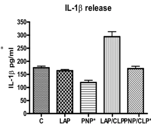
Both surgical procedures, PNP/CLP and LAP/CLP, induced higher TNFĮ, IL-1ȕ and IL-6 in the peritoneal fluid than were found in the control group. In contrast, the peritoneal fluid TNFĮ, IL-1ȕ and IL-6 levels in the pneumoperitoneum (PNP) group were significantly lower than in the other groups (p<0.05). The PNP/CLP rats had a significantly lower elevation of TNFĮ, IL-1ȕ and IL-6 expression in the peritoneal fluid than LAP/CLP rats (p<0.05) (Figures 1,2,3).
Figure 1 - No statistical difference was observed between groups C and LAP. When comparing IL-1ȕ expression in groups LAP/CLP and PNP/CLP**, the difference was significant. (p<0.05).
IL-1E release
C LAP PNP* LAP/CLPPNP/CLP** 0
50 100 150 200 250 300 350
*
IL
-1
E
p
g
/m
Figure 2 - The laparoscopic sepsis group (PNP/CLP) expressed IL-6 significantly lower than laparotomic group (p<0.05). *p<0,05 vs C, PNP/CLP, LAP/CLP
IL-6 release
C LAP PNP* LAP/CLPPNP/CLP** 0
50 100 150 200 250 300 350
IL
-6
p
g
/m
l
Figure 3 - TNF-Į levels were significantly lower in laparoscopic sepsis rats (PNP/CLP) than when sepsis was induced by laparotomy (LAP/CLP) (p<0.05).
TNFD release
C LAP PNP* LAP/CLPPNP/CLP** 0
100 200 300
TN
F
D
pg/
m
L
Gasometry - Arterial blood gas parameters (pH, pO2) in rats from the
control group (C) remained significantly higher than in rats from LAP, PNP and PNP/CLP groups (p<0.01). Rats from PNP/CLP group developed significant hypercarbic acidosis with mean pH of 7.18r0.05 and pCO2 60.7r10.2 when compared to LAP/CLP group. The LAP/CLP rats acidosis was not hypercarbic (Table 1). The pCO2 was significantly higher on PNP/CLP rats than controls (p<0.01). Significantly reduced pCO2 was observed following LAP/CLP, compared to LAP, PNP and PNP/CLP (p<0.01).
Table 1 – Arterial blood gas parameters
GROUP pH pCO2 pO2
C 7.36±0.08* 43.4±4.7§ 88.5±9*
LAP 7.23r0.03 50.1r9.2 62.4r19
PNP 7.22r0.07 48.6r9.6 61.2r15
PNP/CLP 7.18±0.05** 60.7±10.2 63.7±18
LAP/CLP 7.26±0.04 35.45±7.9† 49.4±19
Table 2 – Intraperitoneal exsudate gas parameters
GROUP pH pCO2 pO2
C 7.31±0.16 33.6±7.6 113.3±23
LAP 7.30r0.1 39.7r10 95r31
PNP 7.16r0.16** 47r9.7 102r15
PNP/CLP 6.8±0.25* 86.5±41.3* 82.3±15§
LAP/CLP 7.27±0.11 48.3±9.4 122.3±19
*P<0,01 vs C, LAP, PNP, LAP/CLP; **p<0.01 vs C, LAP; § p<0,05 vs C, PNP/CLP
Intraperitoneal exsudate gas analysis revealed acidosis in the PNP group, and the difference was significant when compared with C and LAP groups (p<0.01). However, the septic rats subjected to CO2pneumoperitoneum (PNP/CLP) developed a profound intraperitoneal acidosis, as a consequence of pCO2 significantly higher (p<0.01) than in all other groups (Table 2). In contrast, the pO2 was significantly lower than in C and LAP/CLP groups (p<0.05).
While subperitoneal exsudate pH (6.5±0.05) following laparoscopic CLP using CO2 (PNP/CLP) was significantly lower (p<0.01) than in C, LAP and LAP/CLP rats, the difference did not reach statistical significance when compared with the acidotic pH (6.7±0.08) of PNP group (Table 3). The CO2 pneumoperitoneum produced a significant increase in pCO2 in the subperitoneal exsudate, when compared with all the other groups (p<0.01).
Table 3 – Subperitoneal exsudate gas parameters
GROUP pH pCO2 pO2
C 7.16±0.2 21±8 146±12
LAP 7.0r0.2 18r9.6 161r20
PNP 6.7r0.08 15r4.4 130r19
PNP/CLP 6.5±0.05* 46±3.8* 122±8.9*
LAP/CLP 7.16±0.1 24±9.8** 165±10.8**
*p<0,01 vs C, LAP, LAP/CLP; **p<0,01 vs PNP/CLP
Leukocytes - White blood cell count in pneumoperitoneum and
laparoscopic CLP groups was similar to those of controls (Table 4). Significantly reduced white cell counts were observed following laparoscopic CLP compared to open CLP (p<0.01)
PNP/CLP, compared with controls (p<0.05). The decrease in LAP/CLP eosinophil was greater than in PNP/CLP rats.
Table 4. Leukocyte blood counts after laparotomy, pneumoperitoneum and cecal ligation and puncture.
LEUCOCYTES C LAP PNP PNP/CLP LAP/CLP
WBC (K/µL) 6.2±0.6 8.8±0.9ɮ 4.8±0.6 4.7±0.6† 10.4±1.9 Neutrophil 51.7±12 49.1±12 51.3±9.5 60.8±11 78.2±8*
Lymphocyte 32.5±10 40±14 36.4±9 23±4.8** 11.7±3
Eosinophils 4±0.5 3.9±0.3 1.7±0.06˩§ 2±0.08 0.8±0.01* C, control; LAP, laparotomy; PNP, pneumoperitoneum; PNP/CLP, pneumoperitoneum/cecum ligation and puncture; LAP/CLP, laparotomy/ cecum ligation and puncture; WBC, white blood count.
p<0.01 vs LAP, LAP/CLP; ɮ p<0.01 vs PNP, PNP/CLP; † p<0.01 vs LAP/CLP; *p<0.01 vs C, LAP, PNP, PNP/CLP; **p<0.01 vs LAP,PNP; §p<0.05 vs C,LAP, LAP/CLP.
Discussion
Because laparoscopic surgery is increasingly used for treating peritonitis and other septic states, a theoretic concern is related to the hypothesis that CO2 pneumoperitoneum may increace bacteremia with adverse effects for the patient. Some studies have reported technical fiseability of laparoscopic appendectomy, perforated peptic ulcer, perforated diverticulitis and other septic surgical situations14,15,16. They are small studies with low numbers of included patients, that can not give strong evidence about improved safety of laparoscopic surgery regarding septicemia. Study in rats showed that pneumoperitoneum causes intestinal ischemia with oxygen free radical production and bacterial translocation, related to mechanical pressure of CO217. Other studies focused on whether a pneumoperitoneum amplifies the extent and severity of peritonitis or of bacteremia in various animal models18,19,20,21. Findings from these investigations are controversial. Whereas some authors reported no increase in bacteremia, intraperitoneal abscess formation, or correlates of sepsis, others reported increased bacterial translocation and severity of peritonitis and sepsis.
Laparoscopic surgical technique requires the maintenance of a continuous positive intraperitoneal pressure in patients (approximately 10-15 mmHg) for visualization and manipulation of the viscera. In cases of peritonitis, viable bacteria and bacterial byproducts (including endotoxin) exist free in the peritoneal cavity. Positive intraperitoneal pressure may increase bacteraemia and endotoxaemia, and thus may worsen clinical sepsis. As experience with laparoscopic surgery increases, its use in more debilitated and critically ill patients is being reported22. These patients often have sepsis and suffer from diffuse peritonitis of unclear aetiology, usually the result of a perforated viscera. It has not been clear whether a laparoscopic approach worsens the septic state or whether minimally invasive surgery is beneficial in these critically ill patients.
Experimental model - Several animal models of peritoneal sepsis have
The convenience of the model of laparoscopic CLP in rats, used in the present experiment, is two-fold. First, the use of a septic animal model magnifies the stress induced by a surgical procedure to more clearly delineate the modifying effects of laparoscopy on the inflammatory response. Second, the combined stressors of bacterial contamination of
the peritoneal cavity and bowel ischemia present following laparoscopic CLP provide an environment analogous to clinical situations in which laparoscopy is used to aid in the diagnosis and treatment of patients with peritonitis. The model used in this study presupposes that CLP caused sepsis and that the injury caused by CLP was equivalent between groups. We established the presence or absence of sepsis in rats by evaluating each rat for the presence or absence of periorbital dark halo, the presence or absence of piloerection and normal or decreased activity. All 12 rats that were subjected to CLP were identified as having clinical sepsis, and all 20 rats considered control, or that had received other procedures, were identified as not having sepsis.
Cytokines - In the present study we observed a significant decrease of
TNFĮ, IL-1ȕ and IL-6 in rats subjected to CO2 pneumoperitoneum with and without sepsis, and these findings coincided with acidosis in arterial blood, in intraperitoneal and subperitoneal exsudates. These data are in agreement with other investigators, who showed inhibition of human peritoneal macrophage cytokine production when these cells were incubated in an acidic extracellular environment, lowered the intracellular pH and attenuated cytokine release27.
Similarly, Carozzi et al28 showed decreased spontaneous release of IL-1, IL-6, IL-8, and TNF when incubations were performed in pH 5.5 medium compared to much higher cytokine levels from cells incubated in medium with a pH of 7.4. In the present study the PNP and PNP/CLP rats exhibited arterial, intraperitoneal and subperitoneal acidosis, suggesting intracellular acidosis. West et al have proposed relative intracellular acidosis as the mechanism by which the decrease in cytokines is exerted29. Redmond et al30 showed that circulating monocytes obtained from patients after laparoscopic cholecystectomy exhibited reduced TNF release compared to those from patients who had open cholecystectomy. They also reported that peritoneal macrophages derived from animals undergoing CO2 laparoscopy released less TNF in response to lipopolyssacharides (LPS) than those undergoing air laparoscopy31. West et al32 have shown that murine peritoneal macrophages exposed to CO2 in vitro exhibit inhibition of LPS-stimulated IL-1 and TNF cytokine release, suggesting that this effect is related to the influence of the CO2 environment. These findings contribute to explain the reduction of cytokine release when animals and patients are operated under effect of CO2 pneumoperitoneum.
Whereas these effects might be considered beneficial from the standpoint of inflammation following elective surgery, one experimental study suggests that CO2 induced immunossuppression might be deleterious in the setting of infection13. The clinical significance of these findings remains unknown. In studies where laparoscopy was compared to open surgery for peritoneal infection such as appendicitis, there was no clear augmentation of infectious complications associated with the use of CO2 pneumoperitoneum33.
Gas analysis - The effect of pneumoperitoneum on hemodynamics and
a lesser degree in acute models of sepsis. In the present study, pneumoperitoneum alone and associated with CLP sepsis induced alterations of the acid-base balance, such as fall of pH, and elevation of pCO2 , in arterial blood, intraperitoneal and subperitoneal exsudate, without correlation to pO2. The decrease in pH, that were more accentuated with CLP, was similar to what has been reported25. These findings were substantiated by other investigators, who found that intraabdominal pH diminishes with application of a CO2 pneumoperitoneum. CO2 used as an insufflation gas appeared to lower peritoneal, blood, and subcutaneous pH more than helium, which induced smaller changes35. Gandara et al36 stated that, in addition to CO2 absorption, this might be a phenomenon of tissue hypoperfusion. Nevertheless, in our study pO2 was not affected by pneumoperitoneum in septic and non septic rats. This finding can be explained by the fact that pneumoperitoneum was performed with 3 mmHg pressure, sufficient to keep a normal and spontaneous respiration in rats. This pressure was used in rats by other investigators37. By the way, Kuntz et al35. showed that, after insufflation with CO2, intraperitoneal pH was inversely related to the intraabdominal pressure.
Leukocytes - All rats subjected to CLP were found at autopsy having
darkish, foul-smelling peritoneal fluid consistent with gross fecal contamination of the abdominal cavity. In the present study the total white cell count and circulating neutrophil were significantly lower following laparoscopic CLP using CO2 than following LAP/CLP. Laparotomic CLP produced a significant reduction in lymphocyte count compared to laparoscopic CLP using CO2, corroborating with data from other authors37. Another work has shown a profound drop in white blood cells in animals subjected to laparotomy or pneumoperitoneum under intraperitoneal inoculation of Escherichia coli, without difference between
them. These data suggest that some results are conflicting, but there is a tendency to leucopenia and lymphopenia after laparoscopic CLP.
Conclusions- In conclusion, this study demonstrated that the CO2
pneumoperitoneum reduced the inflammatory response in an animal model of peritonitis with respect to intraperitoneal cytokines, white blood cell count and clinical correlates of sepsis. The pneumoperitoneum produced hypercarbic acidosis in septic rats.
Address:
Núcleo de Cirurgia Experimental-UFRN Av. Cordeiro de Faria S/N
References
1. Aneman A, Svensson M, Stenqvist O, Dalenback J, Lonnroth H. Intestinal perfusion during pneumoperitoneum with carbon dioxide, nitrogen, and nitric oxide during laparoscopic surgery. Eur J Surg. 2000;166:70–6.
2. Crabtree JH, Fishman A. Videoscopic surgery under local and regional anesthesia with helium abdominal insufflation. Surg Endosc. 1999;13:1035–9.
3. Eisenhauer DM, Saunders CJ, Ho HS, Wolfe BM. Hemodynamic effects of argon pneumoperitoneum. Surg Endosc. 1994; 8:315–320.
4. Fernandez-Cruz L, Saenz A, Taura P, Sabater L, Astudillo E, Fontanals J. Helium and carbon dioxide pneumoperitoneum in patients with pheochromocytoma undergoing laparoscopic adrenalectomy. World J Surg. 1998;22:1250–5.
5. Mann C, Boccara G, Grevy V, Navarro F, Fabre JM, Colson P. Argon pneumoperitoneum is more dangerous than CO2 pneumoperitoneum during venous gas embolism. Anesth Analg. 1997;85:1367–71.
6. Vezakis A, Davides D, Gibson JS, Moore MR, Shah H, Larvin M, McMahon MJ. Randomized comparison between low-pressure laparoscopic cholecystectomy and gasless laparoscopic cholecystectomy. Surg Endosc. 1999;13: 890–3.
7. Gurtner GC, Robertson CS, Chung SCS, Ling TKW, Ip SM, Li AKC. Effect of carcon dioxide pneumoperitoneum on bacteremia and endotoxemia in an animal model of peritonitis. Br J Surg. 1995;82:844-8. 8. Tate JJT, Dawson JW, Chung SCS, Lau WY, Li AKC. Laparoscopic
versus open appendicectomy - prospective randomised trial. Lancet. 1993;342:633-7.
9. Lau JY, Lo SY, Ng EK, Lee DW, Lam YH, Chung SC. A randomized comparison of acute phase response and endotoxemia in patients with perforated peptic ulcers receiving laparoscopic or open patch repair. Am J Surg. 1998;175:325-7.
10.Phillips EH, Franklin M, Carroll BJ, Fallas MJ, Ramos R, Rosenthal D. Laparoscopic colectomy. Ann Surg. 1992; 216: 703-7.
11.Basso N, Chang TM, Howard TJ, Passaro E Jr. Laparoscopic surgery. A difference. Arch Surg. 1992;127:1269-71.
12.Kopernik G, Avinoach E, Grossman Y, Levy R, Yulzari R, Rogachev B, Douvdevani A. The effect of a high partial pressure of carbon dioxide environment on metabolism and immune functions of human peritoneal cells-relevance of carbon dioxide pneumoperitoneum. Am J Obstet Gynecol. 1998;179:1503-10.
13.Chekan EG, Nataraj C, Clary EM, Hayward TZ, Brody FJ, Stamat JC, Fina MC, Eubanks WS, Westcott CJ. Intraperitoneal immunity and pneumoperitoneum. Surg Endosc. 1999;13:1135-8.
15.Druart ML, Van Hee R, Etienne J, Cadiere GB, Gigot JF, Legrand M, Limbosch JM, Navez B, Tugilimana M, Van Vyve E, Vereecken L, Wibin E, Yvergneaux JP. Laparoscopic repair of perforated duodenal ulcer. A prospective multicenter clinical trial. Surg Endosc. 1997;11:1017-20.
16.Benoit J, Cruaud P, Lauroy J, Boutelier P, Champault G. Does laparoscopic treatment of abdominal infections generate bacteremias? Prospective study: 75 cases. J Chir (Paris). 1995;132:472-7.
17.Eleftheriadis E, Kotzampassi K, Papanotas K, Heliadis N, Sarris K.. Gut ischemia, oxidative stress, and bacterial translocation in elevated
abdominal pressure in rats. World J Surg. 1996;20:11-6.
18.Erenoglu C, Akin ML, Kayaoglu H, Celenk T, Batkin A. Is helium insufflation superior to carbon dioxide insufflation in bacteremia and bacterial translocation with peritonitis? J Laparoendosc Adv Surg Tech A. 2001;11:69-72.
19.Collet e Silva FD, Ramos RC, Zantut LF, Poggetti RS, Fontes B, Birolini D. Laparoscopic pneumoperitoneum in acute peritonitis does not increase bacteremia or aggravate metabolic or hemodynamic disturbances. Surg Laparosc Endosc Percutan Tech. 2000;10:305-10. 20.Ipek T, Paksoy M, Colak T, Polat E, Uygun N. Effect of carbon dioxide
pneumoperitoneum on bacteremia and severity of peritonitis in an experimental model. Surg Endosc. 1998;12:432-5.
21.Ozguc H, Yilmazlar T, Zorluoglu A, Gedikoglu S, Kaya E . Effect of CO2 pneumoperitoneum on bacteremia in experimental peritonitis. Eur Surg Res. 1996;28:124-9.
22.Nordentoft T, Bringstrup FA, Bremmelgaard A, Stage JG. Effect of laparoscopy on bacteremia in acute appendicitis: a randomized
controlled study.
Surg Laparosc Endosc Percutan Tech. 2000;10:302-4.
23.Aguiar JLA, Moreira IEG, Chaves MM, Lopes SL, Santana V. Peritonite experimental: Modificação técnica do modelo de ligadura do ceco em ratos. An Fac Med Univ Fed Pernamb. 1996; 41: 59-62.
24.Kreimer F , Aguiar JLA, Castro C M M B, Lacerda CM, Reis T, Lisboa Jr F. Resposta terapêutica e inflamatória de ratos com peritonite secundária submetidos ao uso tópico de ampicilina/sulbactam. Acta Cir Bras. 2005;20(suppl 1):31-9.
25.Clary DVM, Bruch SM, Lau CL, Ali A, Chekan EG, Garcia-Oria MJ, Eubanks S. Effects of pneumoperitoneum on hemodynamic and systemic immunologic responses to peritonitis in pigs. J Surg Res. 2002;108:32-8.
27.Douvdevani A, Rapaport J, Konforty A. Intracellular acidification mediates the inhibitory effect of peritoneal dialysate on peritoneal macrophages. J Am Soc Nephrol. 1995; 6:207-.13.
28.Carozzi S, Caviglia PM, Nasini MG, Schelotto C, Santoni O, Pietrucci A. Peritoneal dialysis solution pH and Ca2+ concentration regulate peritoneal macrophage and mesothelial cell activation. ASAIO J. 1994; 40:20-3.
29.West MA, Hackam DJ, Baker J, Rodriguez JL, Bellingham J, Rotstein OD. Mechanism of decreased in vitro murine macrophage cytokine release after exposure to carbon dioxide. Ann Surg. 1997;226:179–90. 30.Redmond HP, Watson RWG, Houghton TO, Condron C, Watson RG,
Bouchier-Hayes D. Immune function in patients undergoing open vs laparoscopic cholecystectomy. Arch Surg. 1994;129:1240-6.
31.Watson RWG, Redmond HP, McCarthy J, Burke PE, Bouchier-Hayes D. Exposure of the peritoneal cavity to air regulates early inflammatory responses to surgery in a murine model. Br J Surg. 1995; 82:1060-5. 32.West MA, Baker J, Bellingham J. Kinetics of decreased LPS-stimulated
cytokine release by macrophages exposed to CO2. J Surg Res. 1996;63:269–74
33.Temple LK, Litwin DE, McLeod RS. A meta-analysis of laparoscopic versus open appencectomy in patients suspected of having acute appendicitis. Can J Surg. 1999;42:377-83.
34.Kheirabadi BS, Tuthill D, Pearson R, MacPhee M, Drohan W, Tuthill D. Metabolic and hemodynamic effects of CO2 pneumoperitoneum in a controlled hemorrhage model. J Trauma. 2001;50:1031-43.
35.Kuntz C, Wunsch A, Bodeker C, Bay F, Rosch R, Winderer J, Herfarth C. Effect of pressure and gas type on intraabdominal, subcutaneous, and blood pH in laparoscopy. Surg Endosc. 2000;14:367-71.
36.Gandara V, De Vega DS, Escrin N, Zorrilla IG. Acid-base balance alterations in laparoscopic cholecystectomy. Surg Endosc. 1997;11:707-10.
3.2.Artigo II - Total gastrectomy with substitution of stomach by jejunal pouch with and without duodenal passage. Study in rats1.
Publicado na Acta Cirúrgica Brasileira 2005;20 (supl1): 107-112.
Total gastrectomy with substitution of stomach by jejunal
pouch with and without duodenal passage. Study in rats
1.
Tertuliano Aires Neto2, Jeancarlo Fernandes Cavalcante2, José Brandão-Neto3, Irami Araújo Filho3, Maria das Graças Almeida3, Adriana Augusto
de Rezende3, Eryvaldo Sócrates Tabosa Egito3, Ítalo Medeiros de Azevedo4, Laísa Araújo Mohana Pinheiro5, Aldo da Cunha Medeiros3.
1.Study from Núcleo de Cirurgia Experimental, Universidade Federal do Rio Grande do Norte (UFRN), Natal, Brazil.
2.Postgraduate student (doctorade) from Programa de Pós-graduação em Ciências da Saúde-UFRN.
3.From Programa de Pós-graduação em Ciências da Saúde-UFRN. 4.Statistics undergraduate student-UFRN.
5.Medical student from Programa de Iniciação Científica-UFRN, PIBIC-CNPq.
_______________________________________________________________
ABSTRACT - Purpose: A comparison was done between the F. Paulino jejunal pouch (FP) and a jejunal pouch (JP) as esophagus-duodenum interpositional graft, for replacing the stomach after total gastrectomy. It was investigated the effect of the two procedures on esophagus histology, nutritional state and serum gastrin in rats. Methods: Male Wistar rats weighing 282r17g were randomly submitted to sham operation (S), FP and JP after total gastrectomy. After eight weeks the rats were killed with overdose of anesthetic and tissue was taken from the distal esophagus for histology. Serum levels of total proteins, albumin, iron, transferring, folate, cobalamine, calcium, as well as serum gastrin were determined. Survival was considered. Results: Fourty six rats were operated and thirty survived for eight weeks. Five (33.3%) died after FP and 11 (52.3%) after JP (p<0.05). Postoperative esophagitis occurred in 6 JP rats. At 8th week, no difference was observed on body weight when compared FP and JP rats (p>0.05). The JP rats had a significant decrease in serum albumin, glucose, transferrin, iron, folate and calcium, compared to sham (p<0.05). Serum gastrin, iron and calcium were significantly higher in JP rats than in FP rats (p<0.05). In FP rats, transferrin and cobalamine showed significant decrease comparing the preoperative with 8th week levels (p<0.05).
Conclusion: F. Paulino pouch in rats had lower mortality than JP, and esophagitis was not detected in it. JP rats had serum gastrin, iron and calcium unaffected, possibly because of preservation of duodenal passage.
_______________________________________________________________
Introduction
There are more than 50 described operations for intestinal reconstruction following total gastrectomy1. However, the optimal reconstruction of the gastrointestinal tract following total gastrectomy has not been conclusively identified. Patient quality of life will be dependent on the severity of symptoms that develop postoperatively. The optimal reconstruction should be designed to function in a manner akin to the non-operated gut. The questions that have to be investigated are how to keep nutritional status, determine the benefit of preserving the duodenal food passage as well as the repercussion on the level of gastrinemia, in order to identify which procedures may be advantageous for the patient. Clinical trials comparing various methods of intestinal reconstruction following total gastrectomy have been reported2. Unfortunately, there has been no uniform operation performed and several different outcome variables have been compared. The search for a superior method of reconstruction has been hampered historically by the reliance on retrospective analyses of small groups of patients1. Actually, there is no general agreement with regard to the ideal reconstruction type after total gastrectomy. The importance of the duodenal passage3, and the need for pouch reconstruction2.
The objective of the present study was to examine the benefits and technique of a distal isoperistaltic jejunojejunal stomach replacement pouch (Pernando Paulino pouch) versus interposition of jejunal pouch. Which technique offer the best preconditions related to the importance of the duodenal passage and determine physiologic regulation of postoperative nutrition, mortality, reflux and serum gastrin in rats.
Methods
Male Wistar rats weighing 282r17g were used for experiments. Rats were housed under controlled conditions of illumination (12/12 hours light/dark cycle), humidity (60–70%), and temperature (21°C). The International guidelines for the care and use of laboratory animals were followed throughout the study.
Surgical Procedures
a)Sham: (S) Sham operation was performed by 3 cm midline laparotomy under
ketamine hydrochloride (100 mg/kg) and xylazine (15 mg/kg) anesthesia. The stomach and the intestine were covered with saline-moistened gauze for 40 minutes, which corresponds to the time period required for the other surgical procedures.
b) Fernando Paulino jejunal pouch: (FP) The total gastrectomy was done and
Figure 1. Isoperistaltic Fernando Paulino pouch.
c) Jejunal pouch interposition graft: (JP) After total gastrectomy the jejunum was
divided 5 cm distal to the ligament of Treitz. The digestive reconstruction method included interposition of a jejunal pouch reservoir between the esophagus and duodenum, preserving the duodenal passage (Figure 2).
Figure 2. Jejunal pouch interposition graft.
After the operations the rats had an infusion of Ringer 10ml/Kg intraperitoneal and free access to oral glucose 10% was permitted. Rats resumed to normal diet on third postoperative day. If some animal died before the 60th postoperative day, it was substituted in order to complete 10 rats in each group and the mortality was computed. They were weighed on the same scale each two weeks. A recovery period of eight weeks was allowed for all operated animals before the following experiments were commenced.
Histological procedures
embedded in paraffin, cut at 4µm, and stained with hematoxylin and eosin. The following parameters were considered: 1- loss of surface epithelium; 2- neutrophil infiltration; 3 - increased height of the basal cell layer of the squamous epithelium; 4 - increased depth of the papillae. Diagnosis of esophagitis was positive when 2 or more of these parameters were present. Duodenal mucosa was examined in regard to height of cripts, celularity and depth of papillae.
Laboratory tests
Laboratory measurements were performed before and eight weeks after the operations. Serum levels of total proteins, albumin, iron, transferring, folate, cobalamine, and calcium were determined with an autoanalyzer (Weiner Lab BT Plus 3000). Gastrinemia was measured by a double-antibody liquid phase radioimmunoassay.
Statistical analysis
Datas were analysed by one way variance ANOVA complemented with Newman-Keuls tests. Differences were considered significant at p<0.05 in the two-tailed tests.
Results
Forty six rats were operated. Thirty survived for eight weeks. Five (33.3%) died soon in the postoperative period in the FP group, and 11 (52.3%) of group JP died before the fourth week (Table 1). The difference in mortality was significant (p<0.05).
Postoperative esophagitis occurred in 6 survived rats of the group JP. Loss of surface epithelium, neutrophil infiltration and, increased depth of the papillae were found in 2 rats of this group. Increased height of the basal cell layer of the squamous epithelium and neutrophil infiltration were found in 4 rats. The FP rats had low increasing in height of the basal cell layer, and the sham rats had no signs of histological esophagitis. In group FP it was observed atrophy of duodenal mucosa and, the JP rats showed no mucosal pathological signs.
Table 1 – Operative mortality
Groups Operated rats
(n)
Mortality n(%) Survived after 8 weeks
FP 15 5 (33.3%) 10
JP 21 11(52.3%)* 10
Sham 10 0 (0.0%) 10
Total 46 16 (34.7%) 30
* p<0.05 compared to FP group. Values are expressed as mean±SEM; FP, Fernando Paulino pouch; JP, jejunal pouch interposition graft.
Weight Evolution 0 50 100 150 200 250 300 350 400
1ª 2ª 3ª 4ª 5ª 6ª 7ª 8ª
Week
Weight
F. Paulino
Jejunal pouch
"Sham"
Figure 3 – Weight evolution of rats subjected to total gastrectomy and Fernando Paulino pouch, jejunal pouch and sham. No difference was observed between FP and JP groups (p>0,05).
The JP interposition graft was associated with a significant decrease in serum albumin, glucose, transferrin, folate and cobalamine concentration in the 8th postoperative week, when compared to sham operated rats (p<0.05); calcium and iron was unaffected (table 2). JP rats in the preoperative period, as compared to 8th week, displayed a significant decrease in serum albumin, glucose, transferrin, folate and cobalamine (p<0.05). Glucose and cobalamine serum concentrations showed significantly reduced in FP rats when compared to S rats in 60th postoperative day (p<0.05). In FP rats, only transferrin showed significant decrease comparing the preoperative with 8th week postoperative levels (p<0.05). The serum gastrin levels showed significantly reduced in FP rats compared to JP and sham rats (p<0.05).
Table 2 – Laboratory findings at preoperative and eight weeks.
Laboratory
parameter Group Preoperative 8
th week
Albumin (g/dL) ShamFP JP 3.6±0.5 3.4±0.2 3.4±0.4** 3.8±0.3 3.1±0.2 2.7±0.3* Glucose (mg/dL) Sham FP JP 102±13.1 96±10.3 100±14.3** 99±6.3 86±23.7* 80±12.1* Transferrin (mg/dl) Sham FP JP 236±20.4 224±22.8§ 231±19** 229±24.1 160±15** 163±21.3*
Folate (ng/ml) Sham FP JP 8.5±3.1 8.1±2.0 8.1±2.4** 8.2±1.9 7.5±2.5 7.2±0.7*
Cobalamine (pmol/L) ShamFP JP 221±18 234±22§ 219±23.2** 214±20.4 163±14* 161±12.7* Calcium (mg/dL) Sham FP JP 8.4±0.5 9.1±0.8 8.9±1.6** 9.7±1.8 6.6±3.3 8.5±2.0* Gastrin (pg/mL) Sham FP JP 128.5±7.8 122.3±9.0** 117.8±10.4** 133.7±10.2 17.4±3.1* 86.4±8.2*,§
Values are expressed as means±SEM; FP, Fernando Paulino pouch; JP, jejunal pouch interposition graft.
* p<0.05 compared to sham 8th week; § p<0.05 compared to FP 8th week; ** p<0.05 compared to JP 8th week.
Discussion
It has been hypothesised that passage of food across the duodenum, resulting in the mixture of chyme with biliary and pancreatic secretions, aids in digestion, absorption, and the stimulation of the remaining intestinal tract4,5. These processes should result in better calcium and iron absorption with improved lipid and protein digestion7. In fact, in the present study calcium and iron showed high serum levels when compared the pouch with duodenal passage to interpositional pouch. The formation of an appropriate replacement gastric reservoir, to simulate pre-operative gastric volume, is considered important. Construction of an enteric pouch is thought to enable the patient to consume larger, more customary, and satisfying meals4,6. A pouch should therefore improve the patients quality of life, allow them to ingest more calories, and help to prevent malabsorption and weight loss. Compared to sham, weight loss occurred in the two pouches tested in our study, and no difference was observed between them. Paulino and Roselli8 reported, in 1973, notably satisfying results in three patients with a distal jejunojejunal pouch, tested in the present work. They described the use of either an isoperistaltic or antiperistaltic side-to-side attachment of the proximal afferent to the efferent jejunum, at the Roux-en-Y level, together with an end-to-end esophagojejunal anastomosis. The isoperistaltic technique was used in the present study.
with bile and pancreatic juice and better physiologic regulation of gastrointestinal hormones, thereby offering substantial advantages4. The ideal reconstruction should supply the patient with a sufficiently large reservoir to accommodate more extensive meals. It should also act as a reflux barrier to avoid reflux esophagitis and should enable optimum utilization of the administered substrates. These practical demands are theoretically best served by the formation of a pouch with preservation of the duodenal passage, as that described by Nakayama16.
Advantages of pouch reconstructions9,10, compared to the Roux-en-Y reconstruction and the advantages of restoration of the duodenal passage11 have been repeatedly described, but definitive, statistically significant proof of the superiority of this method has not yet been presented in aprospective randomized study. Total gastrectomy patients suffer from a weight loss of 15% to 20% 12, which is less if the duodenal passage is preserved13. In the present work the levels of serum iron and calcium were preserved in the JP rats, the group where the duodenal passage was preserved and the iron levels showed similar to the sham group As the iron and calcium is absorbed in duodenum, possibly this physiologic characteristic possibly turned this fact possible. In present study no difference was observed in the weight loss when compared the reconstruction tested.
After total gastrectomy iron levels are low in up to 90% of patients14. Blood glucose regulation is disturbed after gastrectomy12,15. Pathologic glucose tolerance develops if the duodenal passage is eliminated. Glucose was unaffected in FP and JP rats of the present study. Alkaline esophagitis occurs after any method of postgastrectomy reconstruction that allows reflux of bile and pancreatic secretions into the distal part of the esophagus. A Roux-en-Y esophagojejunostomy eliminates esophagitis if the length of the jejunum, between the esophagealenteric and distal jejunojejunal anastomoses, is at least 40 to 45 cm . In fact, in the present study F. Paulino procedure eliminated esophagitis, because the referred anastomosis was very distant to esophagus in our experiment.
References
1. Lawrence W Jr. Reconstruction after total gastrectomy: what is preferred technique? Journal of Surgical Oncology 1996;63:215-20.
2. Troidl H, Kusche J, Vestweber KH, Eypasch E, Maul U. Pouch versus esophagojejunostomy after total gastrectomy: a randomized clinical trial. World Journal of Surgery 1987;11:699-712.
3. Cuschieri, A.: Jejunal pouch reconstruction after gastrectomy for cancer: experience in 29 patients. Br. J. Surg. 1990;77:421-4.
4. Schwarz A, Buchler M, Usinger K. Importance of the duodenal passage and pouch volume after total gastrectomy and reconstruction with the Ulm pouch: prospective randomized clinical study. World J Surg 1996; 20: 60–6.
5. Fujiwara Y, Kusunoki M, Nakagawa K. Evaluation of J-pouch reconstruction after total gastrectomy: rho-double tract vs J-pouch double tract. Dig Surg 2000; 17: 475–81.
6 Kalmar K, Cseke L, Zambo K, Horvath OP. Comparison of quality of life and nutritional parameters after total gastrectomy and a new type of pouch construction with simple Roux-en-Y reconstruction: preliminary results of a prospective, randomized, controlled study. Dig Dis Sci 2001; 46: 1791–6.
7 Horvath OP, Kalmar K, Cseke L. Nutritional and life-quality consequences of aboral pouch construction after total gastrectomy: a randomized, controlled study. Eur J Surg Oncol 2001; 27: 558–63.
8 Paulino F, Roselli A. Carcinoma of the stomach. With special reference to total gastrectomy. Curr Probl Surg 1973; 1:3-72.
9. Lygidakis NJ. Total gastrectomy for gastric carcinoma: a retrospective study of different procedures and an assessment of the new technique of gastric reconstruction. Br J Surg 1981;68:649-52.
10. Troidl H, Kusche J, Vestweber KH, Eypasch E, Maul U. Pouch versus esophagojejunostomy after total gastrectomy: a randomized clinical trial. World J Surg 1987;11:699-702.
11. Del Gaudio A, Marzo C. Interposition of the first jejunal loop for reconstruction after total gastrectomy. Int Surg 1991;76:91-4.
12. Siewert JR, Schattenmann G, Ebert R. Importance of the duodenal passage following gastrectomy. In Gastric Cancer, C. Herfarth, P. Schlag, editors. Berlin, Springer-Verlag, 1979, p. 237.
13. Cuschieri A. Jejunal pouch reconstruction after gastrectomy for cancer: experience in 29 patients. Br J Surg 1990;77:421-4.
14. Adams JF. The clinical and metabolic consequences of total gastrectomy. I. Morbidity, weight and nutrition. Scand J Gastroenterol 1967;2:137-9.
15. Butters M, Bittner R, Kieninger G, Hornung A, Schmetzer M, Beger HG. Reconstruction procedures and glucose homeostasis. Nutrition 1988;4:309-12. 16. Nakayama K. Evaluation of the various operative methods for total gastrectomy. Surgery 1956;40:488-91.
17. Hassler H, Bochud R, Nothiger F, Stafford A. Total gastrectomy: is the early postoperative morbidity and mortality influenced by the choice of surgical procedure? World J Surg 1986;10:128-31.
18.McGuigan J, Greider MH. Correlative immunochemical and light microscopic studies of the gastrin cell of the antral mucosa. Gastroenterology 1971;60:223-26.
20.Berson SA, Yalow RS. Nature of immunoreactive gastrin extracted from tissues of gastrointestinal tract. Gastroenterology 1971;60:215-19.
21. Lichtenberger L, Miller LR. Effect of pentagastrin on adult rat duodenal cells in culture. Gastroenterology 1973;65:242-8.
_______________________________________________________________ RESUMO
Objetivo: Estudo comparativo foi realizado entre a bolsa jejunal de Fernando Paulino (FP) e uma bolsa jejunal (JP) interposta entre o esôfago e duodeno, para substituir o estômago após gastrectomia . Foi investigado o efeito dos dois procedimentos na histologia do esôfago, estado nutricional e gastrinemia sérica em ratos. Métodos: Quarenta e seis ratos Wistar pesando 282r17g foram aleatoriamente submetidos a sham operation (S), FP e JP após gastectomia
total. Decorridas 8 semanas, foi colhido sangue por punção cardíaca para dosagem de proteínas totais, albumina, ferro, transferrina, folato, cobalamina, calcio, e gastrina. Os animais receberam dose letal de anestésico e tecido do esôfago terminal foi retirado para histologia. Foi observada a mortalidade operatória dos animais. Resultados: Quarenta e seis ratos foram operados e 30 sobreviveram por 8 semanas. Cinco (33,3 %) morreram após FP e 11 (52,3%) após JP (p<0.05). Esophagitis pós-operatória ocorreu em 6 ratos JP. Na 8ª semana o peso corporal foi maior nosratos submetidos a FP do que JP (p>0.05). Os ratos submetidos a JP tiveram uma diminuição significativa na albumina, glucose, transferrina, ferro, folato e cálcio, comparado com o sham
(p<0.05). Os níveis de gastrina sérica, ferro e calcio mostraram-se significantemente maiores nos ratos submetidos a JP do que nos FP (p<0.05). Nos ratos FP a transferrina e a cobalamina estiveram significantemente diminuídas comparando-se os níveis do pré-operatório com a 8ª semana (p<0.05). Conclusão: A bolsa jejunal de F. Paulino, em ratos, resultou em mortalidade operatória e incidência de esofagite de refluxo menor do que a interposição de JP. A JP não afetou a dosagem sérica de gastrina, ferro e cálcio, provavelmente devido à preservação da passagem dos alimentos pelo duodeno.
DESCRITORES: Gastrectomy total. Bolsa jejunal. Nutrição. Gastrina. Refluxo. _______________________________________________________________
Correspondence: Aldo Cunha Medeiros
3.3.Artigo III
:
ASSOCIATION OF ADVANCED GASTRIC CARCINOMAWITH Helicobacter pylori INFECTION
Submetido para publicação em Arquivos de Gastroenterologia (Indexada no MEDLINE)
ASSOCIATION OF ADVANCED GASTRIC CARCINOMA WITH
Helicobacter pylori
INFECTION
Irami ARAÚJO-FILHO1, José BRANDÃO-NETO2, Eryvaldo Sócrates Tabosa EGITO2, Adriana Augusto de REZENDE2, Laíza Araújo Mohana PINHEIRO3, Aldo Cunha MEDEIROS1.
CORRESPONDING AUTOR:
Prof. Aldo Cunha Medeiros – Av. Miguel Alcides Araújo, 1889 – Cidade Jardim -59078-270 Natal, RN, Brazil. Fax: 84-32176075; 32154250. E-mail:
aldo@ufrnet.br
________________________________________
1Department of Surgery, Federal University of Rio Grande do Norte (UFRN); 2Postgraduate Program in Health Sciences-UFRN; 3Medical Student from
Scientific Initiation Program- UFRN/CNPq.
Adress for correspondence: Prof. Aldo Cunha Medeiros – Av. Miguel Alcides Araújo, 1889 – Cidade Jardim -59078-270 Natal, RN, Brazil. E-mail:
aldo@ufrnet.br
_______________________________________________________________
ABSTRACT - Background - There is substantial evidence that infection with
Helicobacter pylori (H pylory) plays a role in the development of gastric cancer
and that it is rarely found in gastric biopsy of atrophic gastritis and gastric cancer. On advanced gastric tumors, the bacteria can be lost from the stomach.
Objectives - To analyze the incidence of H pylori in operated advanced gastric
carcinomas and adjacent non-tumor tissues, their morphological features, localization, relationships between the infection and clinical-pathologic characteristics of advanced gastric carcinomas. Methods - A prospective
controlled study enrolled 56 patients from Hospital Universitário-UFRN with advanced gastric cancer treated from 2000 february to 2003 march. Immediately after gastrectomy, the resected stomach was opened and several mucosal biopsy samples were taken from the gastric tumor and from the adjacent mucosa within 4 cm distance from the tumor margin. Tissue sections were stained with hematoxyllin and eosin. Lauren‘s classification for gastric cancer was used. The H pylori infection status was assessed by the urease
rapid test, IgG by ELISA and histopathological evaluation by Giemsa staining.