Electrophysiological effects of guanosine and MK-801 in a quinolinic acid-induced
seizure model
Felipe V. Torres
a, Manoel da Silva Filho
b, Catiele Antunes
a, Eduardo Kalinine
a, Eduardo Antoniolli
a,
Luis V.C. Portela
a, Diogo O. Souza
a, Adriano B.L. Tort
a,c,d,aDepartment of Biochemistry, Federal University of Rio Grande do Sul, Porto Alegre, RS 90035, Brazil bDepartment of Physiology, Federal University of Pará, Belém, PA 66075, Brazil
cEdmond and Lily Safra International Institute of Neuroscience of Natal, Natal, RN 59066, Brazil dFederal University of Rio Grande do Norte, Natal, RN 59066, Brazil
a b s t r a c t
a r t i c l e
i n f o
Article history:
Received 28 August 2009 Revised 21 October 2009 Accepted 14 November 2009 Available online 4 December 2009
Keywords:
EEG Seizure Epilepsy Theta Gamma Neuroprotection AED
Quinolinic acid (QA) is anN-methyl-D-aspartate receptor agonist that also promotes glutamate release and inhibits glutamate uptake by astrocytes. QA is used in experimental models of seizures studying the effects of overstimulation of the glutamatergic system. The guanine-based purines (GBPs), including the nucleoside guanosine, have been shown to modulate the glutamatergic system when administered extracellularly. GBPs were shown to inhibit the binding of glutamate and analogs, to be neuroprotective under excitotoxic conditions, as well as anticonvulsant against seizures induced by glutamatergic agents, including QA-induced seizure. In this work, we studied the electrophysiological effects of guanosine against QA-induced epileptiform activity in rats at the macroscopic cortical level, as inferred by electroencephalogram (EEG) signals recorded at the epidural surface. We found that QA disrupts a prominent basal theta (4–10 Hz) activity during peri-ictal periods and also promotes a relative increase in gamma (20–50 Hz) oscillations. Guanosine, when successfully preventing seizures, counteracted both these spectral changes. MK-801, an NMDA-antagonist used as positive control, was also able counteract the decrease in theta power; however, we observed an increase in the power of gamma oscillations in rats concurrently treated with MK-801 and QA. Given the distinct spectral signatures, these results suggest that guanosine and MK-801 prevent QA-induced seizures by different network mechanisms.
© 2009 Elsevier Inc. All rights reserved.
Introduction
Quinolinic acid-induced seizures is one of the variety of models in rodents for epilepsy research (Bradford, 1995; Stone, 2001). These models have improved our comprehension about the pathophysiol-ogy of epilepsy and anticonvulsant drug effects (Bradford, 1995; Lewis et al., 1997). Quinolinic acid (QA) is a product of tryptophan metabolic route, and acts as anN-methyl-D-aspartate (NMDA) receptor agonist, with indirect action as a glutamate releaser and inhibitor of astrocytic glutamate uptake (Connick and Stone, 1988; de Oliveira et al., 2004; Stone, 2001; Tavares et al., 2002, 2005). Given these characteristics, QA is one of the substances used in models studying the effects of overstimulation of the glutamatergic system in the brain. QA has also been suggested to be a neurotoxic endogenous substance involved in the etiology of epilepsy (Nakano et al., 1993), as well as in other diseases like Huntington disease (Ramaswamy et al., 2007) and HIV-associated dementia (Guillemin et al., 2005).
The guanine-based purines (GBPs), namely the nucleotides GTP, GDP and GMP, and the nucleoside guanosine have been shown to modulate the glutamatergic system when administeredin vivo(Lara et al., 2001; Roesler et al., 2000; Schmidt et al., 2000, 2005, 2007, 2008, 2009; Tort et al., 2004; Vinade et al., 2005). Although the exact mechanisms of action underlying these effects remain unclear, they do not seem to involve a direct modulation of G-proteins (reviewed in
Schmidt et al., 2007). GBPs were shown to inhibit the binding of glutamate and analogs (Baron et al., 1989; Paas et al., 1996; Paz et al., 1994), to be neuroprotective under excitotoxic conditions (Frizzo et al., 2002; Malcon et al., 1997; see alsoCiccarelli et al., 2001), as well as anticonvulsant against seizures induced by glutamatergic agents, including QA-induced seizures (de Oliveira et al., 2004; Lara et al., 2001; Schmidt et al., 2000, 2005). Of note, the effects of some GBPs have been shown to depend on their conversion to guanosine (Saute et al., 2006; Schmidt et al., 2005; Soares et al., 2004). In line with these anti-glutamatergic effects, it has been shown that guanosine stimulates astrocytic glutamate uptake (Frizzo et al., 2001, 2003; Frizzo et al., 2002), which is the main mechanism of glutamate
removal from the synaptic cleft (Anderson and Swanson, 2000;
Danbolt, 2001; Beart and O'Shea, 2007). Corresponding author. Edmond and Lily Safra International Institute of
Neurosci-ence of Natal, Natal, RN 59066, Brazil. Fax: +55 84 4008 0554.
E-mail address:adrianotort@gmail.com(A.B.L. Tort).
0014-4886/$–see front matter © 2009 Elsevier Inc. All rights reserved. doi:10.1016/j.expneurol.2009.11.013
Contents lists available atScienceDirect
Experimental Neurology
Although the behavioral effects of guanosine in preventing QA-induced seizures have been well described (de Oliveira et al., 2004; Lara et al., 2001; Schmidt et al., 2000, 2005), little is known about its electrophysiological effects in the brain. In fact, neither the electro-physiological effects of QA nor guanosine when administered alone have been well characterized to date. In this work, we sought to determine the electrophysiological effects of guanosine against QA-induced epileptiform activity at the macroscopic level. We have thus recorded and analyzed epidural electroencephalogram (EEG) record-ings of rats under appropriate treatment conditions. We found that QA disrupted a prominent basal theta (4–10 Hz) activity during peri-ictal periods, and that guanosine, when successfully preventing seizures, counteracted this effect. We also observed that MK-801, a known NMDA-antagonist used as positive control, presented different spectral effects than guanosine in rats concurrently treated with QA.
Materials and methods
Animals
Forty male adult Wistar rats weighting 230–280 g were used.
Animals were kept in temperature-regulated room (22 ± 1 °C), on a 12 h light/12 h dark cycle (light on at 7:00 am), one per cage, with food and waterad libitum. Our institutional protocols for experiments with animals (“Guidelines for Animal Care”), designed to avoid suffering and limit the number of animals sacriଏced, were followed throughout.
Chemicals
Guanosine and quinolinic acid were obtained from Sigma Chemicals (St. Louis, MO, USA). 5-methyl-10-11-dihydro-5Hdibenzo [a,b]cyclohepta-5-10-imine maleate (MK-801) was obtained from RBI-Research Biochemicals International (Natick, MA, USA).
Guano-sine was prepared in NaOH 10̀M (buffered to pH 7.4) and the
concentration was limited to 100 mM due to its poor water solubility. The anesthetic ketamine was obtained from Vetbrands (Jacareí, SP, Brazil) and xylazine was obtained from Coopers Brasil Ltda (Cotia, SP, Brazil).
Surgical procedure
Animals were anesthetized with ketamine (80 mg/kg) and
xylazine (10 mg/kg) i.p. The head of the animal was ଏxed in a
stereotaxic instrument, and the skin covering the skull was cut with a 3-cm-long rostro-caudal incision in the midline. After exposure of the skull bone surface, a 27-gauge 9-mm guide cannula was unilaterally placed at 0.9 mm posterior to bregma, 1.5 mm right from the midline and 1.0 mm above the right lateral brain ventricle. The cannula was implanted 2.6 mm ventral to the superior surface of the skull through a 2-mm hole made in the cranial bone. In addition,ଏve stainless steel screw electrodes (1.0 mm diameter) were placed over the dura mater through holes in the skull bone made with a dental drill. Four electrodes were used as positive electrodes (2.0 mm lateral, right or left, 1.0 mm anterior or 5.0 mm posterior to bregma). The reference (negative) electrode was placed at 4 mm anterior and 2.0 mm right from the midline. The positioning of the cannula and electrodes was ଏxed with dental acrylic cement, and a screw used forଏxation of the dental acrylic helmet to the bone was used as ground.
Experimental design
One week after the surgical procedure, rats were (individually) transferred to an observation cage (Plexiglas chambers). Electrodes were connected to a digital data-acquisition system (Nihon-Kohden, Japan). After an accommodation period of 10 min, a basal EEG activity
was recorded for 5 min. After this, rats were pretreated i.p. with one of these: guanosine (10 ml/kg, 0.75 mg/ml), NaCl 0.09% (10 ml/kg), MK-801 (0.5 mg/kg), and had their EEG recorded for 30 min. Next, the animals received an i.c.v. injection of QA (4̀l, 9.2 mM); for this, animals were gently hand-restrained and infusions were made using a 30-gauge injection cannula that was inserted into the implanted guide cannula and connected by a polyethylene tube to a 5-̀l Hamilton microsyringe. The animals were then submitted to further EEG monitoring until 10 min after the i.c.v. injection. During this period, rats were observed for the occurrence of wild running, clonic, tonic or tonic–clonic seizures lasting more than 5 s. Animals not displaying seizures during these 10 min were considered“protected”. Immedi-ately after this observation, 2% methylene blue was injected i.c.v, followed by anesthesia and sacriଏce by decapitation. Brain was sliced to determine the location of the cannula tip, and animals without dye in the lateral brain ventricle were discarded. The epidural EEG recordings wereଏltered at 0.01–100 Hz and digitally stored at 1 kHz sampling resolution in a computer hard drive for off-line analysis.
Data analysis
All analyses were done using built in and custom written routines in MATLAB (Mathworks, Inc). Both the power and coherence spectra were estimated by means of the Welch periodogram method using a 50% overlapping Hamming window with a length of 1024 points (i.e.
1.024 s), which were obtained using the pwelch and mscohere
functions, respectively, from the Signal Processing Toolbox. These analyses were performed in EEG epochs without motor seizures (i.e., peri-ictally for seizing animals). The time–frequency decompositions used 1024 points sliding windows with 50% overlap, which was obtained using thespectrogramfunction from the Signal Processing Toolbox. Theଏltering was done by means of a linearଏnite impulse response (FIR)ଏlter, which was obtained using theeegଏltroutine
from the EEGLAB toolbox (Delorme and Makeig, 2004). The phase
time series of a ଏltered signal was computed from the Hilbert
transform, which was obtained using thehilbert routine from the
Signal Processing Toolbox. The phase difference ̀ϕ between two
signals was obtained by using the formulàϕ=arg(exp(i(ϕ1−ϕ2))),
where ϕ1 and ϕ2 correspond to the phases of signals 1 and 2,
respectively. Signals that were too noisy or that contained too much movement artifact were excluded from the analysis. These were inferred by visual inspection of the traces along with the presence of abnormally high power in lower frequencies (0–3 Hz).
Statistical analysis
Comparisons of coherence and power values were done using at -test or ANOVA followed by Tukey's -test, as appropriate. Fisher's exact
test was used to compare proportions. A value of pb0.05 was
considered statistically signiଏcant.
Results
Basal epidural EEG traces exhibit prominent theta oscillations
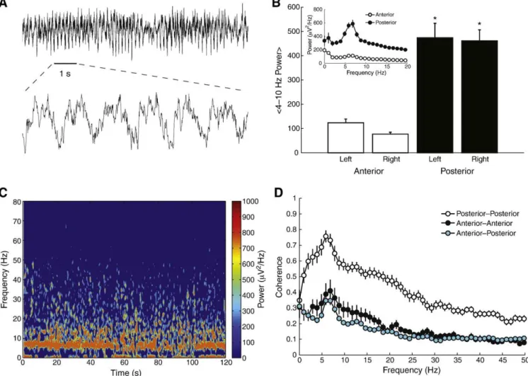
As shown in Fig. 1, we found prominent theta (4–10 Hz)
oscillations in the epidural EEG traces of the animals during the basal period, i.e., before the administration of any drug. Such oscillations could be detected by simple visual inspection (Fig. 1A) and were apparently more pronounced in the posterior electrodes (Fig. 1B; but read below). Fig. 1C shows a representative time– frequency decomposition obtained during the basal period, which depicts a clear, relatively steady theta rhythm. Similar to the power spectrum, the coherence spectra between electrodes also exhibited a clear peak at the theta band (Fig. 1D), which was also apparently more pronounced between the posterior electrodes. However, we note that 297
our reference electrode was located anterior to bregma (seeMaterials and methods), and, by changing the reference electrode to a posterior location, we observed a reversion of theseଏndings; that is, when using a posterior reference, we found that the anterior electrodes present higher theta power and coherence levels than posterior electrodes (not shown). For the rest of this work, we only show results of analyses performed on EEG traces obtained from the posterior electrodes using the anterior reference conଏguration.
Quinolinic acid disrupts theta and increases gamma oscillations
In Fig. 2A we show a representative EEG trace obtained immediately after QA i.c.v. infusion, along with its time–frequency decomposition. We observed that the basal theta activity becomes extremely reduced both before and after seizure, i.e., during the
peri-ictal period (Figs. 2A and B), whereas the seizure event was
characterized by a great increase in power of low frequencies, which spilled over higher frequencies (Fig. 2A). The coherence levels between electrodes following QA injection were also dramatically reduced (Fig. 2C). However, although both the overall theta power and coherence levels were reduced, the peak theta frequency exhibited a slight increase following QA infusion (Figs. 2B and C). In addition to theseଏndings, we found that QA treatment induced bursts
of gamma (20–50 Hz) activity occurring concurrently with the
reduction of theta oscillations (Figs. 2A and D). However, it should be noted that the coherence at the gamma band was actually reduced after QA injection when compared to baseline levels (Fig. 2C). In
accordance with these ଏndings, an analysis of phase-differences
between bilateral electrodes (i.e., inter-hemispheric) revealed dis-rupted phase-locking at both theta and gamma bands following QA treatment (Fig. 2E), including when only periods of higher gamma activity were taken into account (Fig. 2E, bottom panel).
Fig. 1.Epidural EEG traces exhibit prominent theta oscillations. (A) Representative raw signal obtained from a posterior electrode. (Bottom) Zoomed in view (1-s period corresponding to the horizontal black line). (B) Mean theta power levels for each electrode location. Error-bars denote SEM.pb0.01 compared to anterior electrodes (ANOVA followed by Tukey's test;n= 10–12 per group). Inset shows the mean power spectra over all anterior (white) and posterior (black) electrodes. (C) Time–frequency decomposition of a representative signal. (D) Mean coherence spectra between pair of electrodes. Error-bars denote SEM (n= 13 pairs per group).
299
Intraperitoneal infusion of guanosine does not alter EEG spectral content
We next studied the effects of i.p. guanosine on the spectral
content of epidural EEGs. As shown in Fig. 3A, we found that
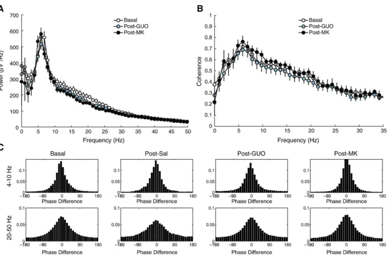
guanosine did not cause any meaningful spectral alteration in the EEG traces in comparison to baselineଏndings. The i.p. injection of a saline control also did not cause any alteration in the power spectrum. The coherence spectrum after saline or guanosine administration was also similar to baseline (Fig. 3B), as well as the phase-difference analysis at both theta and gamma bands (Fig. 3C). Of note, we also did notଏnd meaningful alterations in any of these analyses after MK-801 administration (Figs. 3A–C).
Guanosine reduces the incidence of quinolinic acid-induced seizures
In agreement with previous reports (Lara et al., 2001; Schmidt et al., 2000), QA induced seizures in all animals pre-treated with saline (n= 8 rats; Fig. 4A), whereas seizure episodes were completely prevented by the pre-administration of the NMDA antagonist MK-801 (n= 6 rats). In addition, we found that guanosine was able to protect seizures induced by QA in around 50% of the cases (protectedn= 10 rats; non-protectedn= 9 rats), which is also consistent with previous ଏndings (Lara et al., 2001; Schmidt et al., 2000, 2005). Animals seizing under guanosine or saline pre-treatment did not differ in the duration of the seizure episode (Fig. 4B).
Animals successfully protected by guanosine present lower reduction in theta power than non-protected animals
In Fig. 5A we show the power spectrum densities of seizing animals that were not protected by guanosine for the three different periods of the experiment (baseline, after guanosine i.p., after QA i.c.v.). As in the case of QA injection alone (seeFig. 2B), we found that seizing animals after the combined treatment with guanosine and QA (the latter administered 30 min after the former) present an overall reduction of the spectral power, and, in particular, at the theta band (Fig. 5A). Interestingly, we found that animals that were successfully protected by guanosine presented a lower decrease in power over multiple frequencies, including the theta
band (Fig. 5B). The difference in theta power levels between
seizing and non-seizing animals after QA administration was statistically signiଏcant (t(23) =−3.343, p= 0.0028). When exam-ining the coherence spectra, we found that both seizing and non-seizing animals presented a reduction of inter-hemispheric coher-ence over a wide range of frequencies; it should be noted, however, that non-seizing animals exhibited a trend towards higher
coher-ence values at the theta band (Fig. 5C). Similarly, the
phase-difference histograms revealed disrupted phase-locking between the right and left hemispheres for both seizing and non-seizing animals (Fig. 5D).
Animals pre-treated with MK-801 present an increase in gamma oscillations after quinolinic acid administration
We have then examined the power spectral densities of animals pre-treated with MK-801 i.p. followed by QA i.c.v. administration 30 min later. As mentioned above, none of these animals presented seizures after QA infusion. We found that the levels of theta power after QA in animals pre-treated with MK-801 did not differ from baseline values, although the peak theta frequency tended to slow down (Fig. 6A). Remarkably, however, we found that the EEG traces of these animals presented a marked increase in gamma oscilla-tions after QA administration (Fig. 6A). The coherence spectrum exhibited a mild decrease of inter-hemispheric theta-coherence (Fig. 6B), which could also be noticed by the phase-difference analysis (Fig. 6C).
The combined pre-treatment with guanosine and MK-801 promotes faster theta oscillations after quinolinic acid administration
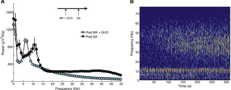
Lastly, we examined the power spectral densities of animals pre-treated with both guanosine and MK-801 i.p. followed by QA i.c.v. administration 30 min later. We found that the average theta power level after the combined administration of guanosine and MK-801 was not different from basal levels (not shown), and, as in the case of MK-801 pre-treatment alone, none of these animals (n= 7) presented motor seizures after QA infusion. Also similarly to the individual pre-treatment with MK-801, we observed a clear increase in gamma oscillations following QA administration (Fig. 7). However, we found that animals pre-treated with both guanosine and MK-801 present a prominent increase in the theta peak frequency following QA infusion (Fig. 7); in fact, as the magnitude of this increase was∼5 Hz, the peak frequency after QA infusion even reached values beyond the deଏnition of theta range (4–10 Hz) employed in this work.
Discussion
We have examined the power spectra of brain electrical activity and signal synchronization (coherence) at baseline as well as in response to a central infusion of QA, which induced acute episodes of motor seizures. In addition to clear EEG changes occurring during the seizure events (Fig. 2A), we found that QA disrupts a prominent basal theta activity during the peri-ictal period; moreover, QA also promoted a relative increase in the power level at the gamma band. These changes in power were accompanied by a reduction of oscillatory coherence between brain hemispheres. Using this same paradigm, we have also examined the electrophysiological effects of a pharmacological intervention that we have previously shown to be neuroprotective against QA-induced seizures, namely the pre-treatment with guanosine. We found that guanosine, when effec-tively preventing seizures, was able to counteract both the decrease in theta power as well as the appearance of gamma oscillatory activity following QA infusion. Additionally, we also studied the electrophysiological effects of the NMDA antagonist MK-801 in the same seizure model, and we found that whereas MK-801 pre-treatment was able to effectively block the decrease in theta power, this drug was associated with the appearance of large gamma oscillations following QA administration. Finally, we also found that the combined pre-treatment with both guanosine and MK-801 in this model led to qualitatively different results than the observed when each drug was administered alone.
There is a striking resemblance of the spectral changes induced by the QA seizure model we described here to oscillatory alterations found in other experimental models of epilepsy. For instance, recent work studying mice subjected to a mesial temporal lobe epilepsy (mTLE) model secondary to hippocampal kainic acid (KA) injection found an abolishment of the theta rhythm in these chronic, epileptic animals (Arabadzisz et al., 2005; Dugladze et al., 2007). Furthermore, it was also shown that KA-induced epileptic animals exhibit an increase in the power of gamma oscillations (Dugladze et al., 2007; Medvedev et al., 2000). Although the QA model employed here is an acute model of seizure, in contrast to the KA model of mTLE, these similarities suggest that a reduction in theta and an increase in gamma could be a hallmark of an epileptic brain. It should also be noted that these similarities were found at different levels of analysis: while we looked at a more macroscopic electrophysiological scale,
Dugladze et al (2007)examined hippocampal localଏeld potentials
(LFPs), that is, a more mesoscopic scale (Young and Eggermont,
2009). Moreover, this same work also reported similar spectral
disruptions at thein vitrocellular level when examining the ଏring
patterns of hippocampal interneurons (Dugladze et al., 2007).
Fig. 3.Neither guanosine nor MK-801 alters EEG spectral content. (A) Mean power spectra. Error-bars denote SEM. (B) Mean coherence spectra. Error-bars denote SEM. (C) Normalized histograms (% of counts) of inter-hemispheric phase difference for theta and gamma oscillations.
301
F.V.
Torres
et
al.
/
Experim
ental
Neurology
221
(2010)
296
–
et al., 2007; Wulff et al., 2009), and a disruption of inhibitory activity is associated with epileptiform activity (Cossart et al., 2001; Dinocourt et al., 2003; Kobayashi and Buckmaster, 2003; Morin et al., 1998). However, whether putative alterations of inhibitory networks are playing a role in the QA seizure model remains to be established, but the similarities between the spectralଏndings suggest that both the KA and QA models may share pathophysiological mechanisms. Of note, these similarities also suggest that the theta oscillations we observed in our epidural recordings were likely volume conducted from the hippocampus.
The work byArabadzisz et al. (2005)also described an overall reduction in the synchronization of bilateral EEG activity in animals subjected to the KA model of mTLE, similarly to what we found in the present study using the QA model of acute seizures (Fig. 2C). In fact, it should be noted that the inter-hemispheric impairments of synchrony were also observed to some extent even in rats successfully protected against seizures under the pre-treatment of either guanosine (Figs. 5C and D) or MK-801 (Figs. 6B and C). Taken together with the fact that non-seizing animals also presented some degree of alteration in the power of the theta band (Figs. 5B and 6A), our study also shows in particular that protected animals have an altered physiology, which is a fact that could not be hinted only by behavioral studies.
In a recent work, it was shown that the subcutaneous administra-tion of either MK-801 or ketamine induces a marked increase in gamma oscillations in rats (Pinault, 2008), which was discussed in the context of cognitive alterations associated with schizophrenia. Here, we found that the intraperitonial injection of MK-801 (0.5 mg/kg) did not cause alterations in the gamma band; however, the concomitant treatment with QA promoted a marked increase in gamma oscillations (Fig. 6A). Note that this increase was observable in the absolute power levels of these traces (Fig. 6A), and was more prominent than the relative increase of gamma power (assessed by the % of power distribution present in the gamma band; seeFig. 2C) seen after QA
administration alone (compare Figs. 6A and 2B). NMDA receptor
blockers such as MK-801 were reported to induce paradoxical increases in glutamatergic transmission mediated by non-NMDA
receptors activation (Adams and Moghaddam, 2001; Moghaddam
et al., 1997; Moghaddam and Adams, 1998), which were postulated to involve disinhibition of glutamatergic principal cells promoted by less activation of GABAergic interneurons(Lopez-Gil et al., 2007; Moghaddam et al., 1997; Schmidt et al., 2009; Tort et al., 2004). It could be therefore that the increase in gamma oscillations induced by MK-801 (Pinault, 2008) is related to non-NMDA receptors activation. In this sense, whereas the MK-801 dose we used (0.5 mg/kg) was unable to promote higher gamma activity, the co-treatment with QA, which can also inଏuence release and uptake of glutamate (de Oliveira et al., 2004; Tavares et al., 2002, 2005), could have synergistically increased the activation of non-NMDA receptors, leading to the
observed increase in gamma oscillations. In fact, Pinault (2008)
described that the appearance of gamma oscillations with MK-801 treatment was accompanied by ataxic behavior; as we did not observe ataxic behavior under our treatment protocol, this suggests that the effective dose of MK-801 reaching the brain was in fact higher in
Pinault (2008)study than in the present work.
Guanosine occurs naturally in the brain and has been reported to present a myriad of biological effects when administered extracellu-larly, including trophic effects on neural cells (Ciccarelli et al., 2001; Rathbone et al., 1999), stimulation of astrocyte proliferation (Ciccarelli et al., 2000; Kim et al., 1991), and modulation of glutamatergic activity (reviewed inSchmidt et al., 2007). Although these effects might be related to its uptake into the intracellular compartment, a consensus has emerged that some of guanosine actions involve its binding to a speciଏc membrane protein (Traversa et al., 2002, 2003), postulated by some to be a G protein-coupled receptor (Ciccarelli et al., 2001; Traversa et al., 2002, 2003). Indeed, guanosine was reported to present high afଏnity membrane sites in the brain (Traversa et al., 2002), although the actual existence of its putative speciଏc receptor has yet to be demonstrated. More certain is the fact that guanosine presents clear anti-glutamatergic properties, as demonstrated in severalin vivoand in vitroapproaches (reviewed inSchmidt et al., 2007), which places it as a new potential neuroprotective strategy against glutamatergic excitotoxicity. The mechanism of action underlying the modulation of Fig. 4.Guanosine and MK-801 are able to protect against QA-induced seizures. (A) Percentage of seizing animals per pre-treatment group.pb0.05 (Fisher exact test compared to saline group). (B) Mean seizure duration for saline pre-treated rats as well as during unsuccessful guanosine pre-treatment. Error-bars denote SEM.
303
glutamatergic activity by guanosine is currently under research in our and other laboratories. It has been suggested that astrocytes are crucially involved, since guanosine was shown to stimulate glutamate uptake by astrocytes (Frizzo et al., 2001, 2002, 2003), which is the main mechanism of glutamate removal from the synaptic cleft (Danbolt, 2001; Beart and O'Shea, 2007). In addition,
recent works have shown that QA decreases glutamate uptakein
vivoand that this effect is reverted by guanosine when successfully acting as anticonvulsant (de Oliveira et al., 2004; Tavares et al., 2002, 2005). Taken together, there is strong evidence that the antigluta-matergic action of guanosine occurs in a fundamentally different way than MK-801, which would explain the different electrophys-iological results observed here in the network (EEG) level. The
ଏnding that both compounds when administered simultaneously
promoted qualitatively different effects than when each was tested individually is also consistent with different inଏuences of these drugs in the network level. Noteworthy, since guanosine alone also prevented the increase in gamma oscillations after QA infusion, this result suggests that it might cause less cognitive side effects than MK-801 in the clinical setting.
It is known that guanosine is protective against QA-induced seizures in a dose-dependent manner (Schmidt et al., 2000, 2005). However, it still remains puzzling why guanosine is not effective in preventing QA-induced seizures in 100% of the cases, even in the highest soluble dose possible. In the present study, we found that the pre-treatment with guanosine was an effective anticonvulsant therapy in around 50% of the cases (Fig. 4A), a fraction that was similar to what we found in previous studies (Lara et al., 2001; Schmidt et al., 2000, 2005). Moreover, we observed that this fraction is not related to individual differences among animals: when the same group of animals is subjected twice to guanosine pre-treatment followed by i.c.v. QA infusion (with one week of interval between experiments), we obtained roughly the same fraction of protected animals, and we observed that the seizing/protected rats in the
second experiment were not necessarily the same as in theଏrst
experiment (Avila TT, Antunes C, Souza DO, unpublished observa-tions). This suggests that dynamic variables within rats are probably determining whether guanosine will successfully prevent seizures or not. It is possible that these variables are related to pharmacokinetic and pharmacodynamic factors (e.g., effective drug distribution), although it could also be related to other inଏuences as the current internal state of the brain. Importantly, here we have demonstrated for theଏrst time that protected and non-protected animals under guanosine pre-treatment can be distinguished electrophysiologically, even before the beginning of the motor seizures. Accordingly, we found that the level of theta power greatly decreased in seizing animals under (unsuccessful) guanosine pre-treatment, both before and after the seizure event (i.e., peri-ictally), similarly to what we observed in saline pre-treated rats. On the other hand, animals successfully protected by guanosine exhibited a statistically higher level of theta power than seizing animals (compareFigs. 5A and B). Although this result adds an extra step towards understanding why guanosine would be anticonvulsive only at times, the factors responsible for these electrophysiological differences are also puz-zling at the moment. At any event, as the decrease in theta activity could also be seen previous to seizure onset, the present results suggest that the monitoring of theta activity could be a useful marker of an epileptic brain.
mechanism of action of guanosine and how it translates into the present network, macroscopicଏndings.
Acknowledgments
This work was supported by CNPq-PRONEX/FAPESPA #2268, FINEP research grant“Rede Instituto Brasileiro de Neurociência
(IBN-Net)” # 01.06.0842-00, and INCT for Excitotoxicity and
Neuro-protection-CNPq. MSF is a CNPq research fellow. A.B.L.T. has received funding from CNPq and CAPES, Brazil. The authors also thank the HCPA Clinical Engineering Section for technical assistance.
References
Adams, B.W., Moghaddam, B., 2001. Effect of clozapine, haloperidol, or M100907 on phencyclidine-activated glutamate efଏux in the prefrontal cortex. Biol. Psychiatry 50, 750–757.
Anderson, C.M., Swanson, R.A., 2000. Astrocyte glutamate transport: review of properties, regulation, and physiological functions. Glia 32, 1–14.
Arabadzisz, D., Antal, K., Parpan, F., Emri, Z., Fritschy, J.M., 2005. Epileptogenesis and chronic seizures in a mouse model of temporal lobe epilepsy are associated with distinct EEG patterns and selective neurochemical alterations in the contralateral hippocampus. Exp. Neurol 194, 76–90.
Baron, B.M., Dudley, M.W., McCarty, D.R., Miller, F.P., Reynolds, I.J., Schmidt, C.J., 1989. Guanine nucleotides are competitive inhibitors ofN-methyl-D-aspartate at its receptor site bothin vitroandin vivo. J. Pharmacol. Exp. Ther. 250, 162–169. Beart, P.M., O'Shea, R.D., 2007. Transporters for L-glutamate: an update on their
molecular pharmacology and pathological involvement. Br. J. Pharmacol 150 , 5–17. Bradford, H.F., 1995. Glutamate, GABA and epilepsy. Prog. Neurobiol. 47, 477–511. Ciccarelli, R., Di Iorio, P., D'Alimonte, I., Giuliani, P., Florio, T., Caciagli, F., Middlemiss, P.J.,
Rathbone, M.P., 2000. Cultured astrocyte proliferation induced by extracellular guanosine involves endogenous adenosine and is raised by the co-presence of microglia. Glia 29, 202–211.
Ciccarelli, R., Ballerini, P., Sabatino, G., Rathbone, M.P., D'Onofrio, M., Caciagli, F., Di Iorio, P., 2001. Involvement of astrocytes in purine-mediated reparative processes in the brain. Int. J. Dev. Neurosci. 19, 395–414.
Connick, J.H., Stone, T.W., 1988. Quinolinic acid effects on amino acid release from the rat cerebral cortexin vitroandin vivo. Br. J. Pharmacol. 93, 868–876.
Cossart, R., Dinocourt, C., Hirsch, J.C., Merchan-Perez, A., De Felipe, J., Ben-Ari, Y., Esclapez, M., Bernard, C., 2001. Dendritic but not somatic GABAergic inhibition is decreased in experimental epilepsy. Nat. Neurosci. 4, 52–62.
Danbolt, N.C., 2001. Glutamate uptake. Prog. Neurobiol. 65, 1–105.
Delorme, A., Makeig, S., 2004. EEGLAB: an open source toolbox for analysis of single-trial EEG dynamics including independent component analysis. J. Neurosci. Methods 134, 9–21.
de Oliveira, D.L., Horn, J.F., Rodrigues, J.M., Frizzo, M.E., Moriguchi, E., Souza, D.O., Wofchuk, S., 2004. Quinolinic acid promotes seizures and decreases glutamate uptake in young rats: reversal by orally administered guanosine. Brain Res. 1018, 48–54.
Dinocourt, C., Petanjek, Z., Freund, T.F., Ben-Ari, Y., Esclapez, M., 2003. Loss of interneurons innervating pyramidal cell dendrites and axon initial segments in the CA1 region of the hippocampus following pilocarpine-induced seizures. J. Comp. Neurol. 459, 407–425.
Dugladze, T., Vida, I., Tort, A.B., Gross, A., Otahal, J., Heinemann, U., Kopell, N.J., Gloveli, T., 2007. Impaired hippocampal rhythmogenesis in a mouse model of mesial temporal lobe epilepsy. Proc. Natl. Acad. Sci. U. S. A. 104, 17530–17535. Frizzo, M.E., Lara, D.R., Dahm, K.C., Prokopiuk, A.S., Swanson, R.A., Souza, D.O., 2001.
Activation of glutamate uptake by guanosine in primary astrocyte cultures. Neuroreport 12, 879–881.
Frizzo, M.E., Lara, D.R., Prokopiuk Ade, S., Vargas, C.R., Salbego, C.G., Wajner, M., Souza, D.O., 2002. Guanosine enhances glutamate uptake in brain cortical slices at normal and excitotoxic conditions. Cell Mol. Neurobiol. 22, 353–363.
Frizzo, M.E., Antunes Soares, F.A., Dall'Onder, L.P., Lara, D.R., Swanson, R.A., Souza, D.O., 2003. Extracellular conversion of guanine-based purines to guanosine speciଏcally enhances astrocyte glutamate uptake. Brain Res. 972, 84–89.
Gloveli, T., Dugladze, T., Rotstein, H.G., Traub, R.D., Monyer, H., Heinemann, U., Whittington, M.A., Kopell, N.J., 2005. Orthogonal arrangement of rhythm-generating microcircuits in the hippocampus. Proc. Natl. Acad. Sci. U. S. A. 102, 13295–13300.
Guillemin, G.J., Kerr, S.J., Brew, B.J., 2005. Involvement of quinolinic acid in AIDS dementia complex. Neurotox. Res. 7, 103–123.
Kim, J.K., Rathbone, M.P., Middlemiss, P.J., Hughes, D.W., Smith, R.W., 1991. Purinergic stimulation of astroblast proliferation: guanosine and its nucleotides stimulate cell division in chick astroblasts. J. Neurosci. Res. 28, 442–455.
Kobayashi, M., Buckmaster, P.S., 2003. Reduced inhibition of dentate granule cells in a model of temporal lobe epilepsy. J. Neurosci. 23, 2440–2452.
Lara, D.R., Schmidt, A.P., Frizzo, M.E., Burgos, J.S., Ramirez, G., Souza, D.O., 2001. Effect of orally administered guanosine on seizures and death induced by glutamatergic agents. Brain Res. 912, 176–180.
Lewis, S.M., Lee, F.S., Todorova, M., Seyfried, T.N., Ueda, T., 1997. Synaptic vesicle glutamate uptake in epileptic (EL) mice. Neurochem. Int. 31, 581–585. Lopez-Gil, X., Babot, Z., Amargos-Bosch, M., Sunol, C., Artigas, F., Adell, A., 2007.
Clozapine and haloperidol differently suppress the MK-801-increased glutamater-gic and serotonerglutamater-gic transmission in the medial prefrontal cortex of the rat. Neuropsychopharmacology 32, 2087–2097.
Malcon, C., Achaval, M., Komlos, F., Partata, W., Sauressig, M., Ramirez, G., Souza, D.O., 1997. GMP protects against quinolinic acid-induced loss of NADPH-diaphorase-positive cells in the rat striatum. Neurosci. Lett. 225, 145–148.
Medvedev, A., Mackenzie, L., Hiscock, J.J., Willoughby, J.O., 2000. Kainic acid induces distinct types of epileptiform discharge with differential involvement of hippo-campus and neocortex. Brain Res. Bull. 52, 89–98.
Moghaddam, B., Adams, B.W., 1998. Reversal of phencyclidine effects by a group II metabotropic glutamate receptor agonist in rats. Science 281, 1349–1352. Moghaddam, B., Adams, B., Verma, A., Daly, D., 1997. Activation of glutamatergic
neurotransmission by ketamine: a novel step in the pathway from NMDA receptor blockade to dopaminergic and cognitive disruptions associated with the prefrontal cortex. J. Neurosci. 17, 2921–2927.
Morin, F., Beaulieu, C., Lacaille, J.C., 1998. Selective loss of GABA neurons in area CA1 of the rat hippocampus after intraventricular kainate. Epilepsy Res. 32, 363–369. Nakano, K., Takahashi, S., Mizobuchi, M., Kuroda, T., Masuda, K., Kitoh, J., 1993. High
levels of quinolinic acid in brain of epilepsy-prone E1 mice. Brain Res. 619, 195–198.
Fig. 7.Rats pre-treated with both guanosine and MK-801 present faster theta oscillations after QA administration. (A) Mean power spectra after guanosine and MK-801 i.p. (cyan) and after QA i.c.v. (black). Error-bars denote SEM (n= 14 traces per group). Guanosine and MK-801 i.p. pre-treatment was followed by QA i.c.v. 30 min later (arrow scheme). (B) Time–frequency decomposition of a representative signal after QA infusion (same pseudocolor scale as inFig. 1C).
305
Paas, Y., Devillers-Thiery, A., Changeux, J.P., Medevielle, F., Teichberg, V.I., 1996. Identiଏcation of an extracellular motif involved in the binding of guanine nucleotides by a glutamate receptor. EMBO J. 15, 1548–1556.
Paz, M.M., Ramos, M., Ramirez, G., Souza, D., 1994. Differential effects of guanine nucleotides on kainic acid binding and on adenylate cyclase activity in chick optic tectum. FEBS Lett. 355, 205–208.
Pinault, D., 2008.N-methyl-D-aspartate receptor antagonists ketamine and MK-801 induce wake-related aberrant gamma oscillations in the rat neocortex. Biol. Psychiatry 63, 730–735.
Ramaswamy, S., McBride, J.L., Kordower, J.H., 2007. Animal models of Huntington's disease. ILAR J. 48, 356–373.
Rathbone, M.P., Middlemiss, P.J., Gysbers, J.W., Andrew, C., Herman, M.A., Reed, J.K., Ciccarelli, R., Di Iorio, P., Caciagli, F., 1999. Trophic effects of purines in neurons and glial cells. Prog. Neurobiol. 59, 663–690.
Roesler, R., Vianna, M.R., Lara, D.R., Izquierdo, I., Schmidt, A.P., Souza, D.O., 2000. Guanosine impairs inhibitory avoidance performance in rats. Neuroreport 11, 2537–2540.
Saute, J.A., da Silveira, L.E., Soares, F.A., Martini, L.H., Souza, D.O., Ganzella, M., 2006. Amnesic effect of GMP depends on its conversion to guanosine. Neurobiol. Learn Mem. 85, 206–212.
Schmidt, A.P., Lara, D.R., Maraschin, J.F., Perla, A.S., Souza, D.O., 2000. Guanosine and GMP prevent seizures induced by quinolinic acid in mice. Brain Res. 864, 40–43. Schmidt, A.P., Avila, T.T., Souza, D.O., 2005. Intracerebroventricular guanine-based
purines protect against seizures induced by quinolinic acid in mice. Neurochem. Res. 30, 69–73.
Schmidt, A.P., Lara, D.R., Souza, D.O., 2007. Proposal of a guanine-based purinergic system in the mammalian central nervous system. Pharmacol. Ther. 116, 401–416. Schmidt, A.P., Tort, A.B., Souza, D.O., Lara, D.R., 2008. Guanosine and its modulatory effects on the glutamatergic system. Eur. Neuropsychopharmacol. 18, 620–622. Schmidt, A.P., Tort, A.B., Silveira, P.P., Bohmer, A.E., Hansel, G., Knorr, L., Schallenberger,
C., Dalmaz, C., Elisabetsky, E., Crestana, R.H., Lara, D.R., Souza, D.O., 2009. The NMDA antagonist MK-801 induces hyperalgesia and increases CSF excitatory amino acids in rats: reversal by guanosine. Pharmacol. Biochem. Behav. 91, 549–553.
Soares, F.A., Schmidt, A.P., Farina, M., Frizzo, M.E., Tavares, R.G., Portela, L.V., Lara, D.R., Souza, D.O., 2004. Anticonvulsant effect of GMP depends on its conversion to guanosine. Brain Res. 1005, 182–186.
Stone, T.W., 2001. Kynurenic acid antagonists and kynurenine pathway inhibitors. Expert Opin. Investig. Drugs 10, 633–645.
Tavares, R.G., Tasca, C.I., Santos, C.E., Alves, L.B., Porciuncula, L.O., Emanuelli, T., Souza, D. O., 2002. Quinolinic acid stimulates synaptosomal glutamate release and inhibits glutamate uptake into astrocytes. Neurochem. Int. 40, 621–627.
Tavares, R.G., Schmidt, A.P., Abud, J., Tasca, C.I., Souza, D.O., 2005. In vivo quinolinic acid increases synaptosomal glutamate release in rats: reversal by guanosine. Neurochem. Res. 30, 439–444.
Tort, A.B., Mantese, C.E., dos Anjos, G.M., Dietrich, M.O., Dall'Igna, O.P., Souza, D.O., Lara, D.R., 2004. Guanosine selectively inhibits locomotor stimulation induced by the NMDA antagonist dizocilpine. Behav. Brain Res. 154, 417–422.
Tort, A.B., Rotstein, H.G., Dugladze, T., Gloveli, T., Kopell, N.J., 2007. On the formation of gamma-coherent cell assemblies by oriens lacunosum-moleculare interneurons in the hippocampus. Proc. Natl. Acad. Sci. U. S. A. 104, 13490–13495.
Traversa, U., Bombi, G., Di Iorio, P., Ciccarelli, R., Werstiuk, E.S., Rathbone, M.P., 2002. Speciଏc [(3)H]-guanosine binding sites in rat brain membranes. Br. J. Pharmacol. 135, 969–976.
Traversa, U., Bombi, G., Camaioni, E., Macchiarulo, A., Costantino, G., Palmieri, C., Caciagli, F., Pellicciari, R., 2003. Rat brain guanosine binding site. Biological studies and pseudo-receptor construction. Bioorg. Med. Chem. 11, 5417–5425. Vinade, E.R., Schmidt, A.P., Frizzo, M.E.S., Portela, L.V., Soares, F.A., Schwalm, F.D.,
Elisabetsky, E., Izquierdo, I., Souza, D.O., 2005. Effects of chronic administered guanosine on behavioral parameters and brain glutamate uptake in rats. J. Neurosci. Res. 79, 248–253.
Wulff, P., Ponomarenko, A.A., Bartos, M., Korotkova, T.M., Fuchs, E.C., Bahner, F., Both, M., Tort, A.B., Kopell, N.J., Wisden, W., Monyer, H., 2009. Hippocampal theta rhythm and its coupling with gamma oscillations require fast inhibition onto parvalbumin-positive interneurons. Proc. Natl. Acad. Sci. U. S. A. 106, 3561–3566.