Gambling
Igor Elman
1,2*
, Tamara V. Gurvits
3, Evelyne Tschibelu
2, Justin D. Spring
3, Natasha B. Lasko
3,
Roger K. Pitman
31Providence VA Medical Center, Harvard Medical School, Cambridge, Massachusetts, United States of America,2Cambridge Health Alliance, Harvard Medical School, Cambridge, Massachusetts, United States of America,3Department of Psychiatry, Massachusetts General Hospital, Harvard Medical School, Charlestown, Massachusetts, United States of America
Abstract
Increased neurological soft signs (NSSs) have been found in a number of neuropsychiatric syndromes, including chemical
addiction. The present study examined NSSs related to perceptual-motor and visuospatial processing in a behavioral
addiction viz., pathological gambling (PG). As compared to mentally healthy individuals, pathological gamblers displayed
significantly poorer ability to copy two- and three-dimensional figures, to recognize objects against a background noise,
and to orient in space on a road-map test. Results indicated that PG is associated with subtle cerebral cortical abnormalities.
Further prospective clinical research is needed to address the NSSs’ origin and chronology (e.g., predate or follow the
development of PG) as well as their response to therapeutic interventions and/or their ability to predict such a response.
Citation:Elman I, Gurvits TV, Tschibelu E, Spring JD, Lasko NB, et al. (2013) Neurological Soft Signs in Individuals with Pathological Gambling. PLoS ONE 8(4): e60885. doi:10.1371/journal.pone.0060885
Editor:Antonio Verdejo Garcı´a, University of Granada, Spain
ReceivedDecember 11, 2012;AcceptedMarch 4, 2013;PublishedApril 4, 2013
This is an open-access article, free of all copyright, and may be freely reproduced, distributed, transmitted, modified, built upon, or otherwise used by anyone for any lawful purpose. The work is made available under the Creative Commons CC0 public domain dedication.
Funding:This work was supported by grant DA#017959 (to IE) from the National Institute on Drug Abuse. This study was also supported with resources and the use of facilities at the Providence VA Medical Center. The funders had no role in study design, data collection and analysis, decision to publish, or preparation of the manuscript.
Competing Interests:The authors have declared that no competing interests exist.
* E-mail: [email protected]
Introduction
Parallel to the ongoing expansion of legalized gambling
activities is an increase in the prevalence of pathological gambling
(PG) [1,2]. Pathological gambling afflicts up to 5% of the general
adult population and it costs American society an estimated $54
billion annually due to crime, decreased productivity, and
bankruptcies [3–7]. These estimates are likely conservative, given
that PG is not a conspicuous addiction, and it is devoid of typical
symptoms of intoxication, needle marks, or overdose. It may only
become noticeable in later stages of the illness, with the emergence
of highly visible behaviors including attempted suicide in up to
24% of untreated individuals [7–9]. To improve prevention and
treatment of PG, it is important to identify its behavioral markers
and their neural correlates.
A relatively consistent finding in functional brain imaging
studies of PG is failure of prefrontal cortical areas to activate when
challenged by cognitive tasks that normally evoke cerebral blood
flow and metabolic responses in these regions [10–17]. Likewise,
neuropsychological impairments are commonly documented in
PG patients [18–20], but their role in the course of the disorder
remains unclear [16], as they do not reliably reflect the severity of
gambling problems [21,22]. The nonspecificity of PG
neuropsy-chological findings may be partially attributable to the
multidi-mensionality of the tests employed [23]. Additionally, some results
may reflect poor motivation and attention [24,25] rather than
PG-related primary neuropathology, which has not yet been well
defined [23].
Neurological assessment paradigms may be of value in revealing
cortical abnormalities in PG. In this regard, neurological soft signs
(NSSs) are reliable [26–28], easily administered and temporally
stable [29,30] markers of neurological compromise, which impose
fewer cognitive demands than neuropsychological tests and are
therefore less influenced by performance confounds [31]. In
contrast to hard neurological signs localizable to a specific brain
site, their soft counterparts are attributed to wider brain regions
and functionally connected neuroanatomical systems, involved in
integrative neurological functions such as sensory perception,
coordination and motor sequencing [32,33]. Neurological soft
signs have been observed in a growing number of neuropsychiatric
syndromes including mood disorders [34–36],
obsessive-compul-sive disorder (OCD) [37–39], post-traumatic stress disorder
[26,27], impulse control disorder [40], schizophrenia [32,34,41],
and attention deficit hyperactivity disorder [42]. Furthermore, an
inverse relationship between NSSs scores and total brain volume
has been noted in psychopathological populations [27,43] adding
support to the generalized rather than localized NSSs’ nature.
In a previous paper, we reported that cocaine dependence is
characterized by the NSS of constructional apraxia [31]. As with
PG, cocaine dependence is classified in the DSM-V draft among
Substance Use and Addictive Disorders [44]. However, in
addition to its representing a behavioral addiction, a substance
addiction to cocaine exerts profound chemical effects on the brain
that may even result in such injuries as
subarachnoid/parenchy-mal hemorrhages [45–56] and infarcts [47,50].
addiction is accompanied by neurological compromise. To our
knowledge, NSSs have not yet been investigated in pathological
gamblers. The presence in PG of obsessive/compulsive and
impulsive features each of which has been previously linked with
NSSs [40,57,58] suggests that NSSs may also be seen in PG.
Accordingly, in this project we assessed three NSSs in PG and
healthy subjects. These were: a) copying two- and
three-dimensional figures (as previously tested in cocaine subjects
[31]); b) filtration of visual signal from noise; and c) left-right
orientation in the form of reading and understanding a simple
road map. These visuospatial and sensory integration tasks were
selected for the present project from our comprehensive NSSs
assessment battery based upon their discriminative ability in
drug-dependent and other psychiatric patients [27,31,59] as well as
their ease of administration as paper-and-pencil tasks. We
hypothesized that patients with PG would be more impaired than
healthy subjects on all three tasks.
Methods
Subjects
Twenty-one subjects who met the Diagnostic and Statistical
Manual of Mental Disorders, Fourth Edition, Text Revision (DSM
IV-TR) criteria for PG, and 10 non-gamblers who did not meet
DSM IV-TR criteria for any disorder, were recruited by
newspaper advertisement for participation in a previous study on
the neurobiology of PG. The biochemical [60] and psychosocial
[61] stress responsivity findings from that study have been
reported elsewhere. After a full explanation of the procedures,
all subjects gave written informed consent to the McLean Hospital
Institutional Review Board-approved protocol. Those with any
cognitive impairment that precluded informed consent based on
clinical interview and the assessments instruments (see below) were
excluded from study participation. Subjects were diagnosed by a
research psychiatrist using a best estimate format utilizing all
available sources of information including clinical history,
interview, and the following psychodiagnostic instruments: the
Structured Clinical Interview for DSM-IV (SCID [62]); the South
Oaks Gambling Screen (SOGS [63]); the DSM PG checklist
(DSMIV-TR [64,65]); and the Addiction Severity Index [66]. All
subjects were right handed as determined by the Edinburgh
Handedness Inventory [67], and scored at least 28 on the Mini
Mental Status Examination (MMSE [68]). Furthermore, they were
in good physical health as ascertained by the Cornell Medical
Index Health Questionnaire [69].
The exclusion criteria included left handedness, lifetime history
of dementia, schizophrenia, or other psychotic disorder, bipolar
disorder, anxiety disorder, current drug or alcohol dependence,
past but not current PG, or major depression with onset prior to
PG. We also excluded potentially confounding neurological
conditions, such as seizure disorder, head trauma accompanied
by loss of consciousness greater than 10 minutes, brain surgery,
multiple sclerosis, and Parkinson’s disease, as well as potentially
confounding medical conditions such as chronic obstructive
pulmonary disease, coronary artery disease, diabetes, obesity
(body mass index
$
30), congestive heart failure, hypertension,
renal diseases, cirrhosis, HIV-positive status and AIDS. Recent
drug and alcohol consumption was ruled out by negative results on
urine toxicology screen and breathalyzer.
Procedures
The three tasks were administered over one session in the
following order: Copy Figure Test (CFT), Detection and
Recognition of an Object Test (DROT) and Road Map Test
(RMT). None of the tasks was timed. Responses on the DROT
and the RMT were recorded by a research assistant seated next to
the subject. Both the subject and the examiner were ‘blind’ to the
study’s hypothesis.
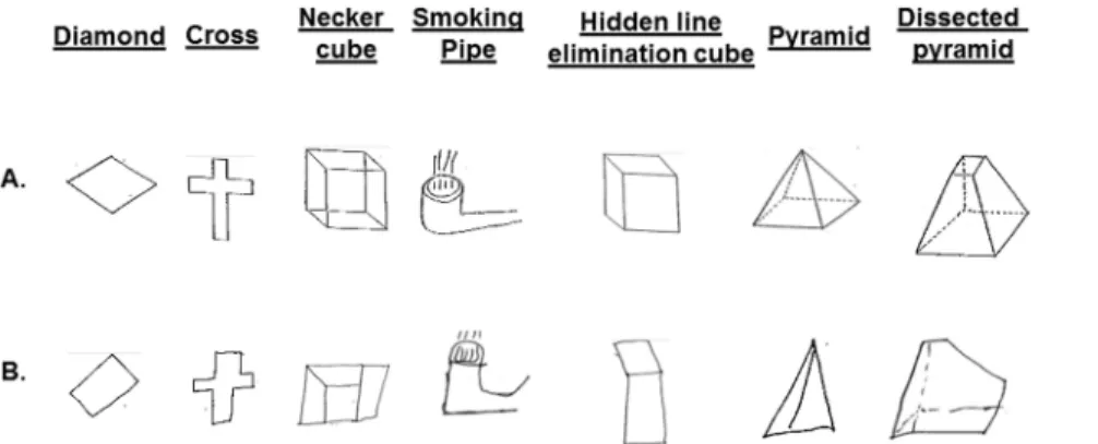
The CFT [70,71] is perceptual-motor in nature and comprises
two-dimensional (diamond and cross) and three-dimensional
(Necker cube, smoking pipe, hidden line elimination cube,
pyramid and dissected pyramid) figures (Figure 1A). Subjects were
instructed to copy each figure exactly as it appeared to them with a
pen. They were allowed to look at each figure as often as needed.
They were further instructed neither to erase any lines nor to draw
any lines that did not appear in the figure they copied.
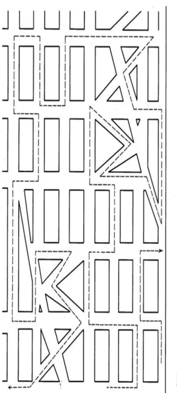
The DROT [72,73] consisted of two sets of the same six images
(Figure 2). Each image depicted a single basic household object,
namely key, shovel, pitcher, eyeglasses, hammer, and kettle.
However, the object recognition was complicated by background
‘‘noise,’’ consisting of a field of black squares of two different
densities, namely 35 and 15 squares per line. Subjects viewed all
six objects with the denser (more difficult) background first,
followed by all six objects with the less dense (less difficult)
background. They were instructed to identify all objects and were
allowed to bring the page as close to the eyes as desired.
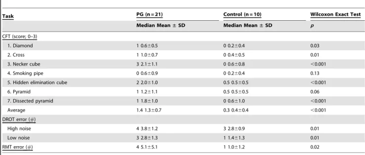
The RMT [74] is designed to evaluate directional sense in
visuospatial processing (Figure 3). Subjects were presented with a
map of an imaginary town, with a delineated route containing 32
intersections. They were instructed to imagine driving this route
and to indicate at each intersection whether the route turned left
or right. The research assistant followed the route with a pencil
and marked R or L in accordance with the verbal response at each
intersection. The map remained in a fixed position in front of the
subject, and they were not allowed to move it. Each subject’s
familiarity with the task was confirmed via a brief practice trial.
The CFT was scored by a dually trained psychiatrist and
neurologist, who not only was blind to diagnosis but had never
seen the subjects, utilizing a four-point scoring convention for each
figure. Zero (0) coded perfect or near perfect reproduction; 1
coded mild distortion or rotation; 2 coded moderate distortion or
rotation, or severe micropsy or a loss of three-dimensionality; and
3 coded gross distortion of the basic gestalt or a virtually
unrecognizable image. On the DROT, number of failed
identifications was scored. On the RMT, number of wrong turns
was scored.
Demographic variables were analyzed by Student’s t-tests or
Fisher’s exact tests as appropriate. Because most of the CFT,
DROT, and RMT data were ordinal and not normally
distributed, they were summarized as both median and mean
6
standard deviation (SD). The univariate nonparametric Wilcoxon
rank-sum test was used to compare groups. Significance was
defined as
p
,
0.05, one-tailed, with more abnormalities predicted
in the PG group.
Results
Table 1 presents demographic and psychometric data for the
two groups. These data demonstrate that pathological gamblers
were not significantly different from healthy controls with respect
to age, race, gender, years of education, performance on the
MMSE, and consumption of alcohol. As planned, there were
conspicuous differences in SOGS score and the number of
DSM-IV TR PG criteria met.
Figure 1B presents examples of mistakes made by PG subjects
on the CFT. Table 2 presents the group medians and means
±
medians, and the results of the group comparisons. With the
exception of the smoking pipe figure and the pyramid figure (for
which there was a trend), all tests revealed significantly poorer
performance in the PG group. Performance on the hidden line
elimination- and Necker cubes was dramatically poorer in the PG
subjects. Notably, the latter test is characterized by ambiguous
front-back orientation necessitating visuospatial ability to shift
attention between two equally plausible figural spatial
represen-tations [75].
Repeating the analyses after excluding ten smokers (all in the
PG group; among them are two subjects with respective cocaine
and alcohol dependence, both in full sustained remission), the
group effect remained significant for the CFT average score
(p = 0.002), for the high (p = 0.03) and low (p = 0.0005) noise
DROT errors and for the RMT errors (p = 0.03).
Discussion
In this study we identified several signs in pathological gamblers
reflecting their diminished ability to recognize and construct
objects and orient them in space. These dysfunctions have not yet
been addressed in literature on neuropsychological disturbances in
PG. In comparison to healthy subjects, pathological gamblers
showed substantially worse performance on copying two- and
three-dimensional figures, recognizing objects against background
noise, and discriminating left from right turns on a map.
Methodological similarities between the present study and our
prior study of cocaine dependence [31] included enrollment of
subjects with addictive disorders and use of a standard copy figure
task. There were differences in the type of addiction and in the
number of tasks performed by subjects. Overall these results
provide further support for subtle neurobiological impairment in a
behavioral addiction that is not confounded by exogenous
chemical use. Our data are also consistent with a substantial body
of literature documenting neuropsychological impairments in PG
patients [18–20], and they extend prior findings by suggesting that
the impairments are not restricted to the cognitive domains
addressed by neuropsychological testing but also generalize to the
sensorimotor domain.
Several brain regions influence the drawing of
three-dimen-sional figures, but as evident from research on cortically damaged
patients [76] and from neuroimaging work [75,77] the most
important of the regions is the parietal cortex. Ventral striatum
and related mesolimbic dopaminergic circuitry are traditionally
considered to be a key component of reward system involved in
addiction [78], and it is commonly hypothesized that changes in
the mesolimbic pathways underlying motivational processes are
responsible for transforming regular drives into heightened
incentive salience assigned to addiction-related cues [79].
How-ever, recent research suggests a novel factor in the mechanisms
underlying incentive sensitization by implicating parietal cortex in
the control exerted over striatal signals of salience via integration
of visuospatial, motor and cognitive (e.g., hedonic value and
categorical boundaries) inputs [80]. In addition to these theoretical
considerations, an abundant clinical literature demonstrates
parietal cortex changes in the context of chronic addictive
behaviors [81,82]. Hence NSSs examination may support the
Figure 1. The two-dimensional (diamond and cross) and three-dimensional (Necker cube, smoking pipe, hidden line elimination cube, pyramid and dissected pyramid) figures copied by the subjects (Panel A). Examples of PG subjects’ performance on the Copy Figure Test (Panel B).
doi:10.1371/journal.pone.0060885.g001
Figure 2. Detection and Recognition of an Object Test (DROT).
‘‘High noise’’ and ‘‘low noise’’ sets were presented separately, with the latter following the former. Subjects were instructed to identify the object embedded in the noise.
need to focus on this important region and on its role in the
pathophysiology of PG.
A limitation of the cross-sectional design employed here is its
inability to resolve the origin of elevated NSSs in PG. One
possibility is that they are preexisting vulnerability markers [83]. A
growing body of work points to compromised cortical function
reflected in NSSs that precedes the emergence of mood, anxiety
[57], psychotic [84–86] and obsessive-compulsive [57,87]
symp-toms. Neurological soft signs are also commonly observed in
mentally healthy relatives of schizophrenic patients [88–91],
further suggesting their preexisting and inheritable trait-like
nature. Notably, as suggested by twin studies, PG has a robust
genetic component ranging from 50 to 60% [92]. Greater
premorbid hyperactivity, impulsivity, and antisociality have been
found in PG subjects [93].
A second possible origin of NSSs in PG is that they are
acquired, e.g., they are a consequence of excessive gambling.
People who gamble lose money, and a consequence of losing
money may be increased stress, possibly leading to brain
alterations. Pathological gambling is indeed associated with an
exaggerated sympathoadrenal tone suggestive of heightened levels
of stress and arousal [94] at baseline [95,96] and while engaged in
gambling [8,9,97–100]. Subjects with PG have greater amygdala
activation in response to the alpha-2 adrenergic antagonist,
yohimbine [60]. Research in laboratory animals [101] and
humans [102,103] has shown that increased sympathetic activity
may cause vasospasm and microthrombosis resulting in
dimin-ished cerebral perfusion. It would be of interest to test whether
antiadrenergic agents (e.g., clonidine or prazosin) might moderate
the NSSs observed here. However, the reversibility of NSSs is
questionable [39], given that this has only been found in some
[104] but not in all OCD patients [105–107], and not in patients
with bipolar disorder [108] or schizophrenia [107,109]. In sum,
resolution of the risk factor vs. acquired origin interpretation of the
observed NSSs in PG, as well as NSSs’ possible response to
treatment and/or their ability to predict [37,110] such a response
(as has been shown for OCD patients) will require prospective
clinical trials.
The present design is unable to inform the question as to
whether the same visual agnosia displayed by the PG subjects on
the DROT is not likewise implicated in their constructional
apraxia on the figure copying task. Disentangling this would
require an exclusively motor processing task that does not involve
visual input [27]. Such tasks are included in the full assessment
battery of previously reported NSSs [27], which assesses motor
coordination and both motor and sensory integration.
In conclusion, the data presented here shed light on the
neurological function of patients with PG and suggest that NSS
examination has heuristic value for illuminating brain
abnormal-ities in this disorder. Pathological gambling offers a unique model
as it represents an addictive behavior in the absence of the
Figure 3. The Money Road Map Test (RMT). The continuous dotted line represents the path followed by the researcher’s pen. Subjects were asked at each successive turn to indicate whether it was right or left. The smaller dotted line in the lower right serves as a practice trial.
doi:10.1371/journal.pone.0060885.g003
Table 1.
Demographic and Clinical Characteristics (Means
6
SDs or Ratios) of Study Participants.
Variable PG (n = 21) Control
(n = 10) T-test (df = 29)
t p
Age (year) 45.569.9 43.6614.2 0.44 0.66
Education (year) 15.062.8 15.161.4 20.16 0.88
MMSE (score) 29.360.9 29.461.1 20.19 0.86
Alcohol (drink/week) 1.063.0 0.661.3 0.44 0.66
DSM-IV-TR PG criteria met 7.361.2 0.060.0
SOGS 13.563.8 0.060.0
Fisher’s exact test
Gender (M/F) 13/8 5/5 0.74
Race (W/B) 10/11 7/3 0.72
potentially confounding pharmacologically neurotoxic effects of
chemical substances. Therefore these findings provide a new
perspective in the exploration of addiction neurobiology.
Acknowledgments
The views expressed in this article are those of the authors and do not reflect the position or policy of the Department of Veterans Affairs or the United States Government.
Author Contributions
TVG ET NBL JDS RKP. Conceived and designed the experiments: IE TVG RKP. Performed the experiments: ET. Analyzed the data: IE JDS RP NBL. Contributed reagents/materials/analysis tools: TVG ET NBL. Wrote the paper: IE TVG JDS RKP.
References
1. Welte JW, Wieczorek WF, Barnes GM, Tidwell MC, Hoffman JH (2004) The relationship of ecological and geographic factors to gambling behavior and pathology. J Gambl Stud 20(4):405–23.
2. Gerstein DR, Murphy SA, Toce MT, Hoffmann J, Palmer A, et al. (1999) Gambling impact and behavior study: Report to the National Gambling Impact Study Commission. Chicago, National Opinion Research Center at the University of Chicago.
3. Potenza MN, Kosten TR, Rounsaville BJ (2001) Pathological gambling. JAMA 286(2):141–4.
4. Shaffer HJ (1997) The most important unresolved issue in the addictions: conceptual chaos. Subst Use Misuse 32(11):1573–80.
5. Cunningham-Williams RM, Grucza RA, Cottler LB, Womack SB, Books SJ, et al. (2005) Prevalence and predictors of pathological gambling: results from the St. Louis personality, health and lifestyle (SLPHL) study. J Psychiatr Res 39(4):377–90.
6. Grinols EL (2004) Gambling in America: Costs and Benefits. Cambridge, U.K.; New York: Cambridge University Press.
7. DeCaria CM, Hollander E, Grossman R, Wong CM, Mosovich SA, et al. (1996) Diagnosis, neurobiology, and treatment of pathological gambling. J Clin Psychiatry; 57 Suppl 8:80–3.
8. Moodie C, Finnigan F. (1996) A comparison of the autonomic arousal of frequent, infrequent and non-gamblers while playing fruit machines. Addiction 100(1):51–9.
9. Meyer G, Schwertfeger J, Exton MS, Janssen OE, Knapp W, et al. (2004) Neuroendocrine response to casino gambling in problem gamblers. Psycho-neuroendocrinology 29(10):1272–80.
10. Potenza MN, Leung HC, Blumberg HP, Peterson BS, Fulbright RK, et al. (2003) An fMRI Stroop task study of ventromedial prefrontal cortical function in pathological gamblers. Am J Psychiatry; 160(11):1990–4.
11. Tanabe J, Thompson L, Claus E, Dalwani M, Hutchison K, et al. (2007) Prefrontal cortex activity is reduced in gambling and nongambling substance users during decision-making. Hum Brain Mapp 28(12):1276–86.
12. Brewer JA, Potenza MN (2008) The neurobiology and genetics of impulse control disorders: relationships to drug addictions. Biochem Pharmacol 75(1):63–75.
13. Topf JL, Yip SW, Potenza MN (2009) Pathological Gambling: Biological and Clinical Considerations. J Addict Med 3(3):111–9.
14. Lawrence NS, Jollant F, O’Daly O, Zelaya F, Phillips ML (2009) Distinct roles of prefrontal cortical subregions in the Iowa Gambling Task. Cereb Cortex 19(5):1134–43.
15. Fineberg NA, Potenza MN, Chamberlain SR, Berlin HA, Menzies L, et al. (2010) Probing compulsive and impulsive behaviors, from animal models to endophenotypes: a narrative review. Neuropsychopharmacology 35(3):591– 604.
16. van Holst RJ, van den Brink W, Veltman DJ, Goudriaan AE (2010) Brain imaging studies in pathological gambling. Curr Psychiatry Rep 12(5):418–25. 17. Dannon PN, Kushnir T, Aizer A, Gross-Isseroff R, Kotler M, et al. (2011) Alternation learning in pathological gamblers: an fMRI Study. Brain Imaging Behav 5(1):45–51.
18. Kalechstein AD, Fong T, Rosenthal RJ, Davis A, Vanyo H, et al. (2007) Pathological gamblers demonstrate frontal lobe impairment consistent with that of methamphetamine-dependent individuals. J Neuropsychiatry Clin Neu-rosci19(3):298–303.
19. Hur JW, Shin NY, Kim SN, Jang JH, Choi JS, et al (2012) Do pathological gambling and obsessive-compulsive disorder overlap? a neurocognitive perspective. CNS Spectr;1–7.
20. Marazziti D, Catena DM, Conversano C, Consoli G, Vivarelli L, et al. (2008) Executive function abnormalities in pathological gamblers. Clin Pract Epidemiol Ment Health 4:7.
21. Forbush KT, Shaw M, Graeber MA, Hovick L, Meyer VJ, et al. (2005) Neuropsychological characteristics and personality traits in pathological gambling. CNS Spectr 13(4):306–15.
22. van Holst RJ, van den Brink W, Veltman DJ, Goudriaan AE (2010). Why gamblers fail to win: a review of cognitive and neuroimaging findings in pathological gambling. Neurosci Biobehav Rev 34(1):87–107.
23. Conversano C, Marazziti D, Carmassi C, Baldini S, Barnabei G, et al. (2012) Pathological gambling: a systematic review of biochemical, neuroimaging, and neuropsychological findings. Harv Rev Psychiatry 20(3):130–48.
24. Carlton PL, Manowitz P, McBride H, Nora R, Swartzburg M, et al. (1987) Attention deficit disorder and pathological gambling. J Clin Psychiatry 48(12):487–8.
Table 2.
Group medians and mean (
6
SDs) for the performance indices on the Copy Figure, Detection and Recognition of an
Object and the Road Map tests.
Task PG (n = 21) Control (n = 10) Wilcoxon Exact Test
Median Mean±SD Median Mean±SD p
CFT (score; 0–3)
1. Diamond 1 0.660.5 0 0.260.4 0.03
2. Cross 1 1.060.7 0 0.460.5 0.01
3. Necker cube 3 2.161.1 0 0.660.8 ,0.001
4. Smoking pipe 0 0.660.9 0 0.260.4 0.13
5. Hidden elimination cube 2 2.061.0 0.5 0.560.5 ,0.001
6. Pyramid 1 1.261.1 0.5 0.560.5 0.06
7. Dissected pyramid 1 1.861.0 0 0.661.0 ,0.001
Average 1.4 1.360.7 0.3 0.460.4 ,0.001
DROT error (#)
High noise 4 3.861.2 3 2.860.9 0.01
Low noise 3 2.861.3 1 1.461.3 0.01
RMT error (#) 4 5.165.1 1 1.061.2 0.02
25. de GM, Enzi B, Prosch U, Gantman A, Tempelmann C, et al. (2010) Decreased neuronal activity in reward circuitry of pathological gamblers during processing of personal relevant stimuli. Hum Brain Mapp 2010 Nov;31(11):1802–12.
26. Gurvits TV, Gilbertson MW, Lasko NB, Tarhan AS, Simeon D, et al. (2000) Neurologic soft signs in chronic posttraumatic stress disorder. Arch Gen Psychiatry 57(2):181–6.
27. Gurvits TV, Metzger LJ, Lasko NB, Cannistraro PA, Tarhan AS, et al. (2006) Subtle neurologic compromise as a vulnerability factor for combat-related posttraumatic stress disorder: results of a twin study. Arch Gen Psychiatry 2006 May;63(5): 571–6.
28. Krebs MO, Gut-Fayand A, Bourdel M, Dischamp J, Olie J (2000) Validation and factorial structure of a standardized neurological examination assessing neurological soft signs in schizophrenia. Schizophr Res 45(3):245–60. 29. Smith RC, Kadewari RP, Rosenberger JR, Bhattacharyya A (1999)
Nonresponding schizophrenia: differentiation by neurological soft signs and neuropsychological tests. Schizophr Bull 25(4):813–25.
30. Buchanan RW, Koeppl P, Breier A (1994) Stability of neurological signs with clozapine treatment. Biol Psychiatry 36(3):198–200.
31. Elman I, Chi WH, Gurvits TV, Ryan ET, Lasko NB, et al. (2008) Impaired reproduction of three-dimensional objects by cocaine-dependent subjects. J Neuropsychiatry Clin Neurosci 20(4):478–84.
32. Bombin I, Arango C, Buchanan RW (2005) Significance and meaning of neurological signs in schizophrenia: two decades later. Schizophr Bull 31(4):962–77.
33. Denckla MB (1985) Revised Neurological Examination for Subtle Signs. Psychopharmacol Bull 21(4):773–800.
34. Woods BT, Short MP (1985) Neurological dimensions of psychiatry. Biol Psychiatry; 20(2):192–8.
35. Goswami U, Gulrajani C, Varma A, Sharma A, Ferrier IN, et al. (2007) Soft neurological signs do not increase with age in euthymic bipolar subjects. J Affect Disord 103(1–3):99–103.
36. Negash A, Kebede D, Alem A, Melaku Z, Deyessa N, et al. (2004) Neurological soft signs in bipolar I disorder patients. J Affect Disord 80(2–3):221–30. 37. Hollander E, Kaplan A, Schmeidler J, Yang H, Li D, et al. (2005) Neurological
soft signs as predictors of treatment response to selective serotonin reuptake inhibitors in obsessive-compulsive disorder. J Neuropsychiatry Clin Neurosci 17(4):472–7.
38. Hollander E, Kim S, Khanna S, Pallanti S (2007) Obsessive-compulsive disorder and obsessive-compulsive spectrum disorders: diagnostic and dimen-sional issues. CNS Spectr 12(2 Suppl 3):5–13.
39. Jaafari N, de la Cruz LF, Grau M, Knowles E, Radua J, et al. (2012) Neurological soft signs in obsessive-compulsive disorder: two empirical studies and meta-analysis. Psychol Med 1–11.
40. Stein DJ, Hollander E, Liebowitz MR (1993) Neurobiology of impulsivity and the impulse control disorders. J Neuropsychiatry Clin Neurosci 5(1):9–17. 41. Heinrichs DW, Buchanan RW (1988) Significance and meaning of
neurolog-ical signs in schizophrenia. Am J Psychiatry 145(1):11–8.
42. Patankar VC, Sangle JP, Shah HR, Dave M, Kamath RM (2012) Neurological soft signs in children with attention deficit hyperactivity disorder. Indian J Psychiatry 54(2):159–65.
43. Dazzan P, Morgan KD, Orr KG, Hutchinson G, Chitnis X, et al. (2004) The structural brain correlates of neurological soft signs in AESOP first-episode psychoses study. Brain 127(Pt 1):143–53.
44. Denis C, Fatseas M, Auriacombe M (2012) Analyses related to the development of DSM-5 criteria for substance use related disorders: 3. An assessment of Pathological Gambling criteria. Drug Alcohol Depend 122(1–2):22–7. 45. Benowitz NL (1992) How toxic is cocaine? Ciba Found Symp 166:125–43. 46. O’Connor AD, Rusyniak DE, Bruno A (2005) Cerebrovascular and
cardiovascular complications of alcohol and sympathomimetic drug abuse. Med Clin North Am 89(6):1343–58.
47. Patrizi R, Pasceri V, Sciahbasi A, Summaria F, Rosano GM, et al. (2006) Evidence of cocaine-related coronary atherosclerosis in young patients with myocardial infarction. J Am Coll Cardiol; 47(10):2120–2.
48. Neiman J, Haapaniemi HM, Hillbom M (2000) Neurological complications of drug abuse: pathophysiological mechanisms. Eur J Neurol 7(6):595–606. 49. Lange RA, Hillis LD use (2001) Cardiovascular complications of cocaine.
N Engl J Med 345(5):351–8.
50. Morales Vidal SG, Hornik A, Morgan C (2012) Cocaine induced hippocampi infarction. BMJ Case Rep.
51. Robledo-Carmona J, Ortega-Jimenez MV, Garcia-Pinilla JM, Cabra B, de TE (2006) Severe cardiomyopathy associated to cocaine abuse. Int J Cardiol 112(1):130–1.
52. Steinhauer JR, Caulfield JB (2001) Spontaneous coronary artery dissection associated with cocaine use: a case report and brief review. Cardiovasc Pathol 10(3):141–5.
53. Hsue PY, Salinas CL, Bolger AF, Benowitz NL, Waters DD (2002) Acute aortic dissection related to crack cocaine. Circulation 105(13):1592–5.
54. Strickland TL, Miller BL, Kowell A, Stein R (1998) Neurobiology of cocaine-induced organic brain impairment: contributions from functional neuroimag-ing. Neuropsychol Rev 8(1):1–9.
55. Nanda A, Vannemreddy P, Willis B, Kelley R (2006) Stroke in the young: relationship of active cocaine use with stroke mechanism and outcome. Acta Neurochir Suppl 96:91–6.
56. Westover AN, McBride S, Haley RW (2007) Stroke in young adults who abuse amphetamines or cocaine: a population-based study of hospitalized patients. Arch Gen Psychiatry 64(4):495–502.
57. Hollander E, DeCaria CM, Aronowitz B, Klein DF, Liebowitz MR, et al. (1991) A pilot follow-up study of childhood soft signs and the development of adult psychopathology. J Neuropsychiatry Clin Neurosci 3(2):186–9. 58. Hollander E, Schiffman E, Cohen B, Rivera-Stein MA, Rosen W, et al. (1990)
Signs of central nervous system dysfunction in obsessive-compulsive disorder. Arch Gen Psychiatry 47(1):27–32.
59. Gurvits TV, Lasko NB, Repak AL, Metzger LJ, Orr SP, Pitman RK (2002) Performance on visuospatial copying tasks in individuals with chronic posttraumatic stress disorder. Psychiatry Res 112(3):263–8.
60. Elman I, Becerra L, Tschibelu E, Yamamoto R, George E, et al. (2012) Yohimbine-induced amygdala activation in pathological gamblers: a pilot study. PLoS One 2012;7(2):e31118.
61. Elman I, Tschibelu E, Borsook D (2010) Psychosocial stress and its relationship to gambling urges in individuals with pathological gambling. Am J Addict 19(4):332–9.
62. First MB, Spitzer RL, Gibbon MM, Williams JBW (2002) User’s Guide for the SCID-I: Structured Clinical Interview for DSM-IV-TR Axis I Disorders (Research version). New York State Psychiatric Institute, New York Biometrics Research Department.
63. Lesieur HR, Blume SB (1987) The South Oaks Gambling Screen (SOGS): a new instrument for the identification of pathological gamblers. Am J Psychiatry 144(9): 1184–8.
64. Wohl MJ, Matheson K, Young MM, Anisman H (2008) Cortisol rise following awakening among problem gamblers: dissociation from comorbid symptoms of depression and impulsivity. J Gambl Stud 24(1):79–90.
65. American Psychiatric Association (2000) Diagnostic and Statistical Manual of Mental Disorders: Text Revision. 4 ed. Washington, DC: American Psychiatric Publishing
66. McLellan AT, Luborsky L, Woody GE, O’Brien CP (1980) An improved diagnostic evaluation instrument for substance abuse patients. The Addiction Severity Index. J Nerv Ment Dis 168(1):26–33.
67. Oldfield RC (1971) The assessment and analysis of handedness: the Edinburgh inventory. Neuropsychologia 9(1):97–113.
68. Folstein MF, Folstein SE, McHugh PR (1975) ‘‘Mini-mental state’’. A practical method for grading the cognitive state of patients for the clinician. J Psychiatr Res 12(3):189–98.
69. Seymour GE (1976) The structure and predictive ability of the Cornell Medical Index for a normal sample. J Psychosom Res 20(5):469–78.
70. Luria AR (1966) High Cortical Functions in Man. New York: Basic Books. 71. Strub RL, Black FW (2000) Constructional Ability. The Mental Status
Examination in Neurology.Philadelphia: FA Davis Company.
72. Tonkonogy IM (1973) Introduction to Clinical Neuropsychology. Leningrad, USSR: Medicine.
73. Vasserman L, Dorofeeva S, Meerson Y (1997) Methods of Neuropsychological Diagnostics, Practice Manual. St. Petersburg, Stroylespechat.
74. Money J, Alexander D, Walker HT (1965) Road Map Test of Directional Sense. Baltimore, Johns Hopkins University Press.
75. Inui T, Tanaka S, Okada T, Nishizawa S, Katayama M (2000) Neural substrates for depth perception of the Necker cube; a functional magnetic resonance imaging study in human subjects. Neurosci Lett 282(3):145–8. 76. Critchley M (1969) The Parietal Lobes. New York, Hafner Publishing
Company.
77. Nishida Y, Hayashi O, Iwami T, Kimura M, Kani K, et al. (2001) Stereopsis-processing regions in the human parieto-occipital cortex. Neuroreport 12(10):2259–63.
78. Koob GF, Volkow ND (2010) Neurocircuitry of addiction. Neuropsychophar-macology 35(1):217–38.
79. Elman I, Borsook D, Lukas SE (2006) Food intake and reward mechanisms in patients with schizophrenia: implications for metabolic disturbances and treatment with second-generation antipsychotic agents. Neuropsychopharma-cology 31(10):2091–120.
80. Freedman DJ, Assad JA (2006) Experience-dependent representation of visual categories in parietal cortex. Nature 2006 443(7107):85–8.
81. Aron JL, Paulus MP (2007) Location, location: using functional magnetic resonance imaging to pinpoint brain differences relevant to stimulant use. Addiction 102 Suppl 1:33–43.
82. Thalemann R, Wolfling K, Grusser SM (2007) Specific cue reactivity on computer game-related cues in excessive gamers. Behav Neurosci 121(3):614–8. 83. Peralta V, de Jalon EG, Campos MS, Basterra V, Sanchez-Torres A, et al. (2010) Risk factors, pre-morbid functioning and episode correlates of neurological soft signs in drug-naive patients with schizophrenia-spectrum disorders. Psychol Med 1–11.
84. Jones P, Rodgers B, Murray R, Marmot M (1994) Child development risk factors for adult schizophrenia in the British 1946 birth cohort. Lancet 344(8934):1398–402.
85. Dazzan P, Lloyd T, Morgan KD, Zanelli J, Morgan C, et al. (2008) Neurological abnormalities and cognitive ability in first-episode psychosis. Br J Psychiatry 2008 193(3):197–202.
87. Grisham JR, Fullana MA, Mataix-Cols D, Moffitt TE, Caspi A, et al. (2011) Risk factors prospectively associated with adult obsessive-compulsive symptom dimensions and obsessive-compulsive disorder. Psychol Med 1–12. 88. Rieder RO, Nichols PL (1979) Offspring of schizophrenics. III. Hyperactivity
and neurological soft signs. Arch Gen Psychiatry 36(6):665–74.
89. Chen YL, Chen YH, Mak FL (2000) Soft neurological signs in schizophrenic patients and their nonpsychotic siblings. J Nerv Ment Dis 188(2):84–9. 90. Ismail B, Cantor-Graae E, McNeil TF (1998) Minor physical anomalies in
schizophrenic patients and their siblings. Am J Psychiatry 155(12):1695–702. 91. Egan MF, Hyde TM, Bonomo JB, Mattay VS, Bigelow LB, et al. (2001)
Relative risk of neurological signs in siblings of patients with schizophrenia. Am J Psychiatry 158(11):1827–34.
92. Lobo DS, Kennedy JL (2009) Genetic aspects of pathological gambling: a complex disorder with shared genetic vulnerabilities. Addiction 104(9):1454– 65.
93. Langenbucher J, Bavly L, Labouvie E, Sanjuan PM, Martin CS (2001) Clinical features of pathological gambling in an addictions treatment cohort. Psychol Addict Behav 15(1):77–9.
94. Goudriaan AE, Oosterlaan J, de BE, van den Brink W (2004) Pathological gambling: a comprehensive review of biobehavioral findings. Neurosci Biobehav Rev 28(2):123–41.
95. Bergh C, Eklund T, Sodersten P, Nordin C (1997) Altered dopamine function in pathological gambling. Psychol Med 27(2):473–5.
96. Roy A, Adinoff B, Roehrich L, Lamparski D, Custer R, et al. (1998) Pathological gambling. A psychobiological study. Arch Gen Psychiatry 45(4):369–73.
97. Krueger TH, Schedlowski M, Meyer G (2005) Cortisol and heart rate measures during casino gambling in relation to impulsivity. Neuropsychobiology 52(4):206–11.
98. Meyer G, Hauffa BP, Schedlowski M, Pawlak C, Stadler MA, et al. (2000) Casino gambling increases heart rate and salivary cortisol in regular gamblers. Biol Psychiatry 48(9):948–53.
99. Sharpe L, Tarrier N, Schotte D, Spence SH (1995) The role of autonomic arousal in problem gambling. Addiction 90(11):1529–40.
100. Sharpe L (2004) Patterns of autonomic arousal in imaginal situations of winning and losing in problem gambling. J Gambl Stud 20(1):95–104. 101. Tuor UI (1990) Local distribution of the effects of sympathetic stimulation on
cerebral blood flow in the rat. Brain Res 529(1–2):224–31.
102. Tschuor C, Asmis LM, Lenzlinger PM, Tanner M, Harter L, et al. (2008) In vitro norepinephrine significantly activates isolated platelets from healthy volunteers and critically ill patients following severe traumatic brain injury. Crit Care 12(3):R80.
103. Ibrahim GM, Macdonald RL (2012) Electrocardiographic changes predict angiographic vasospasm after aneurysmal subarachnoid hemorrhage. Stroke 43(8):2102–7.
104. Mergl R, Mavrogiorgou P, Juckel G, Zaudig M, Hegerl U (2004) Effects of sertraline on kinematic aspects of hand movements in patients with obsessive-compulsive disorder. Psychopharmacology (Berl) 171(2):179–85.
105. Thienemann M, Koran LM (1995) Do soft signs predict treatment outcome in obsessive-compulsive disorder? J Neuropsychiatry Clin Neurosci 7(2):218–22. 106. Caramelli P, de Lima MA, Stip E, Bacheschi LA (1996) Neurological
examination in obsessive-compulsive disorder. Sao Paulo Med J 114(5):1255–8. 107. Karadag F, Tumkaya S, Kirtas D, Efe M, Alacam H, et al. (2011) Neurological soft signs in obsessive compulsive disorder with good and poor insight. Prog Neuropsychopharmacol Biol Psychiatry 35(4):1074–9.
108. Cherian A, Kuruvilla K (1989) Prevalence of neurological ‘‘soft signs’ in affective disorder and their correlation with response to treatment. Indian J Psychiatry 31(3):224–9.
109. Chan RC, Xu T, Heinrichs RW, Yu Y, Wang Y (2010) Neurological soft signs in schizophrenia: a meta-analysis. Schizophr Bull 36(6):1089–104.