Is Not Associated with Decreased Survival or with Worse
Neurological Outcome
Jean-Francois Brasme1,2,3*, Jacques Grill1,2, Francois Doz4,5, Brigitte Lacour6, Dominique Valteau-Couanet1,2, Stephan Gaillard7,8, Olivier Delalande9, Nozar Aghakhani2,10, Ste´phanie Puget5,11,
Martin Chalumeau3,5,12
1Department of Pediatric and Adolescent Oncology, Institut Gustave Roussy, Villejuif, France,2Universite´ Paris Sud, Le Kremlin Biceˆtre, France,3Inserm U953, Epidemiological Research Unit on Perinatal Health and Women’s and Children’s Health, Hoˆpital Saint-Vincent-de-Paul, Paris, France,4Department of Pediatric and Adolescent Oncology, Institut Curie, Paris, France,5Universite´ Paris Descartes, Paris, France,6French National Registry of Childhood Solid Tumors, CHU Nancy, Vandoeuvre-le`s-Nancy, France,7Department of Neurosurgery, Hoˆpital Foch, Suresnes, France,8Universite´ Paris Ile-de-France Ouest, Guyancourt, France,9Department of Pediatric Neurosurgery, Fondation Rothschild, Paris, France, 10Department of Neurosurgery, Centre Hospitalier de Biceˆtre, AP-HP, Le Kremlin Bicetre, France, 11Department of Pediatric Neurosurgery, Hoˆpital Necker-Enfants Malades, AP-HP, Paris, France,12Department of Pediatrics, Hoˆpital Necker-Enfants Malades, AP-HP, Paris, France
Abstract
Background:The long time to diagnosis of medulloblastoma, one of the most frequent brain tumors in children, is the source of painful remorse and sometimes lawsuits. We analyzed its consequences for tumor stage, survival, and sequelae.
Patients and Methods:This retrospective population-based cohort study included all cases of pediatric medulloblastoma from a region of France between 1990 and 2005. We collected the demographic, clinical, and tumor data and analyzed the relations between the interval from symptom onset until diagnosis, initial disease stage, survival, and neuropsychological and neurological outcome.
Results:The median interval from symptom onset until diagnosis for the 166 cases was 65 days (interquartile range 31–121, range 3–457). A long interval (defined as longer than the median) was associated with a lower frequency of metastasis in the univariate and multivariate analyses and with a larger tumor volume, desmoplastic histology, and longer survival in the univariate analysis, but not after adjustment for confounding factors. The time to diagnosis was significantly associated with IQ score among survivors. No significant relation was found between the time to diagnosis and neurological disability. In the 62 patients with metastases, a long prediagnosis interval was associated with a higher T stage, infiltration of the fourth ventricle floor, and incomplete surgical resection; it nonetheless did not influence survival significantly in this subgroup.
Conclusions:We found complex and often inverse relations between time to diagnosis of medulloblastoma in children and initial severity factors, survival, and neuropsychological and neurological outcome. This interval appears due more to the nature of the tumor and its progression than to parental or medical factors. These conclusions should be taken into account in the information provided to parents and in expert assessments produced for malpractice claims.
Citation:Brasme J-F, Grill J, Doz F, Lacour B, Valteau-Couanet D, et al. (2012) Long Time to Diagnosis of Medulloblastoma in Children Is Not Associated with Decreased Survival or with Worse Neurological Outcome. PLoS ONE 7(4): e33415. doi:10.1371/journal.pone.0033415
Editor:Robert S. Phillips, University of York, United Kingdom
ReceivedNovember 30, 2011;AcceptedFebruary 8, 2012;PublishedApril 2, 2012
Copyright:ß2012 Brasme et al. This is an open-access article distributed under the terms of the Creative Commons Attribution License, which permits unrestricted use, distribution, and reproduction in any medium, provided the original author and source are credited.
Funding:The authors have no support or funding to report.
Competing Interests:The authors have declared that no competing interests exist. * E-mail: jfbrasme@gmail.com
Introduction
Brain tumors are the leading cause of solid cancers in children [1]. Medulloblastoma, one of the most common types [1], has a 10-year survival rate of 50% [2–5], and many survivors have neurological and cognitive sequelae [6,7]. The time to diagnosis for brain tumors is one of the longest of all childhood cancers, with a median ranging from 2 to 5 months [8–24]. We showed recently that the time from symptom onset to diagnosis of medulloblastoma is long (median 65 days), and we analyzed the causes of this lengthy interval. In particular, it was significantly
associated with the presence of apparently psychological symp-toms [25].
[13]. Nonetheless several factors limit the usefulness of these results: the limited numbers (,100) of pediatric patients [13,17], pooled analyses for pediatric and adult patients [13], single-center recruitment subject to selection bias [13], incomplete initial disease staging for some patients that increases the likelihood of classification bias [13], a study period partially preceding the availability of CT and MRI [13], and a lack of multivariate analyses despite the presence of potential confounders [13,17]. Finally, none of these studies analyzed the relation between time to diagnosis and either local tumor stage, complete surgical resection or neurological and cognitive sequelae.
Our objective was therefore to analyze, in a pediatric population-based study, the consequences of the time to diagnosis of medulloblastoma on initial tumor stage, survival, and neuropsychological and neurological outcome, while taking confounding factors into account.
Methods
Patients
We conducted a multicenter historic population-based cohort study that included all patients in one French region (Ile-de-France, the Paris metropolitan region) who were younger than 15 years when diagnosed with a histologically-confirmed medullo-blastoma from 1990 through 2005. The study has been described in detail elsewhere [25]. The geographic exhaustiveness of the recruitment was verified from the French National Registry of Childhood Solid Tumors [29,30].
Data collected
We collected the following data from each medical file in the neurosurgery and oncology departments: age, sex, symptoms, the radiologic and pathology characteristics of the tumor (standard or nodular/desmoplastic histology [31,32], the latter referred to hereafter as desmoplastic), local and metastatic staging, postoper-ative status (posterior fossa syndrome), vital status and neurological outcome at last follow-up (full scale intelligence quotient (IQ) score and neurological examination, classified into 3 groups: strictly normal neurological examination; moderate and unilateral dysmetria without functional consequence; or neurological disability). Pathologists at each hospital (sites of the neurosurgery and oncology departments) confirmed all histological diagnoses of biopsy or resection samples within days of the radiological diagnosis. The time to diagnosis, expressed in days, was defined as the interval between the first symptom attributable to the disease and the date of diagnosis (date of brain imaging). When ambiguous (for 2% of patients), it was independently evaluated by 3 of the authors, who reached a consensus.
Tumor stage was assessed: (1) by cerebral MRI, before and shortly after surgery, to determine the completeness of the resection, (2) pre- or postoperative spinal MRI, and (3) a postoperative lumbar puncture to look for metastasis. All patients had a complete staging work-up, except one who died within 24 hours of surgery. Based on the surgical report and early postoperative imaging, the initial disease stage was classified by the T stage according to the Chang-Harisiadis classification [33,34] and according to the classification of risk groups recognized by the International Society of Pediatric Oncology: standard risk
Statistical analyses
First, we described the demographic and tumor characteristics and the time to diagnosis. Second, we studied the relation between this prediagnosis interval and initial severity factors (metastasis, tumor volume, T stage and completeness of resection) by univariate (Kruskal-Wallis test) and multivariate analysis, taking into consideration the cofactors of interest, by either stratification or adjustment in a backward stepwise logistic regression model. Third, we studied the relations between time to diagnosis, survival, and neurological and neuropsychological outcome. These analyses were performed first for the entire population (Kaplan-Meier method and logrank test), then after stratification for the identified prognostic factors, and finally by adjustment for the cofactors of interest in a Cox model. One of the Cox models was constructed by adjusting the relation between time to diagnosis and survival by a propensity score for a long prediagnosis interval [38], calculated by the logistic regression equation produced at stage 2. The time to diagnosis was used as a binary variable after dichotomization around the median or as a continuous variable (after testing linearity). Age was dichotomized around 5 years, in view of the difference in treatment around this age [4,5]. Statistical analyses were performed with STATA (StataCorp.).
Ethics Statement
The Institutional Review Committee (Comite´ de Protection des Personnes Ile-de-France III) stated that ‘‘this research was found to conform to scientific principles and ethical research standards and to the laws and regulations of France’’ and specifically approved this study. Written informed consent of the patients or their parents was not judged necessary for this kind of retrospective study. Data were anonymized before the clinical records were included.
Results
Population, distribution of time to diagnosis
Examination of the diagnostic code lists showed 170 patients eligible for this study. Of these 170 eligible files, 4 (2%) were lost or incomplete. The analysis therefore concerned 166 patients, 72% of whom were boys. Median age at the first symptom was 6 years (interquartile range (IQR): 4–9) and 31% were younger than 5 years.
The patients had a local tumor in 62% (standard risk 40%, local high risk 22%) and a metastatic tumor in 38% of cases. The median tumor volume was 33 cm3(IQR 22–42). Staging showed 2% of the children had T1 local tumors, 27% T2, 15% T3A, 39% T3B, and 17% T4. The surgeon observed fourth ventricle floor invasion in 47% of the 161 cases for which surgery was performed. Resection was complete in 52% of cases. The histologic type was standard for 78%, desmoplastic for 22%, and large-cell anaplastic for one patient (pooled with the ‘‘standard histology’’ patients for subsequent analysis).
Table 1.Relation between time to diagnosis (TtD) and clinical and tumor characteristics.
short TtD long TtD
Characteristics n median TtD p1 ,65 days .65 days
OR [95% CI]¤ p± ORa [95% CI]¤¤ p{
(total = 166) (days) (n = 83) (n = 83)
Age at diagnosis,5 years 51 55 0.03 30 21 1.7 [0.8, 3.4] 0.13 1.3 [0.6, 2.7] 0.5
.5 years 115 77 53 62
Psychological signs**: impaired school performance, depression, behavioral problems, anxiety
absent 122 60 0.001 68 54 2.4 [1.1, 5.3] 0.01 2.5 [1.1, 5.6] 0.03
present 44 91 15 29
Tumor characteristics
standard histology 129 61 0.01 70 59 2.4 [1.03, 5.5] 0.03 2 [0.8, 4.7] 0.12
desmoplastic 36 112 12 24
metastatic tumor 62 31 ,1024 49 13 8 [3.6, 18]
,1026 7.6 [3.6, 16.4]
,1023
local tumor 103 91 33 70
missing data* 1 1 0
1Degree of significance of nonparametric test (Mann Whitney or Kruskal-Wallis) of the distribution of time to diagnosis. ¤Odd Ratio [95% confidence interval].
6Degree of significance of the chi-2 test or Fisher’s exact test.
¤¤adjusted Odds Ratio [95% confidence interval].
{Degree of significance of the coefficient of the logistic regression test.
*One patient died within 24 h of surgery, before spinal MRI, and was excluded from the analysis. **The data concerning psychological signs have been previously published [25].
doi:10.1371/journal.pone.0033415.t001
Time
to
Diagnosis
of
Medulloblas
toma
in
ONE
|
www.plos
one.org
3
April
2012
|
Volume
7
|
Issue
statistically associated with an age older than 5 years (p= 0.13).
Relations between time to diagnosis, tumor stage, and cofactors
The children with metastatic disease had a significantly shorter prediagnosis interval than those with local tumors (median 31 vs 91 days, p,1024
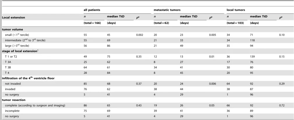
; figure 1, table 1). Overall, tumor volume was significantly larger for the patients with a long time to diagnosis (median tumor volume 34 vs 27 cm3,p= 0.002; table 2). We did not find a statistically significant (p.0.2) association between time to diagnosis and any of the other factors related to local extension (T stage and fourth ventricle floor invasion) or completeness of surgical resection.
For the 103 patients (62%) without metastatic disease (table 2), we did not find a significant association between time to diagnosis and local extension (volume, T stage, or fourth ventricle floor infiltration). Similarly there was no association with the complete-ness of surgical resection among the 102 who had surgery (that is,
prediagnosis intervals: the tumor volume was larger, the T stage more advanced, and fourth ventricle floor infiltration more frequent (table 2). Of the 58 patients with metastatic disease who had surgery, the median prediagnosis interval was 26 days for those with complete resection and 41 days for incomplete resection (p= 0.05).
Table 2.Relation between time to diagnosis (TtD) and local extension.
all patients metastatic tumors local tumors
Local extension n median TtD p* n median TtD p* n median TtD p*
(total = 166) (days) (total = 62) (days) (total = 103) (days)
tumor volume
small (,1sttercile) 55 45 0.002 20 23 0.005 34 71 0.10
intermediate (2ndto 3rdtercile) 55 69 21 33 34 118
large (.3rdtercile) 56 86 21 49 35 94
stage of local extension{
T 1 or T2 49 75 0.35 12 13 0.01 36 139 0.15
T 3A 25 62 8 27 17 76
T 3B 64 61 34 41 30 80
T 4 28 84 8 45 20 95
infiltration of the 4thventricle floor
not invaded 85 68 0.37 20 24 0.006 64 92 0.29
invaded 76 62 38 44 38 87
no surgery 5 41 4 29 1 96
tumor resection
complete (according to surgeon and imaging) 86 65 0.43 19 26 0.05 66 92 0.72
incomplete 75 69 39 41 36 89
no surgery 5 41 4 29 1 96
*Degree of significance of nonparametric test (Mann-Whitney or Kruskal-Wallis) of the distribution of time to diagnosis.
{Local extension stages according to Chang-Harisiadis classification [33,34]. doi:10.1371/journal.pone.0033415.t002
Time
to
Diagnosis
of
Medulloblas
toma
in
ONE
|
www.plos
one.org
5
April
2012
|
Volume
7
|
Issue
Relations between time to diagnosis, survival, and cofactors
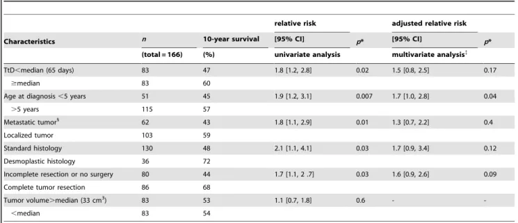
No patients were lost to follow-up. At the last follow-up, 96 patients (58%) were still alive. The median follow-up was 7 years (IQR 5–12 years, range 3–17 years). All survival rates reported below are 10-year rates. The survival of patients with a long time to diagnosis was significantly better than that of patients with a short prediagnostic interval (60 vs 47%, relative risk (RR) = 1.8 [1.2, 2.8], p= 0.02; figure 2). Survival rates were significantly better for patients older than 5 years, those without metastatic disease, those with desmoplastic tumors and those with complete tumor resection (table 3).
After stratification by age, survival rates for the children younger than 5 years were best for those with a long time to diagnosis (67 vs 29%, RR = 3.6 [1.4, 8.9],p= 0.03); for those older than 5 years, survival did not differ significantly according to the length of this interval (57 vs 56%, RR = 1.1 [0.6, 2.1], p.0.2). Stratification by metastatic disease showed that survival for the patients with metastatic tumors was 66% among those with a long interval vs 38% for those with shorter ones, but this difference was not statistically significant (RR = 2.2 [0.8, 6.4], p= 0.12); for patients without metastatic disease, survival did not differ significantly according to prediagnosis interval (62 vs 55%, RR = 1.1 [0.6, 2.3], p.0.2). After stratification by histologic type, survival was better in the group with standard tumor histology when their time to diagnosis was longer than the median (55 vs 42%, RR = 1.8 [1.1, 3.1], p= 0.02); in the group with a desmoplastic tumor (36 patients), survival did not differ signifi-cantly according to this duration (72 vs 71%, RR = 1 [0.3, 3.7], p.0.2). After stratification by completeness of the resection, survival among the patients with incomplete resection or no surgery was better for those with a long interval (54 vs 36%, RR = 2.0 [1.1, 3.8], p= 0.02); for the patients with complete resection, survival did not differ significantly according to time to diagnosis (65 vs 57%, RR = 1.5 [0.73, 3.1], p.0.2).
After adjustment in a Cox model for the covariables of interest associated with survival in the univariate analysis with a p value,0.20 (i.e., time to diagnosis, age, metastasis, histology, and complete resection), only age older than 5 years was independently associated with survival (adjusted relative risk (RRa) = 1.7 [1.0, 2.8],p= 0.04). Survival was not significantly associated with time to diagnosis, metastasis, complete resection, or histologic type (table 3). In two other models, supplementary adjustment for the use of radiotherapy (upfront, delayed or omitted in some children under 5), or for the propensity score did not significantly change the relation between time to diagnosis and survival (RRa = 1.6 [0.9, 2.8], p = 0.10 and RRa = 1.5 [0.8, 2.5], p = 0.17, respective-ly). Using time to diagnosis as a continuous variable did not significantly change the results either.
Relations between time to diagnosis, IQ score, and neurological disability
The presence of postoperative posterior fossa syndrome was associated with a significantly shorter time to diagnosis (median 50 vs 75 days, p = 0.02). The time to diagnosis was not significantly different (p.0.2) between patients with normal neurological findings, with moderate and unilateral dysmetria without func-tional consequence, or with neurological disability (median 87 vs 72 vs 72 days, respectively). The IQ score was significantly associated with the prediagnosis interval, both after linear regression (figure 3, p = 0.01), following transformation with a
fractional polynomial, and after adjustment for age, radiotherapy dose, and the covariables mentioned above (p,0.05).
Discussion
Main results
We found complex and often inverse relations between a longer time to diagnosis of medulloblastoma in children, the initial severity factors, and survival. A long prediagnosis interval was
Table 3.Survival according to age, tumor characteristics, and time to diagnosis (TtD).
relative risk adjusted relative risk
Characteristics n 10-year survival [95% CI] p* [95% CI] p*
(total = 166) (%) univariate analysis multivariate analysis{
TtD,median (65 days) 83 47 1.8 [1.2, 2.8] 0.02 1.5 [0.8, 2.5] 0.17
$median 83 60
Age at diagnosis,5 years 51 45 1.9 [1.2, 3.1] 0.007 1.7 [1.0, 2.8] 0.04
.5 years 115 57
Metastatic tumor1
62 43 1.8 [1.1, 2.9] 0.01 1.3 [0.7, 2.2] 0.4
Localized tumor 103 59
Standard histology 130 48 2.1 [1.1, 4.1] 0.03 1.7 [0.9, 3.4] 0.12
Desmoplastic histology 36 72
Incomplete resection or no surgery 80 44 1.7 [1.1, 2 .7] 0.03 1.6 [0.9, 2.6] 0.09
Complete tumor resection 86 68
Tumor volume.median (33 cm3) 83 53 1.1 [0.7, 1.8] 0.6 -
-,median 83 54
*Degree of significance of the Logrank test.
{Cox model with adjustment for the following covariables: TtD less than or more than the median of 65 days, age older or younger than 5 years, desmoplastic or not histology, metastatic or localized tumor.
1One patient died within 24 h of surgery, before spinal MRI, and was thus excluded from the analysis. doi:10.1371/journal.pone.0033415.t003
was significantly associated with IQ score among survivors. No significant relation was found between the time to diagnosis and neurological disability. In the 62 patients with metastatic disease, a long prediagnosis interval was associated with a more advanced T stage, fourth ventricle floor invasion, and incomplete surgical resection; it nonetheless did not influence survival significantly in this subgroup.
Hypothesis
The inverse relation between time to diagnosis and severity of disease may be explained by the type of tumor progression. Rapidly growing and metastatic tumors might produce swift and intense clinical signs, leading to rapid consultation and diagnosis, but for a very advanced tumor. Inversely, local tumors that grow slowly might cause relatively mild and very progressive clinical signs that would lead to a long period of development before diagnosis. Recent studies of patients with medulloblastoma indicate that outcome is determined much more by underlying molecular biology [39] than clinical factors such as time to diagnosis.
Study limitations
Our population-based cohort allowed us to avoid the recruit-ment bias of single-center studies. The principal limitation of the study is its retrospective nature. Nonetheless the data came from multiple sources and the time to diagnosis was ambiguous for only 2% of patients. Disease extension was determined in all but one patient (,1%), based on standardized measurement methods (imaging). No patients were lost to follow-up. Most studies of time to diagnosis are retrospective, which may lead to some bias, but it is non-differential bias.
Our results are consistent with those of a previous study about the relation between short prediagnostic intervals and metastasis [13]: we confirmed their results in this more recent, larger and exclusively pediatric population-based study that took confounding factors into account and analyzed the consequences for initial tumor stage and survival. Finally, we found the same demograph-ic, clinical, tumor, and prognostic characteristics as in other series of medulloblastoma in children [9–15,40].
Ethical and legal issues
The results of our study suggest that the time to diagnosis of medulloblastoma is related more to the properties of the tumor
the medulloblastoma, a delay sometimes attributable to their psychological or banal nature [23,25]. Parents, in their quest to determine the origin of the disease, feel remorse and guilt for having neglected the initial symptoms, especially when the disease outcome is unfavorable [26]. The diagnosis delay associated with the physician is equally the source of painful regret [26] as well as lawsuits [28]. Our study suggests that there is no simple causal relation between time to diagnosis and harm or damage, even in cases of metastasis, incomplete resection, or large tumor volume. This finding is highly pertinent in a medical malpractice system based on tort law [28], even if the complaint alleges specific acts, of negligence for example, by the doctor rather than the length of the delay alone. Moreover, the length of the diagnosis delay depends on two separate factors: the patient delay, that is, the time interval between the onset of symptoms and the first presentation to a healthcare professional, and the doctor delay, that is, the time interval between the first medical consultation and the final diagnosis. In our study, the median duration of the latter was 30 days and accounted for slightly under half (46%) of the total delay. However, because these data were available for only 40% of the files, they were not analyzed in detail. Finally, the conclusions about the individual consequences of a given diagnosis delay may well differ from the conclusions drawn from the analysis of a cohort of patients.
Conclusion
Although a longer time to diagnosis may not be related to inferior prognosis, we cannot and do not claim that a ‘‘longer than necessary’’ time to diagnosis will not lead to high-risk disease, need for more intensive therapy, and possibly a worse outcome. The inverse association between time to diagnosis and prognosis in a group of patients does not mean that a delay in diagnosis can have no consequences for an individual patient. Moreover, a rapid diagnosis shortens children’s suffering and helps prevent parents’ loss of confidence in the health care system [26].
Author Contributions
Conceived and designed the experiments: J-FB MC JG. Performed the experiments: J-FB MC. Analyzed the data: J-FB MC JG. Contributed reagents/materials/analysis tools: FD DV-C BL SG OD NA SP. Wrote the paper: J-FB MC JG FD.
References
1. Desandes E, Clavel J, Berger C, Bernard JL, Blouin P, et al. (2004) Cancer incidence among children in France, 1990–1999. Pediatr Blood Cancer 43: 749–757. 2. Desandes E, Berger C, Tron I, Demeocq F, Bellec S, et al. (2008) Childhood
cancer survival in France, 1990–1999. Eur J Cancer 44: 205–215.
3. Verlooy J, Mosseri V, Bracard S, Tubiana AL, Kalifa C, et al. (2006) Treatment of high risk medulloblastomas in children above the age of 3 years: a SFOP study. Eur J Cancer 42: 3004–3014.
4. Grill J, Sainte-Rose C, Jouvet A, Gentet JC, Lejars O, et al. (2005) Treatment of medulloblastoma with postoperative chemotherapy alone: an SFOP prospective trial in young children. Lancet Oncol 6: 573–580.
5. Oyharcabal-Bourden V, Kalifa C, Gentet JC, Frappaz D, Edan C, et al. (2005) Standard-risk medulloblastoma treated by adjuvant chemotherapy followed by reduced-dose craniospinal radiation therapy: a French Society of Pediatric Oncology Study. J Clin Oncol 23: 4726–4734.
6. Grill J, Renaux VK, Bulteau C, Viguier D, Levy-Piebois C, et al. (1999) Long-term intellectual outcome in children with posterior fossa tumors according to
8. Reulecke BC, Erker CG, Fiedler BJ, Niederstadt TU, Kurlemann G (2008) Brain tumors in children: initial symptoms and their influence on the time span between symptom onset and diagnosis. J Child Neurol 23: 178–183. 9. Haimi M, Peretz Nahum M, Ben Arush MW (2004) Delay in diagnosis of children
with cancer: a retrospective study of 315 children. Pediatr Hematol Oncol 21: 37–48. 10. Pollock BH, Krischer JP, Vietti TJ (1991) Interval between symptom onset and
diagnosis of pediatric solid tumors. J Pediatr 119: 725–732.
11. Saha V, Love S, Eden T, Micallef-Eynaud P, MacKinlay G (1993) Determinants of symptom interval in childhood cancer. Arch Dis Child 68: 771–774. 12. Thulesius H, Pola J, Hakansson A (2000) Diagnostic delay in pediatric
malignancies–a population-based study. Acta Oncol 39: 873–876.
13. Halperin EC, Watson DM, George SL (2001) Duration of symptoms prior to diagnosis is related inversely to presenting disease stage in children with medulloblastoma. Cancer 91: 1444–1450.
Canadian geographic study. Neurosurgery 51: 365–372; discussion 372– 363.
17. Kukal K, Dobrovoljac M, Boltshauser E, Ammann RA, Grotzer MA (2009) Does diagnostic delay result in decreased survival in paediatric brain tumours? Eur J Pediatr 168: 303–310.
18. Dang-Tan T, Franco EL (2007) Diagnosis delays in childhood cancer: a review. Cancer 110: 703–713.
19. Wilne SH, Ferris RC, Nathwani A, Kennedy CR (2006) The presenting features of brain tumours: a review of 200 cases. Arch Dis Child 91: 502–506. 20. Keene DL, Hsu E, Ventureyra E (1999) Brain tumors in childhood and
adolescence. Pediatr Neurol 20: 198–203.
21. Loh AH, Ha C, Chua JH, Seow WT, Chan MY, et al. (2009) Delays in diagnosis of pediatric solid tumors in Singapore. J Pediatr Hematol Oncol 31: 734–738. 22. Klein-Geltink J, Pogany L, Mery LS, Barr RD, Greenberg ML (2006) Impact of age and diagnosis on waiting times between important healthcare events among children 0 to 19 years cared for in pediatric units: the Canadian Childhood Cancer Surveillance and Control Program. J Pediatr Hematol Oncol 28: 433–439.
23. Edgeworth J, Bullock P, Bailey A, Gallagher A, Crouchman M (1996) Why are brain tumours still being missed? Arch Dis Child 74: 148–151.
24. Dang-Tan T, Trottier H, Mery LS, Morrison HI, Barr RD, et al. (2008) Delays in diagnosis and treatment among children and adolescents with cancer in Canada. Pediatr Blood Cancer 51: 468–474.
25. Brasme JF, Chalumeau M, Doz F, Lacour B, Valteau-Couanet D, et al. (2011) Interval between onset of symptoms and diagnosis of medulloblastoma in children: distribution and determinants in a population-based study. Eur J Pediatr 171: 25–32.
26. Dixon-Woods M, Findlay M, Young B, Cox H, Heney D (2001) Parents’ accounts of obtaining a diagnosis of childhood cancer. Lancet 357: 670–674. 27. Hoven E, Anclair M, Samuelsson U, Kogner P, Boman KK (2008) The
influence of pediatric cancer diagnosis and illness complication factors on parental distress. J Pediatr Hematol Oncol 30: 807–814.
28. Najaf-Zadeh A, Dubos F, Pruvost I, Bons-Letouzey C, Amalberti R, et al. (2010) Epidemiology and aetiology of paediatric malpractice claims in France. Arch Dis Child 96: 127–130.
29. Sommelet D, Clavel J, Lacour B (2005) [Contribution of national paediatric cancer registries to survey and research]. Arch Pediatr 12: 814–816. 30. Lacour B, Guyot-Goubin A, Guissou S, Bellec S, Desandes E, et al. (2010)
Incidence of childhood cancer in France: National Children Cancer Registries, 2000–2004. Eur J Cancer Prev 19: 173–181.
31. Kleihues P, Burger PC, Scheithauer BW (1993) The new WHO classification of brain tumours. Brain Pathol 3: 255–268.
32. Kleihues P, Sobin LH (2000) World Health Organization classification of tumors. Cancer 88: 2887.
33. Chang CH, Housepian EM, Herbert C, Jr. (1969) An operative staging system and a megavoltage radiotherapeutic technic for cerebellar medulloblastomas. Radiology 93: 1351–1359.
34. Harisiadis L, Chang CH (1977) Medulloblastoma in children: a correlation between staging and results of treatment. Int J Radiat Oncol Biol Phys 2: 833–841.
35. Lannering B, Rutkowski S, Doz F, Pizer B, Gustafsson G, et al. (2010) HIT -SIOP PNET4- A randomised multicentre study of hyperfractionated versus standard radiotherapy in children with standard risk medulloblastoma. Pediatr Blood Cancer 55: abstract O78.
36. Dufour C, Couanet D, Figarella-Branger D, Carrie C, Doz F, et al. (2008) Sequential high-dose chemotherapy with autologous stem cell rescue for children with high-risk medulloblastoma and supratentorial primitive neuroec-todermal tumors. Neuro-Oncology 10: 488 (abstract).
37. Dufour C, Couanet D, Figarella-Branger D, Carrie C, Doz F, et al. (2008) Sequential high-dose chemotherapy and reducued craniospinal irradiation in young children with metastatic medulloblastoma. Neuro-Oncology 10: 423 (abstract).
38. D’Agostino RB, Jr. (2007) Propensity scores in cardiovascular research. Circulation 115: 2340–2343.
39. Northcott PA, Korshunov A, Witt H, Hielscher T, Eberhart CG, et al. (2011) Medulloblastoma Comprises Four Distinct Molecular Variants. J Clin Oncol: 29(11): 1408–1414.