Widespread Shortening of 3’ Untranslated
Regions and Increased Exon Inclusion Are
Evolutionarily Conserved Features of Innate
Immune Responses to Infection
Athma A. Pai1☯*, Golshid Baharian2,3☯, Ariane Page´ Sabourin3, Jessica
F. Brinkworth3,4,5, Yohann Ne´de´lec2,3, Joseph W. Foley6, Jean-Christophe Grenier3, Katherine J. Siddle7, Anne Dumaine3, Vania Yotova3, Zachary P. Johnson8,9, Robert
E. Lanford10, Christopher B. Burge1,11, Luis B. Barreiro3,4*
1Department of Biology, Massachusetts Institute of Technology, Cambridge, MA, USA,2Department of Biochemistry, Faculty of Medicine, Universite´ de Montre´al, Montreal, QC, Canada,3Sainte-Justine Hospital Research Centre, Montreal, QC, Canada,4Department of Pediatrics, Faculty of Medicine, University of Montreal, Montreal, QC, Canada,5Department of Anthropology, University of Illinois, Urbana-Champaign, IL, USA,6Department of Pathology, Stanford University School of Medicine, Stanford, CA, USA,
7Department of Organismic and Evolutionary Biology, FAS Center for Systems Biology, Harvard University, Cambridge, MA, USA,8Yerkes National Primate Research Center, Emory University, Atlanta, GA 30322, USA,9Department of Human Genetics, Emory University School of Medicine, Atlanta, GA 30322, USA, 10Texas Biomedical Research Institute, Southwest National Primate Research Center, San Antonio, TX, USA,11Department of Biological Engineering, Massachusetts Institute of Technology, Cambridge, MA, USA
☯These authors contributed equally to this work.
*[email protected](AAP);[email protected](LBB)
Abstract
The contribution of pre-mRNA processing mechanisms to the regulation of immune responses remains poorly studied despite emerging examples of their role as regulators of immune defenses. We sought to investigate the role of mRNA processing in the cellular responses of human macrophages to live bacterial infections. Here, we used mRNA sequencing to quantify gene expression and isoform abundances in primary macrophages from 60 individuals, before and after infection withListeria monocytogenesandSalmonella typhimurium. In response to both bacteria we identified thousands of genes that signifi-cantly change isoform usage in response to infection, characterized by an overall increase in isoform diversity after infection. In response to both bacteria, we found global shifts towards (i) the inclusion of cassette exons and (ii) shorter 3’ UTRs, with near-universal shifts towards usage of more upstream polyadenylation sites. Using complementary data collected in non-human primates, we show that these features are evolutionarily conserved among primates. Following infection, we identify candidate RNA processing factors whose expression is associated with individual-specific variation in isoform abundance. Finally, by profiling microRNA levels, we show that 3’ UTRs with reduced abundance after infection are significantly enriched for target sites for particular miRNAs. These results suggest that the pervasive usage of shorter 3’ UTRs is a mechanism for particular genes to evade repression by immune-activated miRNAs. Collectively, our results suggest that dynamic
a11111
OPEN ACCESS
Citation:Pai AA, Baharian G, Page´ Sabourin A, Brinkworth JF, Ne´de´lec Y, Foley JW, et al. (2016) Widespread Shortening of 3’ Untranslated Regions and Increased Exon Inclusion Are Evolutionarily Conserved Features of Innate Immune Responses to Infection. PLoS Genet 12(9): e1006338. doi:10.1371/journal.pgen.1006338
Editor:Christine A. Wells, The University of Queensland, AUSTRALIA
Received:June 24, 2016
Accepted:September 2, 2016
Published:September 30, 2016
Copyright:©2016 Pai et al. This is an open access article distributed under the terms of theCreative Commons Attribution License, which permits unrestricted use, distribution, and reproduction in any medium, provided the original author and source are credited.
Data Availability Statement:Data generated in this study have been submitted to the NCBI Gene
Expression Omnibus (GEO;http://www.ncbi.nlm.
nih.gov/geo/) under the accession number GSE73765, which comprises of the mRNA-seq data (GSE73502) and small RNA-seq data (GSE73478).
changes in RNA processing may play key roles in the regulation of innate immune responses.
Author Summary
Changes in gene regulation have long been known to play important roles in both innate and adaptive immune responses. While transcriptional responses to infection have been well-characterized, much less is known about the extent to which co-transcriptional mech-anisms of mRNA processing are involved in the regulation of immune defenses. In this study, we sought to investigate the role of mRNA processing in the cellular responses of human macrophages to live bacterial infection. Using primary human macrophages derived from whole blood samples from 60 individuals, we sequenced mRNA both before and after infection with two live bacteria. We show that immune responses to infection are accompanied by pervasive changes in mRNA isoform usage, with systematic shifts towards increased cassette exon inclusion and shortening of Tandem 3’ UTRs post-infec-tion. These patterns are conserved in nonhuman primates, supporting their functional importance across evolutionary time. Complementary microRNA profiling revealed that shortened 3’ UTRs are enriched for target sites of macrophage-expressed miRNAs, many of which are specifically activated after infection to regulate the innate immune response. Our results therefore provide the first genome-wide empirical support for the idea that actively regulated shifts towards shorter 3’ UTRs might allow specific genes to evade repression by immune-activated miRNAs.
Introduction
Innate immune responses depend on robust and coordinated gene expression programs involving the transcriptional regulation of thousands of genes [1–3]. These regulatory cascades start with the detection of microbial-associated products by pattern recognition receptors, which include Toll Like Receptors (TLRs), NOD-like receptors and specific C-type lectins [4,5]. These initial steps are followed by the activation of key transcription factors (e.g., NF-κB and interferon regulatory factors) that orchestrate the inflammatory and/or antiviral response signals involved in pathogen clearance and the subsequent development of appropriate adap-tive immune responses [4].
Although much attention has been devoted to characterizing transcriptional changes in response to infectious agents or other immune stimuli, we still know remarkably little about the contribution of co-transcriptional changes–specifically, changes in alternative pre-mRNA processing–to the regulation of the immune system [6–8]. Alternative splicing can impact cel-lular function by creating distinct mRNA transcripts from the same gene, which may encode unique proteins that have distinct or opposing functions. For instance, in human and mouse cells, several alternatively spliced forms of genes involved in the TLR pathway have been shown to function as negative regulators of TLR signaling in order to prevent uncontrolled inflammation [9–11]. Additionally, several in-depth studies of individual genes suggest that alternative splicing plays an important role in increasing the diversity of transcripts encoding MHC molecules [12] and in modulating intracellular signaling and intercellular interactions through the expression of various isoforms of key cytokines [13,14], cytokine receptors [9,15,16], kinases [17] and adaptor proteins [18–20]. Yet, the extent to which changes in
Natural Sciences and Engineering Research Council of Canada (RGPIN/435917-2013), and the Canada Research Chairs Program (950-228993) (to LBB). Computational resources at MIT supported by the National Science Foundation (Grant No. 0821391). AAP. was supported by a Jane Coffin Childs postdoctoral fellowship. GB was supported by a postdoctoral fellowship from the Fonds de la Recherche en Sante´ du Que´bec. The funders had no role in study design, data collection and analysis, decision to publish, or preparation of the manuscript.
isoform usage are a hallmark of immune responses to infection remains largely unexplored at a genome-wide level [8,21]. Moreover, we still do not know which RNA processing mechanisms contribute most to the regulation of immune responses, or how these disparate mechanisms are coordinated.
To address these questions, we investigated genome-wide changes in transcriptome patterns after independent infections withListeria monocytogenesorSalmonella typhimuriumin pri-mary human macrophages. Because of the distinct molecular composition of these two patho-gens and the way they interact with host cells, they activate distinct innate immune pathways after infection [22,23]. Thus, our study design allowed us to evaluate the extent to which changes in isoform usage are pathogen-specific or more generally observed in response to bac-terial infection. Furthermore, the large number of individuals in our study allowed us to use natural variation in RNA processing to gain insight into fine-tuned inter-individual regulation of immune responses. Our results provide a comprehensive picture of the role that RNA pro-cessing might play in regulating early innate immune responses to bacterial infection in human antigen-presenting cells.
Results
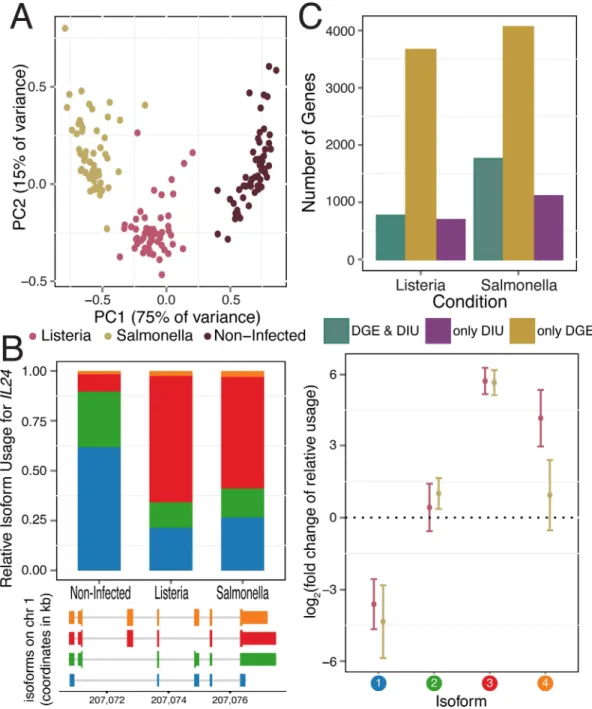
Infection with eitherListeriaorSalmonellainduces dramatic changes in mRNA expression lev-els. Following 2 hours of infection with each bacteria, we collected RNA-seq data from 60 matched non-infected and infected samples, with an average of 30 million reads sequenced per sample (seeMethods;S1 Table). The first principal component of the complete gene expression data set clearly separated infected from non-infected samples, and both PC1 and PC2 clustered infected samples by pathogen (i.e.ListeriaorSalmonella;Fig 1A). Accordingly, we observed a large number of differences in gene expression levels between infected and non-infected cells, with 5,809 (39%) and 7,618 (51%) of genes showing evidence for differential gene expression (DGE) after infection withListeriaandSalmonella, respectively (using DESeq2, FDR0.1% and |log2(fold change)|>0.5;S1A Fig,S2 Table). As expected, the sets of genes that responded
to either infection were strongly enriched (FDR1.8×10−6) for genes involved in immune-related biological processes such as the regulation of cytokine production, inflammatory responses, or T-cell activation (S3 Table).
In order to uncover changes in isoform usage in response to infection using our RNA-sequencing data, we applied two complementary approaches for analyzing RNA processing. We first measured abundances of full isoforms to gain a holistic perspective of the transcrip-tome landscape. Second, we used the relative abundances of individual exons within a gene to gain a finer understanding of the specific RNA processing mechanisms likely to be involved in the regulation of immune responses.
Pervasive differential isoform usage in response to infection
DIU after infection (FDR1%;Fig 1Bfor an example of an DIU gene,S1B Fig,S4 Table). Across all DIU genes, we observed a mean of 10% (± 8% s.d.) and 13% (± 10% s.d.) change in rel-ative isoform usage upon infection withListeriaandSalmonella, respectively (defined as the max-imum change upon infection in relative isoform usage across transcripts of each gene,S1C Fig).
Fig 1. Gene expression and isoform proportion differences in response to bacterial infection.(A) Principal component analysis of gene expression data from all samples (PC1 and PC2 on thex-andy-axis, respectively). (B)IL24, a gene with significant changes in isoform usage upon infection withListeriaandSalmonella. For each IL24isoform in response to infection, plotted are the average relative isoform usages across samples (left panel, top) with their isoform structures (left panel,bottom) and corresponding fold changes (right panel; log2scale; with
standard error bars). Isoforms are ordered by relative abundance in non-infected samples, and colored circles (right panel) correspond to colors in barplot (left panel). (C) Number of genes showing only DIU, only DGE, and both DIU and DGE upon infection withListeriaandSalmonella, (11,353 genes tested).
Following infection withListeriaandSalmonella, 6.2% and 10.2%, respectively, of genes with a dominant isoform prior to infection (seemethods) switch to using an alternative dominant iso-form (S1E Fig).
DIU genes were significantly enriched for genes involved in immune responses
(FDR0.01;S3 Table), including several cytokines (e.g.IL1B,IL7,IL20,and IL24in 2B), che-mokines (CCL15), regulators of inflammatory signals (e.g.ADORA3,MAP3K14) and genes encoding co-stimulatory molecules required for T cell activation and survival (e.g.CD28) (S2 Fig). Notably, 86% of genes with DIU upon infection withListeriawere also classified as having DIU after infection withSalmonella, suggesting that changes in isoform usage are likely to be common across a wide variety of immune triggers. To assess the robustness of our findings, we tested for DIU using isoform-specific expression levels calculated with Kallisto, an alignment-free quantification method [25] (see Supplementary Methods inS1 Text). At an FDR of 5%, we observed a 74% (Listeria) and 78% (Salmonella) agreement with DIU genes identified using RSEM, confirming that identification of DIU genes is largely robust to the method used for iso-form quantification.
Considering the large proportion of genes with significant DIU upon infection, we sought to understand whether these changes arose from a shift in the usage of a dominant isoform or increased isoform diversity within a given gene. To do so, we calculated the Shannon diversity index, which measures the evenness of the isoform usage distribution for each gene before and after infection (low values reflect usage of one or few isoforms; high values reflect either the usage of a more diverse set of isoforms or more equal representation of the same set of iso-forms).ΔShannonthus quantifies the change in isoform diversity after infection (S5 Table). The majority of genes (63% inListeriaand 68% inSalmonella) show an increase in isoform diver-sity (ΔShannon>0) after infection (S1D Fig), indicating a shift away from usage of the primary
(pre-infection) isoform after infection and a general increase in the diversity of transcript spe-cies following infection.
Previously, it had been reported that DGE and DIU generally act independently to shape the transcriptomes of different mammalian tissues [26]. In contrast, we found that DIU genes were significantly enriched for genes with DGE (both up- and down-regulated genes, Fisher's exact test,P7.5x10-6for bothListeriaandSalmonella) (S3A Fig). Despite such enrichment, a substantial proportion of genes (47% (690 genes) inListeriainfected samples and 39% (1,105 genes) inSalmonellainfected samples) with significant changes in isoform usage after infection do not exhibit DGE following infection (1C). Even after relaxing the FDR threshold for DGE by 100-fold, 685 (Listeria) and 1096 (Salmonella) DIU genes still exhibited no significant DGE. Thus, a considerable fraction of transcriptome changes in response to infection do occur solely at the level of RNA processing, and independently of changes in mean expression levels.
Directed shifts in 3’ UTR length and alternative splicing following
infection
composition, whereas RIs and SEs correspond to internal splicing events that usually affect the open reading frame.
For each gene or exon within each RNA processing category, we used the MISO software [27] to calculate a "percent spliced in" (PSI orC) value (S6 Table), defined as the proportion of transcripts from a gene that contain the “inclusion” isoform (defined as the longer isoform for RIs, SEs, and TandemUTRs, or use of the exon most distal to the gene for AFEs and ALEs).ΔC
values thus represent the difference between PSI values calculated for the infected versus non-infected samples. Overall, we observed many significant changes in RNA processing
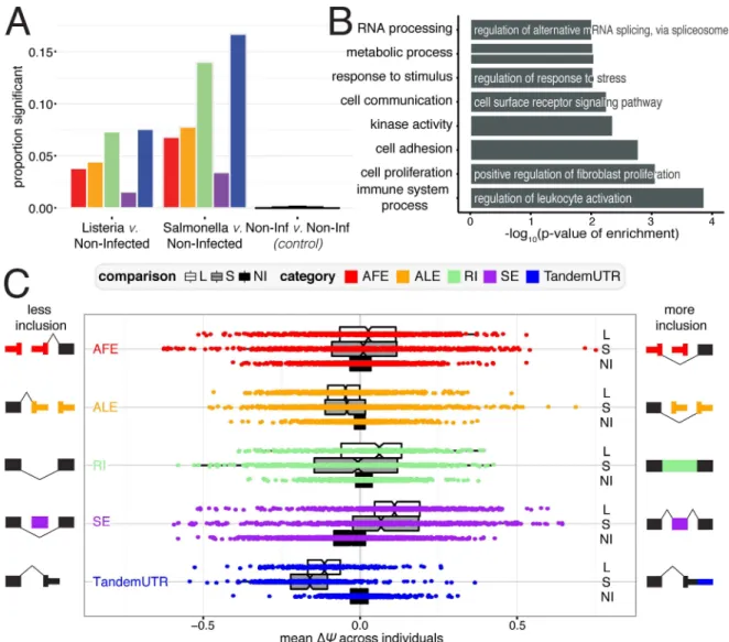
(N1,098) across all categories in both bacteria (significance was defined as Bayes Factor>5
in at least 10% of individuals and |meanΔC|>0.05), compared to null expectations derived
from measuring changes among pairs of non-infected samples (N = 29) (2A,S7 Table). Our criteria for determining significant changes also allows us to choose exons that are more likely to be consistently changing across many individuals; significantly changing exons have lower variance inΔCvalues across individuals than exons that are not changing after infection (S4 Fig). The greatest proportion of changes after infection occurred among retained intron and TandemUTR events. PSI values for 7.3% and 14% of retained introns and 7.6% and 16.7% of TandemUTRs significantly changed after infection withListeriaorSalmonella, respectively. When we considered the set of genes associated with at least one significant change in RNA processing, we observed an over-representation of Gene Ontology categories involved in immune cell processes and the cellular response to a stimulus (Fig 2B,S8 Table).
Genome-wide shifts towards more prevalent usage of inclusion or exclusion isoforms have previously been observed as signatures of cellular stress responses or developmental processes [28–33]. To examine directional shifts in particular RNA processing mechanisms, we used mean
ΔCvalues (defined as the meanΔCacross all individuals for each gene or exon) and observed a striking global shift towards the usage of upstream polyadenylation sites in tandem 3’ UTR regions, indicating pervasive 3’ UTR shortening following infection with eitherListeriaor Salmo-nella(Fig 2C; bothP<2.2×10⁻¹⁶with Mann-Whitney U test; 94.4% and 98% of significant
Tan-demUTRs changes inListeriaandSalmonella). Although less frequent, we also see a general trend towards usage of upstream polyadenylation sites in alternative last exons. Interestingly, we found a striking correlation in the extent to which a given individual has a global shift towards 3’ UTR shortening and towards usage of an upstream ALE (Pearson R = 0.83,P= 4.97 × 10⁻¹⁶for Listeria and Pearson R = 0.78,P= 2.65 × 10⁻¹³for Salmonella,S10A Fig). These correlations sug-gest that the changes in ALEs and TandemUTRs are likely being regulated by similar or coordi-nated mechanisms. Our findings are consistent with previous reports that suggested that ALEs and TandemUTRs could both be regulated by changes in the activities of cleavage and polyade-nylation factors, rather than splicing machinery [34–36]. Additionally, we found a substantial increase in the inclusion of skipped exons after infection (bothP<2.2×10⁻¹⁶with
Mann-Whit-ney U test; 79.8% and 77.3% of significant SE changes inListeriaandSalmonella,S5 Fig). The consistency of these patterns suggests that a dedicated post-transcriptional regulatory program might underlie genome-wide shifts in RNA processing after bacterial infection.
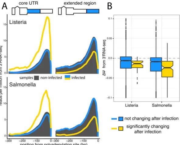
Quantifying changes at the terminal ends of transcripts is challenging due to known biases in standard RNA-seq protocols [37]. Thus, we validated our observation of 3’ UTR shortening upon infection by sequencing RNA from 6 individuals after infection withListeriaand Salmo-nellawith a 3’RNA-seq protocol, which specifically captures the 3’ most end of transcripts (see
Methods). TPM expression values calculated from 3’ RNA-seq data highly correlate with TPMs from RNA-seq data, establishing that this method is able to quantitatively measure of the number of transcripts in a cell (R>0.82 across all conditions,P<2.2 × 10⁻¹⁶,S6 Fig).
polyadenylation site relative to the downstream polyadenylation site, specifically for genes with significantly shorter 3’ UTRs upon infection as assessed by the RNA-seq data (Fig 3A) Finally, to look at 3’ UTR shortening in specific genes, we calculateCvalues for TandemUTRs using the 3’RNA-seq data (see Supplementary Methods inS1 Text) and find again that genes that are significantly changing 3’ UTR usage in the RNA-seq data exhibit a significant shift towards negativeΔCvalues in the 3’RNA-seq data (P<2.2 × 10⁻¹⁶;Fig 3B).
RNA processing changes persist 24 hours after infection
To evaluate whether the strong global shifts we observed were the result of a sustained immune response to the bacteria rather than a short-lived cellular stress response, we sequenced RNA from a subset of our individuals (N = 6) after 24 hours (h) of infection withListeriaor Salmo-nella. Samples sequenced after 24h were paired with matched non-infected controls cultured for
Fig 2. RNA processing changes in response to bacterial infection.(A) Proportion of events for RNA processing category that are significantly changing after infection withListeria(left),Salmonella(middle), or variation between non-infected samples as a control (right). Numbers indicate the number of significant changes per category. (B) Significantly Gene Ontology categories for genes with any significant RNA processing change (FDR10%). (C) Distribution ofΔΨvalues for each RNA processing category. Negative values represent less inclusion, while positive values represent more inclusion, as defined by the schematic exon representations.
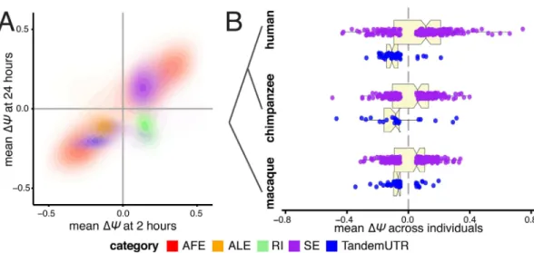
24h to calculateΔCvalues for alternative RNA processing categories using MISO [27]. Across all categories, we found many more significant changes in RNA processing and largerΔCvalues after infection for 24h than after 2h (S7 Fig). These results further support a key role for RNA processing in controlling innate immune responses to infection. For genes or exons that changed significantly after 2h of infection in this set of 6 individuals (seeMethods), the majority of changes (67%) remained detectable at 24h, usually with similar magnitude and direction (Fig 4A). The one notable exception is that introns that are preferentially retained at 2h in infected cells tended to be spliced out at 24h. Our results thus indicate that the splicing of alternative retained introns during the course of the immune response to infection is temporally dynamic.
Shortening of 3’ UTR regions and increased exon inclusion are
evolutionarily conserved responses to infection in primates
Since the innate immune response is ancient, one might expect core aspects of this pathway to be evolutionarily conserved among primates. To explore this question, we used high-depth
Fig 3. 3’RNA sequencing shows increased usage of upstream polyadenylation sites upon infection.(A) Meta-gene distributions of 3’RNA-seq read densities at the upstream polyA sites (core regions,left) and downstream polyA sites (extended regions,right) of Tandem 3’ UTRs after infection withListeriaorSalmonella (topandbottom, respectively). Shown are the read distributions for non-infected samples across all Tandem 3’ UTRs (black) and infected samples at Tandem 3’ UTRs that significantly change after infection (yellow) or show no change after infection (blue), as called by the RNA-seq data. (B) Distribution ofΔΨvalues calculated from 3’RNA-seq data for Tandem 3’ UTRs. We observe significant shifts (P<2.2×10⁻¹⁶for bothListeriaandSalmonella) towards negativeΔΨvalues in Tandem UTRs that are identified as significantly changing in RNA-seq data (yellow) relative to Tandem UTRs without any change after infection (blue).
RNA-seq data from human, chimpanzee, and rhesus macaque primary whole blood cells (N = 6individuals from each species) before and after 4 hours of stimulation with lipopolysac-charide (LPS), the major component of the outer membrane of Gram-negative bacteria. Using an analysis pipeline similar to our analysis of infection after 24 hours of infection withListeria orSalmonella, we calculatedΔCvalues for alternative RNA processing categories using MISO [27] (see Supplementary Methods inS1 Text,S9 Table). As with macrophages, we observed consistent strong shifts towards increased exon inclusion and shorter 3’ UTRs in human whole blood cells stimulated with LPS. Furthermore, we observed similar trends toward increased exon inclusion and significant 3' UTR shortening in macaques and chimpanzees, supporting a conserved response of RNA processing pathways during innate immune responses in primates (Fig 4B). In addition, these data show that the shortening of 3’ UTRs and increased exon inclu-sion are not limited to isolated macrophages but also observed as part of the immune response engaged by interacting primary granulocytes and lymphocytes, more closely mimickingin vivo cellular responses to infection.
Increased exon inclusion is associated with increased gene expression levelsSince we observed strong connections between overall isoform usage and differential gene expression, we explored the relationship between the occurrence of particular RNA processing changes and the direction of gene expression changes in response to infection. We found that genes with significant differences in skipped exon usage pre- and post-infection (up to 80% of which have increased skipped exon inclusion after infection) were more likely to be up-regulated after infection (T-test, bothP1×10−4forListeriaandSalmonella;Fig 5A;S8A Fig). This associa-tion remained when gene expression levels were calculated using reads mapping to constitutive exons only (S8A Fig), eliminating concerns about alternative spliced isoforms impacting gene expression estimates. Our observation is consistent with a recent study that reported increased
Fig 4. Directed shifts in RNA processing persist across time, stimulus conditions, and closely related species.(A) Correlations betweenΔΨvalues after 2 hours of infection (x-axis) andΔΨvalues after 24 hours of infection (y-axis), presented as density plots per RNA processing category in contrasting colors. Plotted are events that are significant after 2 hours of eitherListeriaorSalmonellainfection. (B) Distributions ofΔΨvalues for skipped exons (purple) and TandemUTRs (blue) following infection of whole blood cells with lipopolysaccharide, assessed in human (top), chimpanzee (middle) and rhesus macaque (bottom) individuals(N = 6per species). We observe prominent global shifts in isoform distributions in human and macaque (SEhumanP = 0.003, TandemUTRhuman
P = 2.4×10⁻¹³; SE
macaqueP = 0.05, TandemUTRmacaqueP = 8.8×10⁻⁶) with a more modest trend observed in
chimpanzee, potentially due to poor transcript annotations in the chimpanzee genome (SEchimpanzeeP = 0.26,
TandemUTRchimpanzeeP = 0.002). All p-values were calculated using a Student’s t-test testing deviation from a
mean of zero.
gene expression in tissues with increased inclusion of evolutionarily novel SEs [38]. Genes with other types of splicing changes showed no trend in expression changes.
Genes with significant skipped exon usage were enriched for Gene Ontology categories such as “response to stimulus,” and also for “RNA processing” and “RNA stability” categories (S9A Fig). Included within these latter categories are several members of two major splicing factor families, SR proteins and hnRNPs, which showed increased inclusion of skipped exons and increased gene expression after infection (S9B Fig). The SR protein and hnRNP families are known to function in complex auto- and cross-regulatory networks [39,40], often antago-nizing one another’s effects on splicing [41–43]. Consistent with the slightly higher up-regu-lation of members of the hnRNP family, we observed a consistent enrichment for intronic splicing silencers in upstream intronic regions around significantly excluded skipped exons (S9C Fig). These elements are often bound by hnRNPs to promote exclusion of a cassette exon [42])
Variation across individuals provides insight into putative
trans-regulatory factors
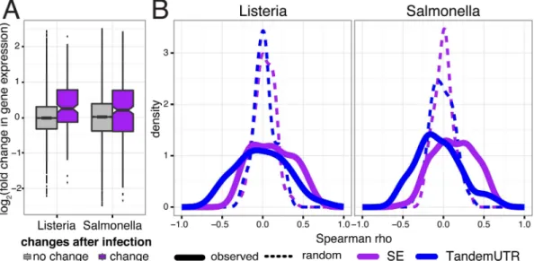
A unique feature of our study design is our sampling of 60 individuals after infection with both ListeriaandSalmonella. By taking advantage of natural variation in the regulation of RNA cessing to infection, we aimed to gain further insight into the connections between RNA pro-cessing and changes in gene expression levels in response to infection. For each gene, we calculated the correlation between the inter-individual variation in RNA processing changes (ΔC) and the fold changes in gene expression levels upon infection. For most categories, we observed shifts in the distribution of correlations between RNA processing and gene expression levels relative to permuted controls (S8B Fig), with skipped exons and TandemUTRs showing the most consistent patterns (Kolmogorov-Smirnov test,P0.005;Fig 5B). Increased skipped
Fig 5. Relationship between RNA processing changes and gene expression changes.(A) Distribution of fold changes in gene expression (y-axis, log2scale) for genes with significant skipped exon changes after infection
(purple) and genes with no change after infection (gray). (B) Distribution of Spearman correlations betweenΔΨ and fold change in gene expression across 60 individuals per gene (solid line), for genes with only one annotated alternative event and significantly changed SE usage (purple; nL= 46 genes and nS= 97 genes) or tandem UTR
usage (blue; nL= 36 genes and nS= 86 genes). Dotted lines show distribution of the correlation coefficients after
permutingΔΨvalues.
exon inclusion correlates with increased gene expression of the gene across individuals in 69% of the genes with significant skipped exons. In addition, preferential expression of shorter alter-native 3’ UTR isoforms tends to be correlated with increased up-regulation of the associated genes in response to infection. Our findings thus suggest that RNA processing changes may directly impact gene expression levels, or at least are commonly regulated with changes in tran-scription or mRNA stability.
Substantial inter-individual variation in the genome-wide average RNA processing patterns also correlated strongly with average gene expression changes upon infection, particularly for changes in 3’ UTR usage. That is, individuals who exhibited more skipped exon inclusion or 3’ UTR shortening also tended to exhibit larger global shifts towards up-regulation of gene expression levels upon infection (S10B Fig).
Next, we sought to exploit our relatively large sample size to identify candidatetrans-factors impacting the amount of skipped exon and TandemUTR changes observed across individuals. Specifically, we examined the relationship between mean changes in exon inclusion and fold change in expression for all expressed genes, across the 60 individuals. When we focus on pro-teins with known RNA binding functions or domains–hypothesizing that these factors are more likely to regulate post-transcriptional mechanisms–we find many significant correlations (FDR1%) for known regulators of the corresponding RNA processing category (S11 Fig). For instance, genes whose changes in expression correlated with individual-specific mean 3’ UTR shortening included several factors previously implicated in regulating polyadenylation site usage (such as cleavage and polyadenylation factors [44], hnRNP H proteins [27], and hnRNP F [45];S11A Fig). Additionally, many known regulators of alternative splicing–includ-ing a number of SR proteins and hnRNPs–have increased expression levels in individuals with larger extents of skipped exon inclusion (S11B Fig).
3’ UTR shortening as a means to evade repression by immune-related miRNAsThe larg-est global shift observed in alternative isoform abundance was for 3’ UTR shortening, where as many as 98% of significantly changing TandemUTRs shifted towards usage of an upstream polyA site post-infection. Previously, global 3’ UTR shortening has been associated with prolif-erating cells, particularly in the context of cellular transitions in development [28], cell differen-tiation [46], cancerous [29] states, or global 3’ UTR lengthening in embryonic development [33]. However, macrophages do not proliferate, which we confirmed with a BrdU labeling assay in both resting and infected conditions (S12 Fig). This result indicates that the pervasive 3’ UTR shortening observed in response to infection is due to an active cellular process that is independent of cell division and might be mechanistically distinct from that leading to 3’ UTR shortening in proliferating cells.
Previous studies in proliferating cells postulated that 3’ UTR shortening can act as a way to evade regulation by microRNAs (miRNAs), since crucial miRNA target sites are most often found in 3’ UTR regions [28]. To evaluate this hypothesis we performed small-RNA sequenc-ing in 6 individuals after 2h and 24h of infection withListeriaandSalmonella. When focusing specifically on miRNAs expressed in macrophages (S10 Table), we found that the extended UTR regions of infection-sensitive 3’ UTRs have a significantly higher density of miRNA target sites compared to TandemUTRs that do not change in response to infection (Fig 6A). This increased density of miRNA target sites was restricted to the extension region, which is subject to shortening after infection: no difference in the density of target sites was observed in the common “core” 3’ UTR regions of the same genes (S13 Fig).
region of significantly shortened 3’ UTRs (with a background distribution matched for sequence composition;S14 Fig; see Supplemental Methods inS1 Text). We found 15 miRNAs with significantly enriched target sites (FDR10% and enrichment1.5 fold) in either the ListeriaorSalmonellaconditions, with 6 miRNAs over-represented in both bacterial condi-tions (Fig 6B). Interestingly, of the miRNAs with the highest enrichment after infection with eitherListeriaorSalmonella, two (miR-146b and miR-125a) have previously been shown to be important regulators of the innate immune response [47–51]. Accordingly, we found that 4 of the miRNAs with the highest enrichment after infection (miR-125a, miR146b, miR-3661, and miR-151b) are all up-regulated following infection, and strongly so after 24h of infection (Fig 6B). In the majority of cases, greater 3’ UTR shortening was associated with a stronger increase in gene expression across individuals, suggesting that shifts towards shorter 3’ UTRs after infection may allow these transcripts to escape from repression by specific immune-induced miRNAs.
Fig 6. Tandem 3’ UTR shortening allows evasion of regulation by miRNAs.(A) Distribution of frequency of miRNA target sites per nucleotide in the extended regions of Tandem UTRs that either show no change after infection (grey) or significantly change after infection (blue). (B) Significantly enriched miRNA target sites (FDR10%, |FC|>1.5) in the extended regions of significantly changing Tandem UTRs after infection withListeria-only (top), withSalmonella-only (middle), or both bacteria (bottom). For each bacteria, the barplots in the left panels show the fold enrichment (x-axis, log2
scale) of target sites in the extended regions. White bars represent non-significant enrichments. Panels on the right show the fold change in miRNA expression (x-axis, log2scale with standard error bars) after either 2 hours of infection (light colors) or 24 hours of infection (dark colors).
Discussion
Taken together, our results provide strong evidence that RNA processing may play a key role in the regulation of innate immune responses to infection. Despite known differences in the regulatory pathways elicited in macrophages byListeriaandSalmonella, our data revealed striking similarities in the overall patterns of RNA processing induced in response to both pathogens. Given the strong overlaps, we chose throughout this study to focus on consistent patterns across the two bacteria.
Though genes that have differential isoform usage in response to infection are more likely to show changes in gene expression, we found that as many as 47% of genes showing differential isoform usage have no evidence for differences in gene expression upon infection. This obser-vation suggests that a considerable proportion of genes are regulated predominantly by post-transcriptional mechanisms, with minimal changes in post-transcriptional regulation. Differential isoform usage is coupled to a prominent increase in the diversity of isoforms following infec-tion. Intriguingly, genes showing the strongest increases in diversity upon infection were strongly enriched among down-regulated genes (S3B Fig). This observation raises the possibil-ity that the increased isoform diverspossibil-ity could reflect a shift toward less stable isoforms, e.g., those targeted by nonsense-mediated decay (NMD), thus reducing transcript levels [52,53].
Upon infection of human macrophages with eitherListeriaorSalmonella, we observed substantial shifts towards increased skipped exon inclusion and shortening of 3’ UTRs. These RNA processing patterns remained prominent when interrogated in human whole blood after an alternative immune stimulus (LPS), despite the fact the number of significant RNA processing changes in whole blood was generally smaller than that observed in purified mac-rophages infected with live bacteria. This reduced effect is likely explained by the fact that in whole blood, we are interrogating a very heterogeneous cell population within which only a fraction (~3–6%) of the cell types are actually responding to LPS stimulation, such as mono-cytes/macrophages [54]. Additionally, transcriptional changes induced by live bacteria such asListeriaorSalmonella, which concomitantly activates multiple innate immune pathways [22,23], are larger than transcriptional changes that can induced by a single purified ligand, such as LPS, which activates primarily the TLR4 pathway [55]. Despite this reduced statistical power, we still observed conserved RNA processing signatures of immune stimulation through the primate lineage, supporting the likely functional importance of these cellular responses and the ubiquity of RNA processing changes after immune stimulation across evolution.
we observe that the target sites in our distal 3’ UTR regions are directly related to miRNAs whose expression specifically changes after infection.
Specifically, our data support previous hypotheses and find evidence for that 3’ UTR short-ening in macrophages is associated with the loss of target sites for a subset of immune-regu-lated miRNAs, including miR-146b, miR-125a, and miR-151b (Fig 6B). These miRNAs target the extended 3’ UTR regions of several critical activators of the innate immune response– including theIRF5transcription factor [57] and the MAP kinasesMAPKAP1andMAP2K4 [58]–all of which shift to usage of shorter 3’ UTRs after infection. It is tempting to speculate that without such 3’ UTR shortening, the binding of one or more of miRNAs to the 3’ UTR region of these genes would compromise their up-regulation and the subsequent activation of downstream immune defense pathways. Given that many miRNAs are involved in either (or both) mRNA degradation and translational efficiency, we cannot know for sure which of these processes is prevented by the 3’ UTR shortening of these genes.
While our results point to the importance of 3’ UTR shortening to allow genes to evade post-transcriptional regulation by miRNAs, this is not generalizable to all miRNAs induced in response to infection. Indeed, some of the miRNAs most highly induced after infection, such as miR-155 and miR-29b, are not enriched for binding sites in the longer 3’UTR regions. Thus, while miRNA regulation is crucial to mammalian immune response [59–62], the mechanisms that dictate what specific 3’UTR are shortened or not will require further investigation. Indeed, it is likely that miRNA targets whose 3’ UTRs are shortened in response to infection to evade miRNA regulation are actually key targets of the same miRNAs in other cellular conditions.
The convergence towards similar isoform outcomes across many disparate genes suggests the activation of factors acting intransto drive global shifts towards inclusion of cassette exons or usage of upstream proximal polyadenyalation sites. Taking advantage of our relatively large sample size, we were able to identify candidatetrans-factors whose up-regulation upon infec-tion might have widespread impacts on RNA processing patterns, including some known regu-lators of alternative splicing or 3’end processing (S11 Fig). Sets of RNA binding factors likely act in combination to influence final transcriptome states [63]. For instance, a general up-regu-lation of splicing machinery following infection [64], allowing the cell to recognize and use additional splice sites, might contribute to increased exon inclusion and greater isoform diver-sity observed following infection. Members of both the hnRNP and SR protein splicing factor families show up-regulation of their overall gene expression levels (S9B Fig), likely contributing to specific instances of exon inclusion as well as exclusion. Correspondingly, changes in the regulation of many canonical cleavage and polyadenylation factors were tied to individual-spe-cific shifts in 3’ UTR shortening. The significant correlation between global shifts in ALE usage and TandemUTRs across individuals further support the notion that cleavage and poladenyla-tion pathways are regulating the changing 3’ UTR landscape upon infecpoladenyla-tion (S10A Fig). Intriguingly, fold changes in hnRNP H1 gene expression have high correlations with both skipped exon inclusion and 3’ UTR shortening, consistent with its known splicing function and suggesting that this factor may play multiple roles in shaping the transcriptome in response to bacterial infection.
increase in nascent RNA observed by Berg et al. during neuronal differentiation [65] (across 60 samples, paired t-test; P = 0.006 for Listeria and P = 4.57 × 10⁻⁶for Salmonella), combined with no changes in the levels of U1 snRNA (relative to 5S rRNA, Supplemental Methods inS1 Text,S15 Fig). Thus it is possible that a delayed response of U1 snRNA could promote the usage of upstream polyadenylation sites in both TandemUTRs and ALEs. Interestingly, Berg et al. also observe increased up-regulation of individual internal exons upon moderate U1 knockdown in neurons [65], which is consistent with our observation of increased skipped exon inclusion.
Substantial inter-individual variation in the genome-wide average RNA processing patterns also correlated strongly with average gene expression changes upon infection, particularly for changes in 3’ UTRs and skipped exons. That is, individuals who exhibited more skipped exon inclusion or usage of upstream polyadenylation sites also tended to exhibit larger global shifts towards up-regulation of gene expression levels upon infection (S10B Fig). Though these pat-terns might result from stochastic variation in levels of one or severaltrans-factors, they are also likely to be influenced by genetic differences among individuals. It will be interesting to ask whether global isoform differences might potentially be reflective of variation in an individ-ual’s susceptibility to infection.
Materials and Methods
Complete details of the experimental and statistical procedures can be found in SI Materials and Methods. Briefly, blood samples from 60 healthy donors were obtained from Indiana Blood Center with informed consent and ethics approval from the Research Ethics Board at the CHU Sainte-Justine (protocol #4022). All individuals recruited in this study were healthy males between the ages of 18 and 55 y old. Blood mononuclear cells from each donor were iso-lated by Ficoll-Paque centrifugation and blood monocytes were purified from peripheral blood mononuclear cells (PBMCs) by positive selection with magnetic CD14 MicroBeads (Miltenyi Biotec). In order to derive macrophages, monocytes were then cultured for 7 days in RPMI-1640 (Fisher) supplemented with 10% heat-inactivated FBS (FBS premium, US origin, Wisent), L-glutamine (Fisher) and M-CSF (20ng/mL; R&D systems). After validating the differentia-tion/activation status of the monocyte-derived macrophages we infected them at a multiplicity of infection (MOI) of 10:1 forSalmonella typhimuriumand an MOI of 5:1 forListeria monocy-togenesfor 2- and 24-hours. For primate comparisons, whole blood was drawn from 6 healthy adult humans (CHU Sainte-Justine), 6 common chimpanzees, (Texas Biomedical Research Institute), and 6 rhesus macaques (Yerkes Regional Primate Center). Human samples were acquired with informed consent and ethics approval from the Research Ethics Board (CHU Sainte-Justine, #3557). Non-human primate samples were acquired in accordance with indi-vidual institutional IACUCC requirements. For all species, 1ml of blood was drawn into a media-containing tube (Trueculture tube, Myriad, US) spiked with ultrapure LPS (Invitrogen, USA) or endotoxin-free water (control). Samples were stimulated with 1ug/ml LPS, at 37C for 4 hours before total blood leukocytes were isolated and RNA collected. Genome-wide gene expression profiles of untreated and infected/treated samples were obtained by RNA-sequenc-ing for both mRNA transcripts and small RNAs. After a series of quality checks (SI Materials and Methods), mRNA transcript abundances were estimated using RSEM [66] and Kallisto [25] and microRNA expression estimates were obtained as previously described[67]. To detect genes with differential isoform usage between two groups of non-infected and infected samples we used a multivariate generalization of the Welch's t-test. Shannon entropy Hsh(also known
the MISO software package (v0.4.9) [27] using default settings and hg19 version 1 annotations. Events were considered to be significantly altered post-infection if at least 10% (n>= 6) of
individuals had a BF>= 5 and the |meanΔC|0.05 (S6 Table). To conduct a targeting sequencing of 3’ ends of transcripts, We used Smart-3SEQ (Foley et al, manuscript in prepara-tion), a modified version of the 3SEQ method [68]. We used a custom gene ontology script to test for enrichment of functional annotations among genes that significantly changed isoform usage in response to infection (SI Materials and Methods). All predicted miRNA target sites within annotated TandemUTR regions were obtained using TargetScan (v6.2) [69].
Ethics statement
Institutional ethics (IRB) approval was obtained by the CHU Sainte-Justine Institutional Review Board (approved project #3557) and all subjects gave written informed consent prior to participation.
Data access
Data generated in this study have been submitted to the NCBI Gene Expression Omnibus (GEO;http://www.ncbi.hlm.nih.gov/geo/) under the accession number GSE73765, which com-prises of the mRNA-seq data (GSE73502) and small RNA-seq data (GSE73478).
Supporting Information
S1 Text. Supplementary Methods and Materials.
(PDF)
S1 Fig. Differential isoform usage after infection. (A)Volcano plots of differential expres-sionafter infection with Listeria and Salmonella in the left and right panels, respectively.–log10 p-values (y-axis) testing for differential expression are plotted against averagelog2fold changes in expression levels (x-axis) for genes that are not differentially expressed (black) and genes that with significant differential expression after infection (FDR0.1% and|log2(fold change)|
0.5;blue).(B)Distribution of p-values for the differential isoform usage test upon infection withListeriaandSalmonella. Expected distribution of p-values under the null hypothesis of no significant difference between the mean relative isoform usage is shown in grey.(C)A compar-ison of DIU effect sizes between DIU genes at 1% FDR (following infection withListeriain dark pinkandSalmonellaingold) and the background of all genes (lighter colors). Effect sizes are defined as maximum change in relative isoform abundances per gene upon infection.(D)
Distributions ofΔShannondiversity index (ΔSDI) after infection withListeriaandSalmonella. Null distribution (black dotted line) was generated by permuting samples across conditions.(E)
The percentage of genes with a dominant isoform before infection (left) and the fraction of these genes where the dominant isoform changes after infection (right).
(PDF)
S2 Fig. Representative examples of immune-related genes with significant DIU after infec-tion.Heatmaps below each example represent the variation in isoform usage across the 60 individuals, where each vertical bar represents one individual. The dominant isoform in non-infected samples is represented ingrey, while the predominant isoform in infected samples is in a color. Darker bars represent increased relative usage, while lighter bars represent decreased relative usage.
S3 Fig. Gene expression and differential isoform diversity after infection. (A)Proportion of genes with differential isoform usage (y-axis) among all genes (grey), differentially expressed genes that are down-regulated after infection (orange), and differentially expressed genes that are up-regulated after infection (green).(B)log2fold changes (with standard error bars) for odds of lowΔShannonand highΔShannonamong the set of all the differentially expressed genes, up-regulated differentially expressed genes, and down-regulated differentially expressed genes, following infection withListeria(dark pink) andSalmonella(gold).
(PDF)
S4 Fig. Variance in RNA processing across 60 individuals.Distribution of coefficient of vari-ation (x-axis) inΔCvalues for each isoform in a given RNA processing category.
(PDF)
S5 Fig. Two representative examples of genes with significant skipped exon changes after infection.Sashimi plots for two genes,PTK2BandSTK40, that have significant changes in skipped exon usage both after 2 hours and 24 hours of infection with either bacteria. (PDF)
S6 Fig. Relationship between 3'RNA-seq and RNA-seq data.Correlations between TPMs from 3' RNA-seq data (y-axis) and TPMs from RNA-seq data (x-axis).
(PDF)
S7 Fig. RNA processing changes 24hr after infection. (A)Proportion of significantly chang-ing events (x-axis) after 2 hours of infection (dark colors) and 24 hours of infection (light col-ors) with eitherListeria(top) orSalmonella(bottom). Numbers indicate the significant events at corresponding timepoints using only the 6 individuals used for these cross-timepoint analy-ses.(B)Distribution ofΔCvalues (x-axis) for significantly changing events in each event type after either 2 hours (top) or 24 hours (bottom) of infection.
(PDF)
S8 Fig. Relationships between RNA processing and gene expression changes after infection. (A)Distributions of overall fold changes in gene expression (y-axis,log2scale) for genes that have significant splicing changes in each event type (colored boxplots) and genes with no splic-ing changes after infection (grey). Gene expression values are calculated using either full tran-script models (top) or only constitutively included exons (bottom).(B)Distribution of spearman correlations between theΔCof an event and the fold change in gene expression, across individuals, for events that are not significantly changing after infection (left) and signif-icantly changing events (right). Solid lines represent the observed values and dotted lines repre-sent a distribution of correlations after permuting the correspondence betweenΔCs and fold changes in gene expression.
(PDF)
and significantly excluded skipped exons (bottom). Shading of the bars indicates the signifi-cance of the enrichment.
(PDF)
S10 Fig. Inter-individual variation of global shifts in alternative splicing. (A)Correlations between the meanΔCvalues per individual for ALEs (x-axis) and TandemUTRs (y-axis).(B)
Spearman correlations between the meanΔCvalue per individual and the mean fold change of gene expression per individual for corresponding genes.
(PDF)
S11 Fig. Identifying RNA binding factors that might be involved intrans-regulation of
RNA processing after infection.For both Tandem UTRs(A)and Skipped exons(B), the top panel is a scatter plot of Spearman correlations between the individual-specific meanΔC val-ues and individual-specific fold change of gene expression valval-ues for all expressed genes (grey) in bothListeria(x-axis) andSalmonella(y-axis). Genes with significant correlations
(FDR1%) in bothListeriaandSalmonellaconditions are plotted in black, and those factors with known RNA-binding properties are colored by their functional category. The bottom pan-els show distributions of the average Spearman correlation values for each of the RNA-binding functional categories with significant correlations.
(PDF)
S12 Fig. Cellular proliferation after bacterial infection.BrdU cell proliferation assay in mac-rophages (top panel) and LCLs (bottom panel) in non-infected cells and in cells infected with ListeriaorSalmonellafor both 2 and 24 hours. BrdU incorporates into newly synthesized DNA and therefore the quantity of BrdU incorporated into cells (x-axis) is a direct indication of cell proliferation. No evidence for cellular proliferation was observed in macrophages, in contrast to the high rates of proliferating cells observed in our positive control population of LCLs. (PDF)
S13 Fig. Density of miRNA target sites in 3’ UTR regions.Barplots in grey indicate Tandem 3’ UTRs that are not changing after infection, while colored barplots indicate Tandem 3’ UTRs that are significantly changing after eitherListeria(pink) orSalmonella(yellow) infections. (PDF)
S14 Fig. Nucleotide composition of Tandem 3’ UTR regions.The distribution of GC content in core (brown) and extended (blue) regions of Tandem 3' UTRs that are either not changing or significantly changing after infection (left panel). While there 3' UTRs that are significantly changing generally have greater overall GC content, this is true for both the core and extended regions of the 3’ UTRs. Thus, the distributions of relative GC content when comparing the regions is distributed around 1 for both significantly changing (yellow) and not changing (blue) Tandem 3' UTRs (right panel).
(PDF)
proliferate either before or after infection (S12 Fig). There are no significant shifts in the distri-bution of 5s rRNA or U1 snRNA fold changes after infection (t-test for null ofμ = 1, both P>0.01for 5s rRNA and bothP>0.2for U1 snRNA), while the distribution of total RNA
fold changes are significantly increased after infection (t-test for null ofμ = 1,P<0.001for
bothListeriaandSalmonellasamples). (PDF)
S1 Table. mRNA-seq statistics.Details of RNA-seq data for samples after infection at 2 hours or 24 hours. Libraries were made as described in Supplementary Section 1.2.1 inS1 Text. (XLSX)
S2 Table. Gene Level Differential Gene Expression.Estimates of differential gene expression for the 14,954 genes tested by DESeq2, as described in Supplementary Section 1.3.2 inS1 Text. Included are thelog2fold change estimates, p-values for differential expression, and Benja-mini-Hochberg adjusted p-values for differential expression between non-infected samples and bothListeriaandSalmonellasamples.
(XLSX)
S3 Table. Gene Ontology Enrichments for Differential Expression.Enriched Gene Ontology categories for genes that are differentially expressed, or show differential isoform usage after infection. Gene Ontology analyses were performed using GSEA, as described in Supplementary Section 1.3.8 inS1 Text.
(XLSX)
S4 Table. Differential Isoform Usage.Estimates of differential isoform usage for the 11,353 genes tested as described in Supplementary Section 1.3.3 inS1 Text. Included are the changes in relative isoform usage for the isoform with the greatest relative change after infection, the p-value and Benjamini-Hochberg FDR for DIU using isoform expression levels calculated using RSEM, and the FDR for DIU using isoform expression levels calculated using Kallisto. (XLSX)
S5 Table. Change in Isoform Diversity after Infection.Estimates of Shannon diversity indi-cies for 11,353 genes both before and after infection with bothListeriaandSalmonella, as described in Supplementary Section 1.3.4 inS1 Text.
(XLSX)
S6 Table. Differential isoform usage across 5 specific RNA processing categories.Estimates ofCandΔCvalues calculated by MISO (Supplementary Section1.3.5 inS1 Text) for each indi-vidual for each for the 5 different RNA processing categories assess in this study: alternative first exons (AFE), alternative last exons (ALE), retained introns (RI), skipped exons (SE), and Tandem 3’ UTRs (TandemUTR). Also included are Bayes Factors testing for differential iso-form usage per event for each individual, meanΔCvalues across all individuals and individuals with significant changes, and a column indicating whether an event was called significant based on the criteria defined in Supplementary Section 1.3.5 inS1 Text.
(XLSX)
S7 Table. Summaries of Significant RNA processing categories.A summary of the propor-tion of annotated events that were detected and determined to be significant (according to the criteria defined in Supplementary Section 1.3.5 inS1 Text) for each condition and RNA pro-cessing category. Included are tables for metrics after both 2 hours of infection and 24 hours of infection.
S8 Table. Gene Ontology Enrichments for significant RNA processing changes.Top gene ontology categories for genes that have significant RNA processing changes after infection with eitherListeriaorSalmonella. All tests were done using an iterative gene ontology analysis as described in Supplementary Section 1.3.5 inS1 Textand gene names refer to UniProt gene accessions.
(XLSX)
S9 Table. Summary of Significant RNA processing categories following infection with LPS, across primate species.A summary of the proportion of annotated evenst that were detected and determined to be significant (according to the criteria defined in Supplementary Section 1.3.5 inS1 Text) for each species and RNA processing category following infection of primary whole blood cells with LPS.
(XLSX)
S10 Table. miRNA mapping and differential expression statistics.Details of short RNA-seq data for 6 samples sequenced after both 2 hours and 24 hours of infection with eitherListeria orSalmonella, as described in Supplementary Sections 1.2.3 and 1.3.10 inS1 Text. Also included are the log2fold change estimates and p-values for differential expression of miRNAs
after both 2 hours and 24 hours of infection, tested by DESeq2 as described in Supplementary Section 1.3.10 inS1 Text.
(XLS)
Acknowledgments
We thank P Freese for a categorized list of RNA binding proteins, G McVicker for an initial script to perform iterative gene ontology analyses and P Sudmant for help parsing primate transcript annotations. We thank Y Katz, JM Taliaferro, P Sudmant, J Tung, and members of the Barreiro and Burge labs for helpful discussions and comments on the manuscript. We thank Calcul Québec and Compute Canada for managing and providing access to the super-computer Briaree from the University of Montreal, used to do many computations reported in the manuscript.
Author Contributions
Conceived and designed the experiments:AAP LBB.
Performed the experiments:APS JFB AD VY.
Analyzed the data:AAP GB YN JCG.
Contributed reagents/materials/analysis tools:JWF KJS ZPJ REL CBB.
Wrote the paper:AAP GB LBB.
References
1. Huang Q, Liu D, Majewski P, Schulte LC, Korn JM, Young RA, et al. The Plasticity of Dendritic Cell Responses to Pathogens and Their Components. Science. 2001; 294: 870–875. doi:10.1126/science. 294.5543.870PMID:11679675
2. Smale ST. Selective Transcription in Response to an Inflammatory Stimulus. Cell. 2010; 140: 833– 844.http://dx.doi.org/10.1016/j.cell.2010.01.037doi:10.1016/j.cell.2010.01.037PMID:20303874
4. Medzhitov R, Janeway CA Jr. Innate immune recognition and control of adaptive immune responses. Seminars in Immunology. 1998; 10: 351–353.http://dx.doi.org/10.1006/smim.1998.0136PMID:
9799709
5. Kawai T, Akira S. The role of pattern-recognition receptors in innate immunity: update on Toll-like receptors. Nat Immunol. Nature Publishing Group; 2010; 11: 373–384. doi:10.1038/ni.1863PMID:
20404851
6. Lynch KW. Consequences of regulated pre-mRNA splicing in the immune system. Nature Reviews Immunology. 2004; 4: 931–940. doi:10.1038/nri1497PMID:15573128
7. Martinez NM, Lynch KW. Control of alternative splicing in immune responses: many regulators, many predictions, much still to learn. Immunological Reviews. Wiley Online Library; 2013; 253: 216–236. doi:
10.1111/imr.12047PMID:23550649
8. Alasoo K, Estrada FM, Hale C, Gordon S, Powrie F. Transcriptional profiling of macrophages derived from monocytes and iPS cells identifies a conserved response to LPS and novel alternative transcrip-tion. bioRxiv. 2015.
9. Rao N, Nguyen S, Ngo K, Fung-Leung W-P. A novel splice variant of interleukin-1 receptor (IL-1R)-associated kinase 1 plays a negative regulatory role in Toll/IL-1R-induced inflammatory signaling. Molecular and Cellular Biology. Am Soc Microbiol; 2005; 25: 6521–6532. PMID:16024789
10. Wells CA, Chalk AM, Forrest A, Taylor D, Waddell N, Schroder K, et al. Alternate transcription of the Toll-like receptor signaling cascade. Genome Biology. BioMed Central Ltd; 2006; 7: R10.
11. O’Connor BP, Danhorn T, De Arras L, Flatley BR, Marcus RA, Farias-Hesson E, et al. Regulation of Toll-like Receptor Signaling by the SF3a mRNA Splicing Complex. Lin X, editor. PLoS Genet. Public Library of Science; 2015; 11: e1004932. doi:10.1371/journal.pgen.1004932PMID:25658809
12. Ishitani A, Geraghty DE. Alternative splicing of HLA-G transcripts yields proteins with primary struc-tures resembling both class I and class II antigens. Proceedings of the National Academy of Sciences. National Acad Sciences; 1992; 89: 3947–3951.
13. Bihl MP, Heinimann K, Rudiger JJ, Eickelberg O, Perruchoud AP, Tamm M, et al. Identification of a novel IL-6 isoform binding to the endogenous IL-6 receptor. American journal of respiratory cell and molecular biology. Am Thoracic Soc; 2002; 27: 48–56. PMID:12091245
14. Nishimura H, Yajima T, Naiki Y, Tsunobuchi H, Umemura M, Itano K, et al. Differential roles of interleu-kin 15 mRNA isoforms generated by alternative splicing in immune responses in vivo. The Journal of experimental medicine. Rockefeller Univ Press; 2000; 191: 157–170. PMID:10620614
15. Goodwin RG, Friend D, Ziegler SF, Jerzy R, Falk BA, Gimpel S, et al. Cloning of the human and murine interleukin-7 receptors: demonstration of a soluble form and homology to a new receptor superfamily. Cell. Elsevier; 1990; 60: 941–951. PMID:2317865
16. Koskinen LL, Einarsdottir E, Dukes E, Heap GA, Dubois P, Korponay-Szabo IR, et al. Association study of the IL18RAP locus in three European populations with coeliac disease. Hum Mol Genet. Oxford Univ Press; 2009; 18: 1148–1155. doi:10.1093/hmg/ddn438PMID:19103669
17. Jensen LE, Whitehead AS. IRAK1b, a novel alternative splice variant of interleukin-1 receptor-associ-ated kinase (IRAK), mediates interleukin-1 signaling and has prolonged stability. Journal of Biological Chemistry. ASBMB; 2001; 276: 29037–29044. PMID:11397809
18. Gray P, Michelsen KS, Sirois CM, Lowe E, Shimada K, Crother TR, et al. Identification of a novel human MD-2 splice variant that negatively regulates Lipopolysaccharide-induced TLR4 signaling. The Journal of Immunology. Am Assoc Immnol; 2010; 184: 6359–6366. doi:10.4049/jimmunol.0903543
PMID:20435923
19. Ohta S, Bahrun U, Tanaka M, Kimoto M. Identification of a novel isoform of MD-2 that downregulates lipopolysaccharide signaling. Biochem Biophys Res Commun. Elsevier; 2004; 323: 1103–1108. PMID:
15381113
20. Janssens S, Burns K, Tschopp J, Beyaert R. Regulation of interleukin-1-and lipopolysaccharide-induced NF-κB activation by alternative splicing of MyD88. Current Biology. Elsevier; 2002; 12: 467– 471. PMID:11909531
21. Rodrigues R, Grosso AR, Moita L. Genome-wide analysis of alternative splicing during dendritic cell response to a bacterial challenge. PLoS One. 2013.
22. Haraga A, Ohlson MB, Miller SI. Salmonellae interplay with host cells. Nature Reviews Microbiology. Nature Publishing Group; 2008; 6: 53–66. PMID:18026123
23. Pamer EG. Immune responses to Listeria monocytogenes. Nature Reviews Immunology. Nature Pub-lishing Group; 2004; 4: 812–823. PMID:15459672
24. Li B, Ruotti V, Stewart RM, Thomson JA, Dewey CN. RNA-Seq gene expression estimation with read mapping uncertainty. Bioinformatics. 2010; 26: 493–500. doi:10.1093/bioinformatics/btp692PMID:
25. Bray NL, Pimentel H, Melsted P, Pachter L. Near-optimal probabilistic RNA-seq quantification. Nature Biotechnology. Nature Publishing Group; 2016. doi:10.1038/nbt.3519
26. Barbosa-Morais NL, Irimia M, Pan Q, Xiong HY, Gueroussov S, Lee LJ, et al. The evolutionary land-scape of alternative splicing in vertebrate species. Science. American Association for the Advance-ment of Science; 2012; 338: 1587–1593. doi:10.1126/science.1230612PMID:23258890
27. Katz Y, Wang ET, Airoldi EM, Burge CB. Analysis and design of RNA sequencing experiments for identifying isoform regulation. Nat Meth. 2010; 7: 1009–1015. doi:10.1038/nmeth.1528
28. Sandberg R, Neilson JR, Sarma A, Sharp PA, Burge CB. Proliferating Cells Express mRNAs with Shortened 3’ Untranslated Regions and Fewer MicroRNA Target Sites. Science. 2008; 320: 1643– 1647. doi:10.1126/science.1155390PMID:18566288
29. Mayr C, Bartel DP. Widespread Shortening of 3´UTRs by Alternative Cleavage and Polyadenylation Activates Oncogenes in Cancer Cells. Cell. 2009; 138: 673–684. doi:10.1016/j.cell.2009.06.016
PMID:19703394
30. Wong JJL, Ritchie W, Ebner OA, Selbach M, Wong JWH, Huang Y, et al. Orchestrated Intron Reten-tion Regulates Normal Granulocyte DifferentiaReten-tion. Cell. Elsevier Inc; 2013; 154: 583–595. doi:10. 1016/j.cell.2013.06.052PMID:23911323
31. Lackford B, Yao C, Charles GM, Weng L, Zheng X, Choi EA, et al. Fip1 regulates mRNA alternative polyadenylation to promote stem cell self-renewal. The EMBO Journal. 2014; 33: 878–889. doi:10. 1002/embj.201386537PMID:24596251
32. Shalgi R, Hurt JA, Lindquist S, Burge CB. Widespread inhibition of posttranscriptional splicing shapes the cellular transcriptome following heat shock. Cell Reports. Elsevier; 2014; 7: 1362–1370. doi:10. 1016/j.celrep.2014.04.044PMID:24857664
33. Ji Z, Lee JY, Pan Z, Jiang B, Bin Tian. Progressive lengthening of 3’ untranslated regions of mRNAs by alternative polyadenylation during mouse embryonic development. Valcarcel J, editor. PNAS. Pro-ceedings of the. . .; 2009; 106: 7028–7033. doi:10.1371/journal.pone.0008419PMID:19372383
34. Takagaki Y, Manley JL. Levels of Polyadenylation Factor CstF-64 Control IgM Heavy Chain mRNA Accumulation and Other Events Associated with B Cell Differentiation. Molecular Cell. 1998; 2: 761– 771. doi:10.1016/S1097-2765(00)80291-9PMID:9885564
35. Di Giammartino DC, Nishida K, Manley JL. Mechanisms and Consequences of Alternative Polyadeny-lation. Molecular Cell. 2011; 43: 853–866. doi:10.1016/j.molcel.2011.08.017PMID:21925375
36. Taliaferro JM, Vidaki M, Oliveira R, Olson S, Zhan L, Saxena T, et al. Distal Alternative Last Exons Localize mRNAs to Neural Projections. Molecular Cell. 2016; 61: 821–833. doi:10.1016/j.molcel.2016. 01.020PMID:26907613
37. Wang Z, Gerstein M, Snyder M. RNA-Seq: a revolutionary tool for transcriptomics. Nat Rev Genet. 2009; 10: 57–63. doi:10.1038/nrg2484PMID:19015660
38. Merkin JJ, Chen P, Alexis MS, Hautaniemi SK. Origins and impacts of new mammalian exons. Cell Reports. 2015; 10: 1992–2005. doi:10.1016/j.celrep.2015.02.058PMID:25801031
39. Huelga SC, Vu AQ, Arnold JD, Liang TY, Liu PP, Yan BY, et al. Integrative Genome-wide Analysis Reveals Cooperative Regulation of Alternative Splicing by hnRNP Proteins. Cell Reports. 2012; 1: 167–178. doi:10.1016/j.celrep.2012.02.001PMID:22574288
40. Pandit S, Zhou Y, Shiue L, Coutinho-Mansfield G, Li H, Qiu J, et al. Genome-wide analysis reveals SR protein cooperation and competition in regulated splicing. Molecular Cell. Elsevier; 2013; 50: 223–235. 41. House AE, Lynch KW. Regulation of Alternative Splicing: More than Just the ABCs. Journal of
Biologi-cal Chemistry. 2008; 283: 1217–1221. doi:10.1074/jbc.R700031200PMID:18024429
42. Wang Z, Burge CB. Splicing regulation: From a parts list of regulatory elements to an integrated splic-ing code. RNA. 2008; 14: 802–813. doi:10.1261/rna.876308PMID:18369186
43. Busch A, Hertel KJ. Evolution of SR protein and hnRNP splicing regulatory factors. WIREs RNA. Wiley Online Library; 2012; 3: 1–12. doi:10.1002/wrna.100PMID:21898828
44. Zheng D, Tian B. RNA-binding proteins in regulation of alternative cleavage and polyadenylation. Sys-tems Biology of RNA Binding Proteins. Springer; 2014. pp. 97–127.
45. Veraldi KL, Arhin GK, Martincic K, Chung-Ganster LH, Wilusz J, Milcarek C. hnRNP F Influences Bind-ing of a 64-Kilodalton Subunit of Cleavage Stimulation Factor to mRNA Precursors in Mouse B Cells. Molecular and Cellular Biology. 2001; 21: 1228–1238. PMID:11158309
46. Lackford B, Yao C, Charles GM, Weng L, Zheng X, Choi EA, et al. Fip1 regulates mRNA alternative polyadenylation to promote stem cell self-renewal. The EMBO Journal. 2014; 33: 878–889. doi:10. 1002/embj.201386537PMID:24596251
48. Perry MM, Williams AE, Tsitsiou E, Larner-Svensson HM, Lindsay MA. Divergent intracellular path-ways regulate interleukin-1β-induced miR-146a and miR-146b expression and chemokine release in human alveolar epithelial cells. FEBS Letters. Federation of European Biochemical Societies; 2009; 583: 3349–3355. doi:10.1016/j.febslet.2009.09.038
49. Boldin MP, Baltimore D. MicroRNAs, new effectors and regulators of NF-κB. Immunological Reviews. 2012; 246: 205–220. doi:10.1111/j.1600-065X.2011.01089.xPMID:22435557
50. Kim SW, Ramasamy K, Bouamar H. MicroRNAs miR-125a and miR-125b constitutively activate the NF-κB pathway by targeting the tumor necrosis factor alpha-induced protein 3 (TNFAIP3, A20). 2012. doi:10.1073/pnas.1200081109/-/DCSupplemental/pnas.201200081SI.pdfPMID:22550173
51. Banerjee S, Cui H, Xie N, Tan Z, Yang S, Icyuz M, et al. miR-125a-5p Regulates Differential Activation of Macrophages and Inflammation. Journal of Biological Chemistry. 2013; 288: 35428–35436. doi:10. 1074/jbc.M112.426866PMID:24151079
52. Lewis BP, Green RE, Brenner SE. Evidence for the widespread coupling of alternative splicing and nonsense-mediated mRNA decay in humans. PNAS. 2003; 100: 189–192. doi:10.1073/pnas. 0136770100PMID:12502788
53. Green RE, Lewis BP, Hillman RT, Blanchette M, Lareau LF, Garnett AT, et al. Widespread predicted nonsense-mediated mRNA decay of alternatively-spliced transcripts of human normal and disease genes. Bioinformatics. Oxford Univ Press; 2003; 19: i118–i121. PMID:12855447
54. Zarember KA, Godowski PJ. Tissue Expression of Human Toll-Like Receptors and Differential Regula-tion of Toll-Like Receptor mRNAs in Leukocytes in Response to Microbes, Their Products, and Cyto-kines. The Journal of Immunology. American Association of Immunologists; 2002; 168: 554–561. doi:
10.4049/jimmunol.168.2.554PMID:11777946
55. Poltorak A, He X, Smirnova I, Liu M-Y, Van Huffel C, Du X, et al. Defective LPS Signaling in C3H/HeJ and C57BL/10ScCr Mice: Mutations in Tlr4 Gene. Science. American Association for the Advance-ment of Science; 1998; 282: 2085–2088. doi:10.1126/science.282.5396.2085PMID:9851930
56. Lianoglou S, Garg V, Yang JL, Leslie CS, Mayr C. Ubiquitously transcribed genes use alternative poly-adenylation to achieve tissue-specific expression. Genes & Development. Cold Spring Harbor Lab; 2013; 27: 2380–2396. doi:10.1101/gad.229328.113
57. Lawrence T, Natoli G. Transcriptional regulation of macrophage polarization: enabling diversity with identity. Nature Reviews Immunology. Nature Publishing Group; 2011; 11: 750–761. doi:10.1038/ nri3088PMID:22025054
58. Arthur JSC, Ley SC. Mitogen-activated protein kinases in innate immunity. Nature Reviews Immunol-ogy. Nature Publishing Group; 2013; 13: 679–692. doi:10.1038/nri3495PMID:23954936
59. Xiao C, Rajewsky K. MicroRNA Control in the Immune System: Basic Principles. Cell. 2009; 136: 26– 36. doi:10.1016/j.cell.2008.12.027PMID:19135886
60. Ma F, Xu S, Liu X, Zhang Q, Xu X, Liu M, et al. The microRNA miR-29 controls innate and adaptive immune responses to intracellular bacterial infection by targeting interferon-[gamma]. Nat Immunol. Nature Research; 2011; 12: 861–869. doi:10.1038/ni.2073
61. Vigorito E, Kohlhaas S, Lu D, Leyland R. miR-155: an ancient regulator of the immune system. Immu-nological Reviews. 2013; 253: 146–157. doi:10.1111/imr.12057PMID:23550644
62. Luo X, Ranade K, Talker R, Jallal B, Shen N, Yao Y. microRNA-mediated regulation of innate immune response in rheumatic diseases. Arthritis Research & Therapy 2013 15:2. BioMed Central; 2013; 15: 210. doi:10.1186/ar4194
63. Brooks AN, Duff MO, May G, Yang L, Bolisetty M, Landolin J, et al. Regulation of alternative splicing in Drosophila by 56 RNA binding proteins. Genome Research. Cold Spring Harbor Lab; 2015;:
gr.192518.115. doi:10.1101/gr.192518.115
64. Munding EM, Shiue L, Katzman S, Donohue JP, Ares M Jr. Competition between Pre-mRNAs for the Splicing MachineryDrives Global Regulation of Splicing. Molecular Cell. Elsevier Inc; 2013; 51: 338– 348. doi:10.1016/j.molcel.2013.06.012
65. Berg MG, Singh LN, Younis I, Liu Q, Pinto AM, Kaida D, et al. U1 snRNP Determines mRNA Length and Regulates Isoform Expression. Cell. 2012; 150: 53–64. doi:10.1016/j.cell.2012.05.029PMID:
22770214
66. Li B, Dewey CN. RSEM: accurate transcript quantification from RNA-Seq data with or without a refer-ence genome. BMC Bioinformatics. BioMed Central Ltd; 2011. p. 323. doi:10.1186/1471-2105-12-323
PMID:21816040