Asthma and LPS-induced Acute Lung Injury
Xinxin Ci., Xiao Chu., Miaomiao Wei, Xiaofeng Yang, Qinren Cai, Xuming Deng*
Institute of Zoonoses, College of Animal Science and Veterinary Medicine, Jilin University, Changchun, Jilin, People’s Republic of China
Abstract
Background:Farrerol, isolated from rhododendron, has been shown to have the anti-bacterial activity, but no details on the anti-inflammatory activity. We further evaluated the effects of this compound in two experimental models of lung diseases.
Methodology/Principal Findings:For the asthma model, female BALB/c mice were challenged with ovalbumin (OVA), and then treated daily with farrerol (20 and 40 mg/kg, ip) as a therapeutic treatment from day 22 to day 26 post immunization. To induce acute lung injury, female BALB/c mice were injected intranasally with LPS and treated with farrerol (20 and 40 mg/kg, i.p.) 1 h prior to LPS stimulation. Inflammation in the two different models was determined using ELISA, histology, real-time PCR and western blot. Farrerol significantly regulated the phenotype challenged by OVA, like cell number, Th1 and Th2 cytokines levels in the BALF, the OVA-specific IgE level in the serum, goblet cell hyperplasia in the airway, airway hyperresponsiveness to inhaled methacholine and mRNA expression of chemokines and their receptors. Furthermore, farrerol markedly attenuated the activation of phosphorylation of Akt and nuclear factor-kB (NF-kB) subunit p65 both in vivo and in vitro. However, farrerol has no effect on the acute lung injury model.
Conclusion/Significance:Our finding demonstrates that the distinct anti-inflammatory effect of farrerol in the treatment of asthma acts by inhibiting the PI3K and NF-kB pathway.
Citation:Ci X, Chu X, Wei M, Yang X, Cai Q, et al. (2012) Different Effects of Farrerol on an OVA-Induced Allergic Asthma and LPS-induced Acute Lung Injury. PLoS ONE 7(4): e34634. doi:10.1371/journal.pone.0034634
Editor:Padraic G. Fallon, Trinity College Dublin, Ireland
ReceivedDecember 29, 2011;AcceptedMarch 2, 2012;PublishedApril 26, 2012
Copyright:ß2012 Ci et al. This is an open-access article distributed under the terms of the Creative Commons Attribution License, which permits unrestricted use, distribution, and reproduction in any medium, provided the original author and source are credited.
Funding:This study was financed by the National Science and Technology Supporting Plan of China (No. 2006BAD31B03-4). The funders had no role in study design, data collection and analysis, decision to publish, or preparation of the manuscript.
Competing Interests:The authors have declared that no competing interests exist.
* E-mail: [email protected]
.These authors contributed equally to this work.
Introduction
The lung is a very complex immunologic organ and responds in a variety of ways to inhaled antigens, infectious materials or saprophytic agents. Pulmonary disorders can be classified according to the immune responses which they induce. Innate immunity includes the presence of PMNs, an increase in procoagulant activity and the secretion of IL-8, which are important mediators in diseases, such as pneumonia, acute lung injury and its more severe form, acute respiratory distress syndrome (ARDS). On the other hand, adaptive immune conditions like asthma are characterized by adaptive responses which including Th1 or Th2, eosinophils, antibody mediated [1]. Airway inflammation is present in acute lung injuries and asthma caused by different responses [2,3]. Different results from experiments with infectious and non-infectious mouse airway inflammatory models show a distinct anti-inflammatory role of chemicals that can later be translated for use in the clinic [4,5].
Mammalian Toll-like receptor (TLR) proteins derive their name from the Drosophila Toll protein, which has ten receptors to date [6]. Some studies have revealed that TLR proteins utilize a similar signaling cascade that ultimately culminates in the activation of NF-kB, activator protein-1, phosphatidylinositol 3-kinase, and mitogen-activated protein (MAP) kinases which play a critical role
inhibited the expressions of inflammatory mediators, NO and PGE2 [15]. Farrerol (structure shown in Fig. 1), a new kind of 2,3-dihydro-flavonoid drug, is also isolated from rhododendron, which belongs to a traditional Chinese herbal medicine [16]. It has been shown to have the bacterial activity, but no details on the anti-inflammatory activity [17].
Inflammation is a hallmark of many human diseases, including pulmonary inflammatory diseases (e.g., chronic obstructive pulmonary disease, and asthma), infectious diseases and cancer. Steroids and cyclooxygenase inhibitors have long been used as the main therapeutical anti-inflammatory agents, but they are frequently associated with significant detrimental effects in patients [18,19]. Thus, there is an urgent need for the development of unique anti-inflammatory drugs. An understanding of the airway inflammation in the lung in infectious and non-infectious lung diseases is critical to the development of new and innovative therapies for allergic and inflammatory lung disease.
Results
Farrerol Suppresses Ovalbumin-induced Inflammatory Cell Recruitment
The inflammatory cell levels (i.e., total cells, eosinophils, neutrophils, macrophages and lymphocytes) in the bronchoalve-olar lavage fluid were significantly elevated in OVA-challenged mice versus control mice. Farrerol (20 and 40 mg/kg) decreased the total cell number and eosinophils in a dose-dependent manner (Fig. 2), as compared with the dexamethasone control, but only slightly decreased the number of infiltrating neutrophils and macrophages.
Farrerol Suppresses Airway Inflammation, Goblet Cell Hyperplasia and Mucus Production
To assess the anti-inflammatory effect of farrerol on the airway, histopathological studies were performed. Inflammatory cell infiltration in the peribronchial and perivascular areas (H&E staining), mucus overproduction and goblet cell hyperplasia (as detected by AB-PAS staining) were observed in OVA-challenged mice (Fig. 3A and 3B). Farrerol treatment (20 or 40 mg/kg) dose-dependently and markedly reduced the degree of inflammatory cell infiltration in the peribronchial and perivascular areas (Fig. 3A and 3C), mucus overproduction and goblet cell hyperplasia (Fig. 3B and 3D).
Farrerol Regulates Ovalbumin-induced Bronchoalvelar Lavage Fluid T Helper 1 and T Helper 2 Cytokine Levels and Serum Immunoglobulin Productionin vivo
OVA-sensitized mice showed a notable increase in IFNc, IL-10, IL-4, IL-5, IL-13 and eotaxin levels in the BAL fluid as compared with a saline aerosol control (Figure 4). Farrerol significantly reduced IL-4, IL-5, IL-13, CCL11 and increased IFNclevels in a dose-dependent manner as compared with the DMSO control.
However, there was no significant difference in IL-10 level which suggest farrerol can not suppress Th2 cytokines via upregulating IL-10. This finding suggested that farrerol can regulate the predominant Th1 and Th2 response in our OVA-induced mouse asthma model.
To further evaluate whether farrerol could modify an ongoing OVA-specific Th2 response in vivo, serum levels of total IgE and OVA-specific IgE were determined using ELISA. Farrerol (20 and 40 mg/kg) significantly suppressed OVA-specific IgE, which was induced by OVA.
Effect of Farrerol on Ovalbumin-induced Airway Hyperresponsiveness
To investigate the effect of farrerol on AHR in response to increasing concentrations of methacholine, we measured both RL and Cdyn in mechanically ventilated mice. OVA-challenged mice developed AHR, typically reflected by a high RL and low Cdyn (Figure 5). Farrerol treatment (20 and 40 mg/kg) dramatically reduced RL and restored Cdyn in OVA-challenged mice in response to methacholine.
Effect of Farrerol on OVA-induced Chemokines and Inflammatory Gene Expression in Allergic Airway Inflammation
Whole-lung mRNA expression was determined using quantita-tive RNA analysis. The levels of the chemokines CCL5 and CCL11 and their receptors CCR1 and CCR3 were greatly enhanced in the OVA group in comparison with the control group. We found that farrerol treatment (20 and 40 mg/kg) significantly reduced the chemokines and the upregulation of their receptors after OVA challenge (Fig. 6).
Figure 1. Chemical structure of farrerol. doi:10.1371/journal.pone.0034634.g001
Figure 2. Effects of farrerol on OVA-induced inflammatory cell recruitment and mucus hyper-secretion.Inflammatory cell counts in BALF obtained from sensitized mice 24 h after the last farrerol treatment. Differential cell counts were identified eosinophil (Eos), macrophage (Mac), neutrophil (Neu) and lymphocyte (Lym).
doi:10.1371/journal.pone.0034634.g002
Based on the above result that farrerol can inhibit the recruitment of inflammatory cells, airway inflammation and mucus hypersecretion, we further investigated the mRNA expression of E-selectin, AMCase, Ym1, Ym2 and Muc5ac, which have been shown to play vital roles in airway inflammation and remodeling. Pretreatment with farrerol also strongly suppress the expression of E-selectin, AMCase, Ym1, Ym2 and Muc5ac in the allergic airway.
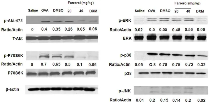
Effect of Farrerol on MAPK, Phosphatidylinositol 3-kinase Activation in vivo
To evaluate the effect of farrerol on the activation of MAPK and PI3K in the mouse asthma model, we homogenized the lung tissue to obtain a total protein lysate. The OVA-challenged mice showed significant upregulation of p-Akt, p-p70S6K, p-p38, pERK, and p-JNK. Farrerol can suppress the OVA-induced upregulation of p-Akt and p-p70 S6K in a dose-dependent manner. However, there was no significant downregulation of MAPK (Fig. 7).
Effect of Farrerol on NF-kB Activation, IkBa
Phosphorylation and Degradation in vivo
To evaluate the effect of farrerol on NF-kB activation in a mouse asthma model, we homogenized the lung tissue to obtain total, nuclear and cytoplasmic proteins. In our study, Fig. 8A showed that OVA-challenged mice showed a notably increased nuclear protein level and a decreased cytoplasmic protein level, compared to PBS-challenged mice. Farrerol treatment significant-ly attenuated these alterations in a dose-dependent manner (*p,0.05, **p,0.01). To gain further insight into the mechanism of farrerol-mediated regulation of NF-kB in vivo, we examined the effect of farrerol on IkBa phosphorylation and degradation. As shown in Fig. 8B, OVA-induced IkBa degradation was significantly blocked by pretreatment with farrerol, which was related to IkBaphosphorylation.
Effect of Farrerol on the Cytokines, PI3K and NF-kB Activation in vitro
To assess whether farrerol treatment could directly affect lymphocyte function, we examined anti-CD3/CD28-challenged Figure 3. Effects of farrerol on lung tissue eosinophilia and mucus production.Histologic examination of lung tissue eosinophilia using HE staining (A) and mucus secretion using AB-PAS staining (B) from: (a) PBS-challenged mice; (b) OVA-challenged mice; (c) OVA-challenged mice treated with farrerol (20 mg/kg); (d) OVA-challenged mice treated with farrerol (40 mg/kg); (e) OVA-challenged mice treated with dexamethasone (2 mg/kg, magnification6400). Quantitative analyses of inflammatory cell infiltration (C) and mucus production (D) in lung sections were performed as previously described [35]. At least 5 different fields for each lung section was performed to score the inflammatory cells and goblet cells. Mean scores were obtained from 5 mice. **P,0.01. vs. OVA.
immune responses in Th2 cells. IL-4, IL-5, and IL-13 concentra-tions in the culture supernatant of Th2 cells were measured by sandwich ELISA (Fig. 9A). Treatment with farrerol (5 and 20 mg/L) can markedly decreased IL-4, IL-5 and IL-13 levels in the cell supernatant which induced by anti-CD3/CD28 (*p,0.05, **p,0.01). we also evaluated the effect of farrerol on p-Akt and the p-IkBaas a marker of NF-kB activation. We found that anti-CD3 stimulation can induce the expression of p-Akt, p-p70S6K and p- IkBa. As shown in Fig. 9B, pretreatment with farrerol downregulated p-Akt, p-p70S6K and IkBa phosphorylation and then block the of IkBadegradation.
Effect of Farrerol on LPS-induced Acute Lung Injury
The total cell counts, differential cell counts, TNF-a, IL-6 and IL-8 in the BALF were evaluated 24 h after LPS administration. As shown in Fig. S1, LPS can markedly increase the number of total cells, neutrophils, macrophages, as well as the levels of TNF-a, IL-6 and IL-8 compared with the control group. However, farrerol does not have any effect on this increase compared to the dexamethasone group. Besides, farrerols can not decrease the upregulation of
NF-kB, p-AKT, and MAPKs (Fig. S2).
Discussion
Pulmonary disorders can be grouped according to whether the primary immune responses are characterized by innate or adaptive immune responses. In recent decades, using molecular, cellular assays together with knockout or transgenic animals, we have got more information about the immunological factors that contribute to the development pulmonary disorders. The immu-nologic features of pulmonary disorders can be used to categorize various conditions and provide focus for potential innovative therapies. Phytochemical and pharmacological studies have identified many potential anti-inflammatory substances, especially those derived from plants used in folk medicine, so natural products are becoming increasingly important as sources of pharmacotherapeutics, either for the treatment of infectious lung diseases or the theatment of noninfectious lung diseases [4,20,21]. In this study, we used two lung disease models: acute lung injury, characterized by innate immune responses and allergic asthma, involving adaptive immune responses, to demonstrate that farrerol can markedly reduce the allergic airway inflammation and AHR for allergic asthma model, but has no effect in an acute lung injury model.
Figure 4. Effects of farrerol on cytokine and chemokine levels in BALF and serum immunoglobulin production in vivo.BALF and blood were collected and centrifuged 24 hours after the last OVA challenge, and the supernatants and serum were measured by ELISA. Results of IgE in serum (mean6SEM n = 10) are expressed as Optical Density values and are representative of at least three independent in vivo experiments. *p,0.05, **p,0.01 vs. OVA.
doi:10.1371/journal.pone.0034634.g004
Airway inflammation is a natural response of the lung to the presence of internal and external substances, which is character-ized by innate immunity and adaptive immunity. Asthma is characterized by AHR and chronic airway inflammation, which includes the infiltration of inflammatory cells into lung tissues, mucus overproduction, allergen-specific IgE, the over-expression of Th2-mediated cytokines, including interleukin (IL)-4, IL-5 and IL-13, and chemokines such as CCL11 (CCL11) and RANTES (CCL5). In contrast, Th1 cytokines such as interferon-c(IFN-c), can inhibit the development of allergic lung inflammation through down-regulating Th2 responses [22]. The predominant inflam-matory cell recruited into asthmatic lung tissue is the eosinophil, which is associated with the production of IL-5. However, macrophages, the minor inflammatory cell recruited into asth-matic lung tissue, can produce IL-10 to suppress Th2 cytokine production and eosinophilia but augments airway reactivity. Acute lung injury is characterized by systemic airway inflammatory response including cytokines (e.g., TNF-a, IL-6, IL-8), chemo-kines, pro-inflammatory mediators and a variety of cells, which regulate the migration and pulmonary infiltration of neutrophils into the interstitial tissue [23]. Neutrophils are an important component of the inflammatory response that characterizes acute lung injury (ALI) [24]. In addition, PI3K contribute to the pathogenesis of asthma or acute lung injury by influencing the recruitment of eosinophils or neutrophils [12]. In previous report, farrerol has been reported to block FBS-induced vascular smooth muscle cells proliferation by acting as a estrogen receptors agonist [25]. In addition, estrogen has been shown to aggravates inflammation in pseudomonas aeruginosa pneumonia in cystic fibrosis mice via increasing the total white blood cell counts and neutrophils, Th1 cytokines (TNF-aand IL-6) Th2 cytokines (IL-5) and extaxin [26]. From our study, we can conclude that farrerol can reduce the number of total inflammatory cells and eosinophils in BAL fluid; IL-4, IL-5 and IL-13 levels in BAL fluid, OVA-specific IgE levels in serum; mRNA levels of CCL5, CCL11, CCR3, CCR5, E-selectin, chitinases, and Muc5ac in lung tissues. Furthermore, as IFN-cwas evoked by farrerol in the lung of mice this cytokine may act to further suppress type 2 pulmonary inflammation. After OVA challenge, farrerol can also inhibit the
expression of p-Akt. However, farrerol does not have any effect on TNF-a, IL-6, and IL-8, which are induced in acute lung injury. So these result suggested that on the one hand, farrerol maybe act as an estrogen receptors agonist to result in aggravating the inflammation in infectious inflammation model; on the other hand, farrerol play an important role in reducing airway inflammation in uninfectious inflammation model. The distinct effect of farrerol on the different airway inflammation maybe depending on its potential as an estrogen receptors or not.
The transcription factor NF-kB regulates the expression of a large number of genes in response to infection, inflammation and other endogenous and exogenous stressors. Under resting conditions, NF-kB is held inactiveby IkB. However, NF-kB can be activated by some stimulation of various receptors including TNF receptor, Toll-like receptors (TLRs) and T-cell receptor (TCR). Persistent activation of NF-kB is central to the pathogenesis of many inflammatory lung disorders including chronic obstructive pulmonary, asthma, pneumonia, and acute lung injury. In our study, farrerol can inhibit NF-kB activation induced by OVA in an allergic asthma model, but not in acute lung injury model, which suggests that the decreased NF-kB activation could account for the inhibition of airway inflamma-tion and AHR.
Corticosteriods have a puissant anti-inflammatory action and have a central role in the treatment for asthma [27]. However, prolonged use of corticosteriods, especially at higher doses, has been accompanied by concerns about both systemic and local side effects [28]. Date in this study indicates a potential therapeutic value for farrerol in the treatment of allergic asthma. In the context of current treatments for allergic asthma, future experimental studies are needed to address if farrerol can reduce corticosteriods dosage using combinations of both drugs.
Materials and Methods
Reagents
The mAbs against mouse CD3, CD28, IL-4, IL-5, IL-13, TNFa, IL-6, IL-8, IFN-cand IL-10 ELISA kits were purchased from Biolegend (California, USA). CCL11 mouse ELISA kit was Figure 5. Effects of farrerol on OVA-induced AHR. Airway responsiveness of mechanically ventilated mice in response to aerosolized methacholine was measured 24 h after the last saline aerosol or OVA aerosol with pretreatment of either DMSO or Farrerol (20 and 40 mg/kg). AHR is expressed as percentage change from the baseline level of (A) dynamic compliance (Cdyn, n = 6 mice) and (B) lung resistance (RL, n = 5 mice). Cdyn refers to the distensibility of the lung and is defined as the change in volume of the lung produced by a change in pressure across the lung. RL is defined as the pressure driving respiration divided by flow. *p,0.05, **p,0.01 vs. OVA.
purchased from Abcam (CA, USA). Western blot antibodies were purchased from Santa Cruz (Santa Cruz, CA, USA) and Cell Signaling Technology Inc. (Beverly, MA). Dimethy sulfoxide (DMSO) and OVA (Grade V) were purchased from Sigma Chemical Co. (St. Louis, MO, USA). Farrerol (analytical grade, purity $98%) was obtained from the National Institute for the Control of Pharmaceutical and Biological Products (Beijing, China).
Animals
Female BALB/c mice, weighing approximately 18 to 20 g, were purchased from Shanghai Jingke Industrial Co., LTD (Certificate SCXK2003-0003) (Shanghai, China) and bred under specific pathogen-free conditions. The mice were housed in microisolator cages and received food and water ad libitum. All animal studies were conducted according to the experimental practices and standards approved by the Animal Welfare and Research Ethics Committee at Jilin University.
Sensitization and Challenge with OVA and Treatment
These mice were divided into six groups (n = 10) and were sensitized with 20mg ovalbumin adsorbed in 100mg/ml of Imject
Alum by i.p. injection on days 0, 7, 14 (general sensitization) in all mice. On day 14, mice are anesthetized and 100mg of OVA in 50ml of PBS administered intranasally in all mice except negative
control sensitized with PBS. Mice are again anesthetized before being challenged with 50mg of OVA in 50ml of PBS on each of days 25–27. On days 25–27, farrerol at 20 and 40 mg/kg and dexamethasone (2 mg/kg) were given by i.p. injection 1 h prior to OVA administration.
Murine Model of LPS-induced ALI
LPS-induced ALI was induced as previously described [29]. Briefly, mice were anesthetized and challenged with intranasal 10mg of LPS in 50ml PBS. Control mice were given 50ml PBS i.n. instillation without LPS. Farrerol at 20 and 40 mg/kg and dexamethasone was i.p. injected 1 h prior to LPS administration.
Collection of Blood and Bronchoalveolar Lavage
At selected times after the last inhalational exposure, mice were anesthetized and bled via the brachial plexus for the collection of blood samples used to estimate the levels of total IgE and OVA-specific IgE. Bronchoalveolar lavage (BAL) was performed twice Figure 6. Effect of farrerol on OVA-induced inflammatory gene expression in allergic airway inflammation.Lung tissues were collected 24 hours after the last OVA aerosol challenge. The mRNA in whole-lung extracts was measured by real-time PCR. Treatment with farrerol (20 and 40 mg/kg) reduced OVA-induced mRNA expression in the lung. The values represent the mean6SEM of 10 animals in each group. **P,0.01. vs. OVA. doi:10.1371/journal.pone.0034634.g006
Figure 7. Effect of farrerol on Akt, P70S6K, and MAPK activation in vivo.Immunoblotting of Akt, P70S6K, and MAPK in proteins extracts of lung tissues isolated from mice 24 hours after the last OVA challenge pretreated with 20 or 40 mg/kg farrerol.b-actin was used as an internal control. Experiments were repeated three times and similar results were obtained.
doi:10.1371/journal.pone.0034634.g007
Figure 8. Effect of farrerol on the activation of NF-kB and IkBaphosphorylation and degradation in vivo.(A) Effect of farrerol treatment on nuclear translocation of NF-kB. Nuclear and cytoplasmic proteins from lung were analyzed by western blot with specific antibodies. (B) Effect of oxytetracycline treatment on IkBaphosphorylation and degradation. Total cellular proteins from lung were analyzed by western blot with specific antibodies.b-Actin was used as an internal control. Experiments were repeated three times and similar results were obtained.
by intratracheal instillation of 500ml of PBS. The lavage fluid was centrifuged and the supernatants were used for cytokine and chemokines measurements. Cell pellets were resuspended in 1 ml of PBS and used for total and differential cell counts as described [30].
Lung Histology
Histopathological evaluations were performed on mice. Left lungs were removed by dissection and fixed in 4% paraformal-dehyde. Lung tissues were sectioned, embedded in paraffin, and cut into 3mm sections. Tissue sections were then stained with
hematoxylin and eosin stain (H&E) for general morphology [31]
and with AB-PAS for the identification of goblet cells in the epithelium [32].
Measurements of Airway Hyperresponsiveness
Airway responsiveness was assessed by methacholine-induced airway resistance (RL) and lung compliance (Cdyn) using a whole-body plethysmograph chamber (Buxco, Sharon, CT) as described [33]. Mice were anesthetized with pentobarbital sodium (100 mg/ kg) and tracheotomy was performed as described [33]. The internal jugular vein was cannulated and connected to a microsyringe for aerosolized methacholine administration. Base-line lung resistance, dynamic compliance, and responses to Figure 9. Effect of farrerol (20 and 40 mg/kg) on the IL-4, IL-5, and IL-13, Akt, P70S6K, and IkBaphosphorylation and degradation in vitro.The supernatant of Th2 cells was measured by sandwich ELISA. The values represent the mean6SEM of three independent in vitro experiments. Th2 cells were cultured with anti-CD3 (5mg/ml) for 1 h (1 mg/L), total cellular proteins were analyzed by western blot with specific antibodies.b-Actin was used as an internal control. Experiments were repeated three times and similar results were obtained.
doi:10.1371/journal.pone.0034634.g009
Table 1.Primer sets for reverse transcriptase-polymerase chain reacion analysis.
Targets Forward Reverse
CCL11 AAACCATAAACAACCTCCTC CAATAATCCCACATCTCCTT
CCL5 GGATAGAGGGTTTCTTGATT GCTGATTTCTTGGGTTTG
CCR1 CACTCACCGTACCTGTAGCC TCTGATGATCCCTGCATAGC
CCR3 TCTGCTGAGATGTCCCAATA TCACCAACAAAGGCGTAG
MUC-5ac GTGTCGGCCGGAGAAAGTTGGT GTCCTGTTGAGCCTGGCCTGTG
Ym-1 CCAGCAGAAGCTCTCCAGAAGCA CAGCTGGTAGGAAGATCCCAGCTGT
Ym-2 TCCACTTTGAACCACATTCCAAGGC CGAGAGACTGAGACAGTTCAGGGA
AMCase-1 TGGACACACCTTCATCCTGA CCTCAGTGGCTCCACTTCTC
E-selecin CCCTTCCACAGAACCTACCA TCAGCAGACATTGCTTCACC
b-Actin CTGTCCCTGTATGCCTCTG ATGTCACGCACGATTTCC
doi:10.1371/journal.pone.0034634.t001
aerosolized saline (0.9% Nacl) were measured first, followed by responses to increasing dose (6.25 to 50 mg/ml) of aerosolized methacholine. Results are expressed as a percentage of the respective basal values.
RNA Preparation and Quantitative RT-PCR
Total RNA was isolated from lungs (5 mice in each group) using the TRIzol reagent (SIGMA-Aldrich, St. Louis, USA). Real-time PCR was performed on cDNA samples using the SYBR Green system (Bio-Rad; Richmond, CA). Primers for inflammatory bio-markers are shown in Table 1. Analysis was performed using the sequence detection software supplied with the instrument. A melting curve analysis was performed to control for the specificity of the amplification products.
CD4+ T Cell Isolation and Th2 Differentiation
To obtain naive CD4+T cells, single cell suspensions were prepared from spleens and treated with monoclonal antibodies to CD4 coupled to magnetic beads according to the protocol provided by the manufacturer. For Th2 differentiation, IL-2 (10 ng/ml), IL-4 (5 ng/ml) and anti-IFN-c(4mg/ml) were added.
Cytokine Assays
Naive CD4+ T cells were stimulated for 72 h with anti-CD3 (5mg/ml) plus anti-CD28 (2.5mg/ml) and Th2 differentiated cells
were restimulated for 24 h with anti-CD3 (5mg/ml) in the presence/absence of farrerol (5 and 20 mg/l), after which supernatants were collected and cytokine concentrations were measured by ELISA using commercially available kits. Control cells were incubated in medium alone for the duration of the experiment. ELISA was also used to determine the levels of Th2 cytokines in BALF.
Western Blot Analysis of MAPK, NF-kB and PI3K
Lung tissues were added to lysis buffer and homogenized. Th2 cells (16106) were plated into 6-well plates and pretreated with 5 and 20 mg/L of farrerol, 1 h prior to a 1 h treatment with
anti-CD3 (5mg/ml). Nuclear and cytoplasmic fractions of lung were prepared as previously described [34]. Whole cell lysates were prepared in 1% Non-diet P-40 lysis buffer with freshly added protease and phosphatase inhibitors. Protein were separated by SDS/PAGE and transferred onto PVDF membrane. Membranes were probed for different antibodies.
Statistical Analysis
All values were expressed as means6the standard error of the mean (SEM). Differences between mean values of normally distributed data were assessed by the two-tailed Student t test. Statistical difference was accepted at P,0.05.
Supporting Information
Figure S1 Effect of farrerol on production of inflamma-tory cytokines TNF-a, IL-6 and IL-8 in the BALF of LPS-induced ALI mice.Mice were given an oral administration of farrerol 1 h prior to an i.n. administration of LPS. BALF was collected at 6, 12 and 24 h following LPS challenge to analyze the inflammatory cytokines TNF-a, IL-6 and IL-8. The values presented are the means6SEM (n = 5 in each group). *p,0.05 vs. LPS group;
(TIF)
Figure S2 Effect of farrerol on Akt, P70S6K, NF-kB and MAPK activation in vivo. Immunoblotting of Akt, NF-kB, P70S6K, and MAPK in proteins extracts of lung tissues isolated from mice 24 hours after the LPS challenge pretreated with 20 or 40 mg/kg farrerol. b-actin was used as an internal control. Experiments were repeated three times and similar results were obtained.
(TIF)
Author Contributions
Conceived and designed the experiments: XXC XMD. Performed the experiments: XXC XC. Analyzed the data: MMW XFY. Contributed reagents/materials/analysis tools: QRC. Wrote the paper: XXC XMD.
References
1. Greenberger PA (2008) 7. Immunologic lung disease. J Allergy Clin Immunol
121: S393–397; quiz S418.
2. Chen H, Bai C, Wang X (2010) The value of the lipopolysaccharide-induced
acute lung injury model in respiratory medicine. Expert Rev Respir Med 4: 773–783.
3. Broide DH, Finkelman F, Bochner BS, Rothenberg ME (2011) Advances in
mechanisms of asthma, allergy, and immunology in 2010. J Allergy Clin Immunol 127: 689–695.
4. Rogerio AP, Andrade EL, Leite DF, Figueiredo CP, Calixto JB (2009)
Preventive and therapeutic anti-inflammatory properties of the sesquiterpene alpha-humulene in experimental airways allergic inflammation. Br J Pharmacol 158: 1074–1087.
5. Opitz B, van Laak V, Eitel J, Suttorp N (2010) Innate immune recognition in
infectious and noninfectious diseases of the lung. Am J Respir Crit Care Med 181: 1294–1309.
6. Kawai T, Akira S (2011) Toll-like receptors and their crosstalk with other innate
receptors in infection and immunity. Immunity 34: 637–650.
7. Roth M, Black JL (2006) Transcription factors in asthma: are transcription
factors a new target for asthma therapy? Curr Drug Targets 7: 589–595.
8. Barnes PJ (2006) Transcription factors in airway diseases. Lab Invest 86:
867–872.
9. Pantano C, Ather JL, Alcorn JF, Poynter ME, Brown AL, et al. (2008) Nuclear
factor-kappaB activation in airway epithelium induces inflammation and hyperresponsiveness. Am J Respir Crit Care Med 177: 959–969.
10. Duan W, Wong WS (2006) Targeting mitogen-activated protein kinases for asthma. Curr Drug Targets 7: 691–698.
11. Rommel C, Camps M, Ji H (2007) PI3K delta and PI3K gamma: partners in crime in inflammation in rheumatoid arthritis and beyond? Nat Rev Immunol 7: 191–201.
12. Park SJ, Min KH, Lee YC (2008) Phosphoinositide 3-kinase delta inhibitor as a novel therapeutic agent in asthma. Respirology 13: 764–771.
13. Ali K, Bilancio A, Thomas M, Pearce W, Gilfillan AM, et al. (2004) Essential role for the p110delta phosphoinositide 3-kinase in the allergic response. Nature 431: 1007–1011.
14. Tian J, Liu J, Hu Z, Chen X (2005) Interaction of wogonin with bovine serum albumin. Bioorg Med Chem 13: 4124–4129.
15. Cao Y, Lou C, Fang Y, Ye J (2002) Determination of active ingredients of Rhododendron dauricum L. by capillary electrophoresis with electrochemical detection. J Chromatogr A 943: 153–157.
16. Choi SE, Park KH, Han BH, Jeong MS, Seo SJ, et al. (2011) Inhibition of Inducible Nitric Oxide Synthase and Cyclooxygenase-2 Expression by Phenolic Compounds from Roots of Rhododendron mucronulatum. Phytother Res. 17. Qiu J, Xiang H, Hu C, Wang Q, Dong J, et al. (2011) Subinhibitory
concentrations of farrerol reduce alpha-toxin expression in Staphylococcus aureus. FEMS Microbiol Lett 315: 129–133.
18. Manson SC, Brown RE, Cerulli A, Vidaurre CF (2009) The cumulative burden of oral corticosteroid side effects and the economic implications of steroid use. Respir Med 103: 975–994.
19. Kleiman A, Tuckermann JP (2007) Glucocorticoid receptor action in beneficial and side effects of steroid therapy: lessons from conditional knockout mice. Mol Cell Endocrinol 275: 98–108.
20. Zhou H, Bian D, Jiao X, Wei Z, Zhang H, et al. (2011) Paeoniflorin protects against lipopolysaccharide-induced acute lung injury in mice by alleviating inflammatory cell infiltration and microvascular permeability. Inflamm Res 60: 981–990.
21. Choi JR, Lee CM, Jung ID, Lee JS, Jeong YI, et al. (2009) Apigenin protects ovalbumin-induced asthma through the regulation of GATA-3 gene. Int Immunopharmacol 9: 918–924.
22. Iwamoto I, Nakajima H, Endo H, Yoshida S (1993) Interferon gamma regulates antigen-induced eosinophil recruitment into the mouse airways by inhibiting the
23. Shields CJ, Winter DC, Redmond HP (2002) Lung injury in acute pancreatitis: mechanisms, prevention, and therapy. Curr Opin Crit Care 8: 158–163. 24. Abraham E (2003) Neutrophils and acute lung injury. Crit Care Med 31:
S195–199.
25. Li QY, Chen L, Zhu YH, Zhang M, Wang YP, et al. (2011) Involvement of estrogen receptor-beta in farrerol inhibition of rat thoracic aorta vascular smooth muscle cell proliferation. Acta Pharmacol Sin 32: 433–440. 26. Wang Y, Cela E, Gagnon S, Sweezey NB (2010) Estrogen aggravates
inflammation in Pseudomonas aeruginosa pneumonia in cystic fibrosis mice. Respir Res 11: 166.
27. Tritar-Cherif F, Ben M’Rad S, Merai S, Djenayah F (2002) [Corticotherapy for asthma in the child]. Tunis Med 80: 1–6.
28. Kos-Kudla B, Pluskiewicz W (1997) Quantitative ultrasound of the heel and serum and urinary cortisol values in assessment of long-term corticotherapy side effects in female bronchial asthma patients. Ultrasound Med Biol 23: 1325–1330.
29. Zhang X, Song K, Xiong H, Li H, Chu X, et al. (2009) Protective effect of florfenicol on acute lung injury induced by lipopolysaccharide in mice. Int Immunopharmacol 9: 1525–1529.
30. Rogerio AP, Fontanari C, Borducchi E, Keller AC, Russo M, et al. (2008) Anti-inflammatory effects of Lafoensia pacari and ellagic acid in a murine model of asthma. Eur J Pharmacol 580: 262–270.
31. McKay A, Leung BP, McInnes IB, Thomson NC, Liew FY (2004) A novel anti-inflammatory role of simvastatin in a murine model of allergic asthma. J Immunol 172: 2903–2908.
32. Edwan JH, Perry G, Talmadge JE, Agrawal DK (2004) Flt-3 ligand reverses late allergic response and airway hyper-responsiveness in a mouse model of allergic inflammation. J Immunol 172: 5016–5023.
33. Bao Z, Lim S, Liao W, Lin Y, Thiemermann C, et al. (2007) Glycogen synthase kinase-3beta inhibition attenuates asthma in mice. Am J Respir Crit Care Med 176: 431–438.
34. Ito K, Jazrawi E, Cosio B, Barnes PJ, Adcock IM (2001) p65-activated histone acetyltransferase activity is repressed by glucocorticoids: mifepristone fails to recruit HDAC2 to the p65-HAT complex. J Biol Chem 276: 30208–30215. 35. Zhou L, Goldsmith AM, Bentley JK, Jia Y, Rodriguez ML, et al. (2005)
4E-binding protein phosphorylation and eukaryotic initiation factor-4E release are required for airway smooth muscle hypertrophy. Am J Respir Cell Mol Biol 33: 195–202.