UNIVERSIDADE FEDERAL DE MINAS GERAIS INSTITUTO DE CIÊNCIAS BIOLÓGICAS DEPARTAMENTO DE BIOQUÍMICA E IMUNOLOGIA
PROGRAMA DE PÓS-GRADUAÇÃO EM BIOQUÍMICA E IMUNOLOGIA
Breakdown of intestinal homeostasis by mucosal infections
triggers adaptive immune responses against
antigens from commensal bacteria
Aluna: Liliane Martins dos Santos Orientador(a): Dr. Yasmine Belkaid (NIH) Co-orientador(a): Dr. Leda Quercia Vieira
2 Trabalho de Tese apresentado ao Programa de Pós-Graduação em Bioquímica e Imunologia da Universidade Federal de Minas Gerais como requisito parcial para a obtenção do título de Doutor(a)
Liliane Martins dos Santos
Breakdown of intestinal homeostasis by mucosal infections triggers adaptive immune responses against antigens from commensal bacteria
Orientador (a): Dra. Yasmine Belkaid (NIH) Orientador (a): Leda Quercia Vieira
3 ACKNOWLEDGEMENTS
Dr. Leda Vieira for having me in her lab for so long, for her support, advices, and great effort in always helping me over these past 10 years;
Dr. Yasmine Belkaid for welcoming me into her lab at the Laboratory of Parasitic Diseases at National Institutes of Health (NIH), for her support and advices;
Dr. Timothy Hand and Dr. Nicolas Bouladoux from the Belkaid Lab for their great help with the experiments and the enriching discussions;
All the other members of the Belkaid Lab for their help and support.
Dr. Michael Grigg for his help with the Toxoplasma gondii parasites;
All the members of the Laboratório de Gnotobiologia e Imunologia (LAGI) at UFMG for making my life as a graduate student a much more enjoyable experience;
The Committee members for accepting our invitation and for their availability;
4 My dear friends Fernanda e Alessandra that contributed to make much easier the rather lonely experience of living and working abroad;
My family, in particular, my sisters for being the best sisters the world has ever seen;
My mother for being the person responsible for molding me into the person I am and, as always, watch and take care of me from wherever she is.
5 SUMMARY
Acknowledgements...i
Summary...iii
List of figures...iv
Acronyms and Abbreviations.………vi
Abstract ………vii
Resumo ……….ix
Citation ……….xi
1. Introduction...1
2. Objectives ...31
3. Materials and Methods ...33
4. Results...44
5. Discussion ...88
6. Conclusion...106
6 LISTA OF FIGURES
Figure 1. Immune responses against T. gondii are down-regulated in the mLNs of ATB mice
………... ..47
Figure 2. Reduced inflammatory immune responses in the small intestine of ATB
treated mice infected with T. gondii
…………...48
Figure 3. Regulatory responses are not affected by depletion of gut microbiota... ...50
Figure 4. Lower parasite burden at day 9 of infection in ATB treated mice infected with
T. gondii
……… …….52
Figure 5. Lower levels of liver enzymes in sera from germfree mice infected with T. gondii
……… ...54
Figure 6. Reduced inflammatory immune responses in the small intestine of germfree
mice infected with T.
gondii……...56
Figure 7. . Germfree mice infected with T. gondii have increased regulatory responses
in the gut
………... .57
Figure 8. Lower parasite burden at day 9 of infection in germfree mice infected with T. gondii... ...59
7
Figure 10. Changes of microbiota during T. gondii
infection... ...62
Figure 11. Temporal immune responses during T. gondii infection... ...64
Figure 12. Systemic translocation of bacteria in T. gondii oral infection ………... ..66
Figure 13. Antibody responses against commensal E. coli after acute parasitic infection...
...69
Figure 14. Antibody responses against commensal E. faecalis after acute parasitic infection... ...70
Figure 15. Antibody responses against commensal P. mirabilis after acute parasitic infection
………... .71
Figure 16. Bacterial FACS for detection of antibodies against commensal E. coli…...72
Figure 17. DTH response to E. coli whole lysate in mice infected with T. gondii... ...74
Figure 18. Antigen-specific CD4 T cell activation and differentiation in response to
commensal antigens during T. gondii infection
………...76
Figure 19. Systemic translocation of gut bacteria in T. gondii oral infection ………...78
Figure 20. Antibody responses against commensal bacteria after E. cuniculi infection... ...80
8 Figure 22. Vaccination of mice against commensal E. coli during T. gondii infection... ...84
Figure 23. Vaccination of mice against commensal E. coli during T. gondii infection... ...85
Figure 24. Pathogenesis after vaccination of mice against commensal E. coli during T. gondii infection... ...86
9 ACRONYMS AND ABBREVIATIONS
GALT: gut associated lymphoid tissues mLn: mesenteric lymph nodes
siLP: small intestine lamina propria Sp: spleen
Pp: Peyer’s patches
IEL: intraepithelial lymphocyte LivLn: liver-draining lymph node T-bet: T-box transcription factor Foxp3: forhead transcription factor 3 Treg cell: T regulatory cell
DC: dendritic cell gfDNA: gut flora DNA TLR: toll like receptor
NOD: nucleotide oligomerization domain IBD: inflammatory bowel disease
sIgA: secretory IgA
10 ABSTRACT
12 RESUMO
O trato gastrointestinal de mamíferos é colonizado por uma diversa comunidade microbiana que co-existe mutualisticamente com seu hospedeiro. Estudos recentes têm demonstrado o papel importante destes microrganismos na modulação de respostas imunes. Por outro lado, bactéria da microbiota podem também contribuir para a patologia no contexto de infecções agudas. Por exemplo, infecções orais com Toxoplasma gondii em certas linhagens murinas levam a uma inflamação intestinal
14 If a man will begin with certainties, he shall end in doubts, but if he will content to begin with doubts, he shall end in certainties
15
16 The intestinal microbiota
Mammalian barrier surfaces host complex microbial communities whose combined membership outnumbers our own cells by at least a factor of ten. Although beneficial bacteria colonize all mammalian epithelial surfaces, the gastrointestinal tract has the largest bacterial burden, with more than 100 trillion individual organisms (Xu and Gordon, 2003). The bacterial communities of the mammalian intestine are also some of the best characterized. For decades, our understanding of the composition of intestinal microbial communities was based on the enumeration and characterization of culturable organisms. However, as most gut organisms are resistant to culture by available methods, this approach left substantial gaps in the catalogue of intestinal bacterial species.
17 Genome Sequencer 454 FLX system, this generally produces around 400,000 reads with average lengths of 250 base pairs (bp) and an average quality score of greater than 99.5% accuracy rate. These read sizes are sufficient to cover most of the variable regions in the 16S rRNA gene (Claesson et al., 2009). These recent high-throughput methods have allowed unprecedented insight into the diversity of intestinal microbial communities, and have even led to the identification of new bacterial species. Currently, new insights into non culturable bacterial communities are placing species estimates from conservative numbers of 1000–2000 to numbers as high as 15,000–40,000 individual members (Hill and Artis, 2010).
18 bacteria, accounting for less than 10% of the total population, belong to the Proteobacteria, Fusobacteria, Actinobacteria, Verrucomicrobia and Spirochaetes phyla and a bacterial group that is closely related to Cyanobacteria. Thus the gastrointestinal tract constitutes an exceptionally diverse and dynamic microbial ecosystem (Wang et al., 2003; Eckburg et al., 2005; Ley et al., 2008).
19 Commensal microbiota can also influence the metabolism of the host. The commensal microorganism Bacteroides thetaiotaomicron, component of the normal mouse and human gut microbiota, was shown to modulate expression of genes involved in several important intestinal functions, including nutrient absorption, mucosal barrier fortification and xenobiotic metabolism (Hooper et al., 2001). Gut microbes can also modulate the bile-acid metabolism (Swann et al., 2011), carbohydrate metabolism, energy production and synthesis of cellular components (Gosalbes et al., 2011). A recent study reported that gut commensal bacteria can positively influence immune responses and protect against the development of inflammatory diseases by regulating the production of short-chain fatty acids (SCFAs), which are produced by fermentation of dietary fiber by intestinal microbiota (Maslowski et al., 2009). Gut microbiota has been shown to also contribute metabolic syndrome cause by deficiency in Toll-like receptor 5 (TLR 5) (Vijay-Kumar et al., 2010).
20 cells (IECs), which line the gut and form a physical barrier between luminal contents and the immune system, have lower expression of Toll-like receptors (TLRs). Interspersed between epithelial cells is a specialized population of T cells known as intraepithelial lymphocytes (IELs). IELs from germ-free mice are reduced in number, and their cytotoxicity is compromised (Lee and Mazmanian, 2010). Germ-free mice also have reduced numbers of γδ T cells and CD4+ T cells in the lamina propria. The development of isolated lymphoid follicles, specialized intestinal structures made of mostly dendritic cells and B cell aggregates, is also dependent on the microbiota (Bouskra et al., 2008; Duan et al., 2010). Therefore, multiple populations of intestinal immune cells require the microbiota for their proper development and function. The absence of microbiota also leads to several extra-intestinal defects, including reduced numbers of CD4+ T cells in the spleen, fewer and smaller germinal centers within the spleen, and reduced systemic antibody levels, which suggests that the microbiota is capable of also shaping systemic immunity (Mazmanian et al., 2005). Beyond development, the microbiota also influences functional aspects of intestinal and systemic immunity, including pathogen clearance. Germ-free mice are more susceptible to infectious agents such as Leishmania, Listeria monocytogenes, Shigella flexneri, Bacillus anthracis (Inagaki et al., 1996; de Oliveira et al., 1999; Smith et al., 2007).
21 Intestinal microbes have been demonstrated to influence the manifestation of gut-associated lymphoid tissues through CCR7 signaling (Pabst et al., 2006). Colonization of germfree mice with the human symbiont Bacteroides fragilis directs the development of the immune system, including the expansion and differentiation of splenic CD4+ T cells. Moreover, treatment of germfree mice with purified polysaccharide A (PSA), a product of B. fragilis, has several immunomodulatory activities such as correcting systemic T cell deficiencies, restoring balance between Th cell subsets and directing lymphoid organogenesis (Mazmanian et al., 2005). Another report revealed that a group of gut microbes, the segmented filamentous bacterium (SFB), a non-culturable Clostridia-related host-specific species that strongly adheres to the ileal mucosa and to Peyer’s patches (Pp), was able to recapitulate the immune-inducing effects of a complete conventional microbiota suggesting, unexpectedly, that only a restricted number of microbiota members have shaped host-immune T cell interactions during evolution (Gaboriau-Routhiau et al., 2009). In return to all these positive effects, resident microorganisms derive benefit from association with their hosts by inhabiting a protected, nutrient-rich environment. Thus, these host-microbial associations constitute a mutually beneficial symbiosis.
22 identified complex pro-inflammatory and immunoregulatory roles for intestinal communities in modulating intestinal cytokine responses and altering resistance to enteric pathogens (Hall et al., 2008; Garner et al., 2009) and in maintaining mucosal homeostasis (Rakoff-Nahoum et al., 2004). Depletion of gut microbiota by treatment with a broad spectrum antibiotic cocktail produces a macroscopically germfree-like phenotype. Macroscopically, germfree mice display hypoplastic secondary lymphoid organs, significantly fewer Peyer’s patches, smaller spleen, enlarged ceca, and reduced epithelial cell turnover (Reikvam et al., 2011). Recent studies have shown that treatment with antibiotics causes changes within the gut microbiota composition. The route of administration as well as the duration of the treatment appear to have different effects (Hill et al., 2010).
23 to resist strongly against stressing environments may allow the bacterium to persist in hospital environments and to survive host defenses. In the last decades, Enterococci have been recognized as one of the most common bacteria involved in hospital-acquired infections. Indeed, these microorganisms can opportunistically invade mucosal tissues and cause sepsis, urinary tract infections, peritonitis and endocarditis. The species E. faecalis is still responsible for the majority of human enterococcal infections (Hanin et al., 2010). Proteobacteria are a major group of gram-negative bacteria that include a variety of pathogens such as Escherichia coli, Salmonella and Helicobacter. The subgroup γ of Proteobacteria comprise the most common human pathobionts. Proteus mirabilis, a facultative anaerobic, urea-hydrolyzing bacterium is a member of the γ-Proteobacteria subgroup and an opportunistic pathogen of the human urinary tract that infects patients with indwelling urinary catheters or with postoperative wound infections (Nielubowicz and Mobley, 2010). Similarly, Bacteroides fragilis is a prominent gram-negative member of the microbiota that closely associates with mucosal surfaces and opportunistically invades intestinal tissues (Redondo et al., 1995). Although E. faecalis, P. mirabilis and B. fragilis are controlled in healthy people, they pose a serious
threat of invasion and disease in immunodeficient individuals.
Probiotics
24 used to describe dietary microorganisms that are beneficial to the health of the host. Many individual or combinations of bacterial species have been shown to help protect against pathogens and ameliorate the symptoms of Inflammatory Bowel Disease (IBD) in humans and mouse models (Round and Mazmanian, 2009). Although many of these probiotic strains decrease toxic microbial metabolic activities, more recent evidence shows that these organisms can modulate intestinal immune responses and protect mice against infections. The probiotic Lactobacillus rhamnosus augmented intestinal IgA responses and protected mice against infection with Escherichia coli O157:H7 (Shu and Gill, 2002). Two strains of Lactobacillus were able to stimulate the immune system and protect mice against a lethal pneumovirus infection (Gabryszewski et al., 2011). Studies by our group have shown that the probiotic candidate Lactobacillus delbrueckii bulgaricus can induce the production of pro-inflammatory cytokines as well as anti-inflammatory cytokines conferring protection against Listeria monocytogenes infection in germfree mice (Dos Santos et al., 2011).
The Intestinal Homeostasis
25 There are several mechanisms that together help contain penetrant bacteria within the mucosal immune compartment that could lead to aberrant responses against commensal antigens and consequently, pathology.
Mechanisms that prevent translocation of intestinal bacteria
The small intestine is organized into crypts and villi to increase the surface area for absorption of nutrients. The entire epithelium is renewed approximately every 5 days in humans because of proliferation and differentiation of the pluripotent stem cells residing in the crypts. The most abundant intestinal surface cell is the enterocyte, an epithelial cell lineage. Enterocyte membranes, together with the tight junctions that they form with their neighboring cells, are essential for preventing bacterial penetration while allowing nutrient flux into host tissues. Besides providing an important physical barrier, enterocytes play a more active role in promoting luminal compartmentalization of symbiotic bacteria by secreting a variety of antimicrobial proteins. These natural antibiotics are members of several distinct protein families such as defensins, cathelicidins, and C-type lectins, and they promote bacterial killing by targeting the integrity of bacterial cell walls (Mukherjee et al., 2009). Antimicrobial proteins are produced either constitutively or are inducible by bacteria (Hooper et al., 2003).
26 layers: the outer layer is colonized with bacteria whereas the inner layer is resistant to bacterial penetration probably due to the antibacterial factors that are secreted by epithelial cells and retained there (Johansson et al., 2008). Paneth cells are specialized cells that harbor secretory granules containing a number of microbicidal proteins including α-defensins, lysozyme and RegIIIγ. When Paneth cells sense bacterial signals, they react by discharging their microbicidal granule contents into the gut lumen (Ayabe et al., 2000). In a recent study genetic ablation of Paneth cells caused increased translocation of bacteria to peripheral tissues indicating that these cells are for controlling mucosal penetration of bacteria (Vaishnava et al., 2008).
27 layer and are in close association with the mucosal surface (Rescigno et al., 2001). The bacteria-laden dendritic cells interact with B and T cells in lymphoid tissues such as the Peyer’s patches (Pp), isolated lymphoid follicles (ILF), or in the intestinal lamina propria inducing B cells to differentiate into plasma cells that produce IgA directed against intestinal bacteria (Macpherson et al., 2000). IgA is essential in maintaining luminal compartmentalization of intestinal bacteria, as shown by the fact that IgA deficiency leads to increased penetration of symbiotic bacteria into the host tissues (Macpherson and Uhr, 2004b). Also, specific pathogen free mice have abundant IgA whereas germfree animals do not, arguing that commensals direct IgA production and that the major role of IgA is in maintaining the balance between the host and its microbiota (Peterson et al., 2007).
28 Finally, symbiotic bacteria that breach the mucosal surface are quickly phagocytosed and killed by macrophages in the lamina propria (Macpherson and Harris, 2004). This is in contrast to pathogens, that actively interfere with macrophage microbicidal mechanisms, allowing survival and replication of these bacteria in host tissues (Sansonetti, 2004). The susceptibility of symbionts to the microbicidal mechanisms of macrophages likely represents an evolutionary coadaptation with their hosts, because suppression or evasion of phagocytic killing would compromise the host health (Macpherson et al., 2005).
Regulation of intestinal balance by innate immunity mechanisms
29 Although various mechanisms are in place to dampen PRR signaling in the healthy gut, there is clear evidence from work in TLR knockout mice that a tonic level of signaling is necessary to maintain intestinal homeostasis. For example, mouse knockouts in TLR9, TLR-4, TLR-2, and the adaptor protein MyD88 all showed increased susceptibility to DSS-induced colitis (Lee et al., 2006). A gut commensal microorganism, Bacteroides fragiles, activates the TLR pathway signaling through TLR2 directly on
Foxp3+ regulatory T cells (Tregs) to promote immunologic tolerance therefore actively suppressing immunity (Round et al., 2011). Additionally, germ-free mice and mice treated with multiple antibiotics to reduce the microbial content of the intestine are also more susceptible to DSS-induced colitis but can be protected by administration of agonists for TLR2 and TLR-4 (Rakoff-Nahoum et al., 2004).
Adaptive immune responses regulating intestinal homeostasis
Several studies revealed the importance of the adaptive immunity for maintaining intestinal homeostasis. It has been reported that in lack of TLR signaling, control of microbial mucosal translocation is impaired. However, commensal-specific serum IgG responses induced in response to these bacteria can restore effective bacterial clearance to wild-type levels (Slack et al., 2009). Thus, innate and adaptive immune mechanisms can complement each other to establish and maintain mutualism.
30 of a nonpathogenic commensal flora (Barnes and Powrie, 2009). The importance of regulatory T (Treg) cells in the regulation of intestinal homeostasis is best illustrated by the finding that these cells can prevent the induction of experimental colitis following transfer into diseased hosts (Powrie et al., 1994a). Foxp3+ Treg cells are abundant in the small intestine and colon, where they control potentially deleterious responses to dietary and microbial stimuli. In addition to thymic-derived Treg cells, the intestine is also a preferential site for TGF-β-dependent induction of Foxp3+ Treg cells from naive CD4+ T cell precursors. Conditioning of gut dendritic cells by food antigen and the gut microbiota induces a CD103+ retinoic acid-dependent dendritic cell that induces Tregs (Sun et al., 2007; Coombes et al., 2007). Intestinal CD103+ DCs are capable of intralymphatic migration through expression of CCR7 (Pabst et al., 2006). Once settled in the mLN, LP-derived CD103+ DCs cooperate with mLN stromal cells to shape a unique environment that critically determines the signature of T cell responses induced here. The antigen-induced Treg cells are further expanded in the lamina propria in models of oral tolerance and can control local and systemic antigen-induced hypersensitivity responses (Izcue et al., 2009; Hadis et al., 2011). In addition, indigenous Clostridia are potent inducers of mucosal and systemic Treg cell responses (Atarashi et al., 2011).
31 required for the differentiation of both populations, the presence of STAT3-mediated signals (such as IL-6 or IL-23) promotes Th17 cells at the expense of Foxp3+ Treg cells (Zhou et al., 2008). The regulation of TGF-β-dependent immune responses seems also to be dependent of retinoic acid that inhibits Th17 immune responses while promoting Foxp3+ cell differentiation and regulating pro- and anti-inflammatory responses (Mucida et al., 2009). Promotion of effector T cell responses by retinoic acid has been shown to occur via the retinoic acid receptor alpha (RARα). Furthermore, immune responses that are abrogated in absence of vitamin A can be rescued by treatment with retinoic acid (Hall et al., 2011). Such mechanisms contribute to keep the balance of immune responses in the gastrointestinal tract. Recent evidence suggests that bacterial components differentially affect this balance, providing potential therapeutic strategies to influence tolerance and immunity in the gut (Littman and Rudensky, 2010).
32 Flagellins have recently been identified as immunodominant antigens of the microbiota, using serologic expression cloning (Lodes et al., 2004). Flagellin is both a potent antigen for an adaptive response and is also able to stimulate innate response through binding its receptor, TLR5, on innate cells (Smith et al., 2003). These microbiota flagellin antigens provide an opportunity to study the host immune response to its microbiota and the role of antigen-specific IgA on intestinal immune homeostasis. A T cell receptor transgenic (Tg) mice specific for one of these flagellins, denoted CBir1 was generated and has been used as a tool for studying specific immune responses against microbiota (Lodes et al., 2004). Studies using this transgenic mouse strain showed that intestinal IgA regulates the activation of peripheral, flagellin-specific CD4+ T cells. Moreover, Tregs control such intestinal IgA B cell responses in an antigen-specific manner, via production of TGF-β. The depletion of CD4+CD25+ Tregs substantially reduced intestinal IgA levels within days, in part due to interruption of survival signals to LP IgA B cells provided by Tregs. These data are consistent with Tregs being the major helper T cells for induction and maintenance of intestinal IgA B cell responses (Cong et al., 2009).
33 instability. For example, high-level T-bet expression in the presence of acute intestinal infection drives Treg cells into an inflammatory IFN-γ-secreting phenotype (Oldenhove et al., 2009).
Intestinal inflammation
34 antibody responses to intestinal bacteria (Sartor, 2008; Hill and Artis, 2010). Patients with IBD have increased numbers of epithelial cell surface-associated bacteria, suggesting a failure of mechanisms that normally limit direct contact between the microbiota and the epithelium (Swidsinski et al., 2005).
Genetic susceptibility loci have been identified for the inflammatory bowel diseases Crohn’s and ulcerative colitis, including mutations in the pattern-recognition receptor nucleotide-binding oligomerization domain-containing protein 2 (NOD2) a component of the innate immune system that is important for immune recognition and responses to intracellular bacteria. These findings implicate altered immune responses to intestinal bacteria in the pathogenesis of IBD (Ogura et al., 2003).
The only microorganism reported to be strongly associated with Crohn’s disease is adherent-invasive E. coli. However, it seems that inflammatory responses in human and experimental IBD are directed towards certain subsets of commensal organisms that have pathogenic potential, such as Helicobacter, Clostridium and Enterococcus species. Nevertheless, these organisms are abundant in the microbiota and are not typically pathogenic. As the microbiota of all mammals contains these potentially harmful species, known as pathobionts, it is not entirely clear why inflammation ensues only in subjects affected by IBD (Scanlan et al., 2006; Round and Mazmanian, 2009).
35 human IBD. However, there is no perfect experimental model, because patients with IBD present a heterogeneous spectrum of pathological features that reflect the participation of a diverse range of innate and adaptive immune effectors. This heterogeneity is further underscored by the recent observations that around 100 distinct genetic loci may contribute to IBD susceptibility, and the key target of these aberrant immune responses, the gut microbiota, is unique to each individual2. It is therefore likely that there will be several etiologies of human IBD, and these may reflect aberrant expression of distinct immune modules (Powrie and Uhlig, 2004).
Thus, the current view is that constitutive sensing of commensals plays an important homeostatic role while active responses against them is believed to be associated to pathogenesis. However, this distinction is clearly not absolute and need to be revisited in light of the observation that healthy human serum normally contains antibodies against commensals. This would suggest that commensal recognition is a common occurrence and in most circumstances, is not associated with pathogenic responses.
Models of intestinal inflammation
Oral infection with Toxoplasma gondii
Toxoplasma gondii is an intracellular parasite that is globally distributed and can
36 occur within gut epithelial cells of the cat, and the products of gamete fusion, the oocysts, are shed in the feces. As in most coccidia, the sexual stages of Toxoplasma are highly specific, occurring in no other known hosts than those of feline species. Nevertheless, in contrast to other coccidia, Toxoplasma has evolved to infect a wide variety of vertebrate species, including humans. In the intermediate host, after infection of intestinal epithelial cells, the infective stages (oocysts or bradyzoies) transform into tachyzoites. When the cells become packed with tachyzoites, the host cell plasma membrane ruptures and parasites are released into the extracellular milieu. The free tachyzoites can then infect virtually any nucleated cell they encounter, and they continue to replicate, spreading throughout host tissues. If not controlled by the immune system, tachyzoites are highly virulent and cause a generalized toxoplasmosis which is always fatal (Denkers and Gazzinelli, 1998).
37 T. gondii infection in inbred mice has become a major model to elucidate the
basis of protective immunity against intracellular pathogens in general and to examine the regulation of immunopathologic. One of the most distinctive immunologic features of T. gondii infection is the strong and persistent cellular-mediated immunity elicited by the parasite, resulting in host protection against rapid tachyzoite growth and consequent pathologic changes. Thus, athymic nude mice, which lack functional T cells, are extremely susceptible to both virulent and avirulent parasite strain. More importantly, adoptive transfer of immune T cells to naive mice protects animals against challenge with virulent T. gondii strains. Immunogenetic studies also point to a major influence of major histocompatibility complex (MHC) class I and II on resistance and susceptibility to the parasite, consistent with the idea that T lymphocytes are crucial in determining the outcome of infection. Virtually all mouse strains develop a strong Th1 immune response to T. gondii, regardless of whether they possess resistant or susceptible MHC haplotypes. Thus, cytokines such as IFN-γ and TNF-α (which activate macrophage functions) are important for controlling tachyzoite replication during both acute and chronic phases of infection (Gazzinelli et al., 1994; Gazzinelli et al., 1996).
38 considering that is very similar to human ileitis with regard to disease localization, histological findings, and immunologic imbalance . Death of mice in this model is not related to an uncontrolled parasite replication but is due to an overwhelming immune response characterized by a Th1-type immunopathology, characterized by a CD4+ T cell-mediated increase in pro-inflammatory mediators including IFN-γ, TNF-α, and NO. Activation of inducible NO synthase (iNOS) by IFN-γ and TNF-α is critical for intestinal pathology (Rachinel et al., 2004).
The gut represents one of the primary sites of exposure to pathogenic microbes. In this environment, the pro-inflammatory properties of commensals can directly contribute to the pathogenesis of mucosal infection. Recent studies have shown that the oral model of T. gondii infection is likely to be one of these scenarios in which pathology is associated with exuberant sensing of commensals. Analysis of the intestinal microflora revealed that T. gondii ileitis is accompanied by increasing bacterial load, decreasing species diversity, and bacterial translocation. Gram-negative bacteria identified as Escherichia coli and Bacteroides/Prevotella spp. accumulated in inflamed ileum at high concentrations. Antibiotic treatment ameliorated immunopathology and reduced intestinal NO and IFN-γ levels indicating that gram-negative bacteria, in this case E. coli in special, aggravated pathogen-induced intestinal Th1-type immunopathology (Heimesaat et al., 2006).
39 Citrobacter rodentium is a natural mouse extracellular enteric pathogen that
mimics human enteropathogenic Escherichia coli (EPEC) and enterohaemorrhagic Escherichia coli (EHEC), all of which use attaching and effacing lesion formation,
initially on gut epithelial cells, as a major mechanism of tissue targeting and infection (Mundy et al., 2005). C. rodentium is highly infectious, causing colitis and transmissible colonic hyperplasia. Following ingestion, C. rodentium colonizes the intestines of mice, residing predominantly in the cecum and colon. Colonization of C. rodentium peaks 10 days after infection and is usually eradicated by day 28 after oral administration in immunocompetent mice. Bacterial colonization is mostly limited to the intestinal mucosa, with a few bacteria reaching systemic sites. The infected mice exhibit a loss of body weight and diarrhea in association with crypt hyperplasia, a loss of goblet cells, and mucosal infiltration of the epithelium with lymphocytes, macrophages, neutrophils, and mast cells.
40 cell and B cell deficient mice also fail to eradicate C. rodentium infection. Among the factors produced by these cells, antibacterial IgG is particularly important. In contrast, mice lacking CD8+ T cells, IgA, secreted IgM and proteins required for transport of IgA and IgM into the lumen (polymeric Ig receptor and J chain) clear C. rodentium normally (Maaser et al., 2004).
Recent analysis of 16S rRNA 454 pyrosequencing revealed that C. rodentium infection is followed by alterations in the Proteobacteria, Deferribacteres, Clostridia, and Lactobacillus lineages and their distributions differ in time post-infection and in space within the gut (Hill et al., 2010). Another study showed that antibiotic treatment alters the colonic mucus layer and predisposes the host to exacerbated C. rodentium-induced colitis (Wlodarska et al., 2011). These data suggest that the gut microbiota may have a role during C. rodentium infection.
Encephalitozoon cuniculi infection
41 host immunity against these infectious agents. E. cuniculi infects epithelial and endothelial cells, fibroblasts, and macrophages in a variety of mammals. In murine E. cuniculi infection, ascitis develops and then clears in immunocompetent mice (Moretto
et al., 2004a). Initial studies showed that cytokines released by sensitized T cells activate macrophages to kill E. cuniculi in vitro (Didier, 2000). It is now known that E. cuniculi infection results in a strong burst of IL-12 production by host macrophages or
dendritic cells. Early IL-12 release leads to polarization towards Th1 cytokines manifested by high levels of IFN-γ in the circulation and tissues, immune response that is required for protection against oral as well as intraperitoneal (IP) challenge. Indeed, treatment of E. cuniculi-infected mice with neutralizing antibody against IFN-γ or IL-12 resulted in increased mortality for these animals. γδ T cells, which are increased at early stages of infection, are probably important sources of IFN-γ production (Khan and Moretto, 1999; Khan et al., 2001; Moretto et al., 2004b).
Adaptive immune responses against commensal bacteria
42 recognition and tightly controlled innate and adaptive immune responses (Sansonetti, 2011). It is believed that lymphoid structures such as mesenteric lymph nodes (mLns) are heavily influenced by the milieu of commensal bacterial molecules that penetrate host tissues, although penetration of live commensal bacteria and the induction of adaptive immune responses are absent or limited in immunocompetent hosts (Macpherson and Uhr, 2004a).
43 However, all the recent data demonstrating that disruption of intestinal homeostasis by infection with mucosal pathogens alter the composition and numbers of indigenous microbiota and that this microbiota can aggravate such infections suggest that mucosal infections could lead to a breakdown of the systemic immune ‘ignorance’ towards commensal bacteria. In fact, it has long been reported by several groups that the systemic adaptive immune system can indeed be primed against gut bacterial antigens. Reports date from 1966 when it was detected significant amounts of immunoglobulins G and M reactive with gram negative bacteria in human adult sera (Cohen and Norins, 1966). Several others studies have demonstrated the presence of bacteria-specific antibodies in human and mice (Scott et al., 1985; Allan et al., 1995; Manukyan et al., 2008; Haas et al., 2011). Bacteria-specific T cells were found in normal intestine and increased in inflamed IBD intestine (Duchmann et al., 1996; Duchmann et al., 1999). Moreover, it has also been shown that anti-commensal microbe IgG responses initiated in mice with deficiencies in innate immunity can also have a protective role (Slack et al., 2009)
45
46 General objective:
The aim of the present study was to investigate the consequences of the disruption of the homeostatic relationship between the host and the microbiota influence systemic immune responses.
Specific objectives:
• To examine the contribution of gut bacteria to immune responses during the acute phase of Toxoplasma gondii infection using an animal models of depletion or absence of intestinal microbiota;
• To identify the contribution of commensal bacteria to the immunopathology during T. gondii infection using of germfree mice;
• To determine the role of the microbiota in the clearance of T. gondii parasites during the acute phase of infection;
• To investigate whether T. gondii infection affects the diversity of the gut microbial community;
• To determine whether T. gondii infection induces systemic translocation of commensal bacteria;
47
• To evaluate whether a mild mucosal infection such as Encephalitozoon cuniculi infection induce systemic translocation of gut bacteria;
• To examine humoral immune responses against gut bacterial antigens during E. cuniculi infection;
• To determine whether Citrobacter rodentium infection activates microbiota-specific adaptive humoral responses in the host;
48
49 Mice
C57BL/6 (WT) and B6.SJL specific pathogen free (SPF) mice were purchased from Taconic Farms and Jackson Laboratory or bred in house. Germfree mice were purchased from Taconic Farms or bred in house. CBir1 transgenic mice were obtained from Dr. Charles Elson (Cong et al., 2009). All mice were bred and maintained at an American Association for the Accreditation of Laboratory Animal Care-accredited animal facility at the NIAID-NIH and housed in accordance with the procedures outlined in the Guide for the Care and Use of Laboratory Animals under an animal study proposal approved by the NIAID Animal Care and Use Committee. Germfree mice were housed in semi-rigid isolators (Park Bioservices LLC, Groveland, MA, USA). Sterility was confirmed once a week by microbiological methods. Mice between 8 and 12 weeks of age were used. Mice were gender, vendor and age-matched for each experiment.
Antibiotic treatment
50 Parasite and infection protocol
ME-49 type II strain (ATCC # 50840) (American Type Culture Collection, Manassas, VA, USA) of T. gondii was used for production of tissue cysts in C57BL/6 mice. Parental ME-49 was electroporated with RFP and selected for red fluorescence. ME-49 C1 clone were used throughout this study. Tissue cysts used in experiments were obtained from mice that were inoculated 1–3 months previously with ten cysts by gavage. Animals were sacrificed, the brains were removed and homogenized in 1 ml of phosphate buffer saline (PBS), pH 7.2, and tissue cysts were counted on the basis of 3 or more aliquots of 20 µL. For all studies mice used were 8 to 12 week old. Mice were infected by oral route.
51 Quantitation of parasite tissue loads
Parasite load was quantitated by using two different methods. Human fibroblast (Hs27; ATCC nº. CRL-1634) cultures were used for parasite burden quantification as previously described (Roos et al., 1994). Briefly, tissue single-cell suspensions (104 to 106 cells) were added to the fibroblast monolayer cultures and titrated by plaque formation. The results of these titrations are reported in Plaque Forming Units (PFUs).
Alternately, parasite burden quantificantion was evaluated by number of cells expressing red fluorescein protein (RFP) which indicates number of cells infected with the ME49 C1 strain of T. gondii. Cells from spleen (Sp), mesenteric lymph nodes (mLNs) and small intestine lamina própria (siLP) were collected and single-cell suspensions were incubated with the LIVE/DEAD Fixable Blue Dead cell stain kit (Invitrogen, Carlsberg, CA, USA) for exclusion of dead cells. Cell acquisition was performed on an LSRII machine using FACSDiVa software (BD Biosciences, San Diego, CA, USA). For each sample, at least 300,000 events were collected. Data were analyzed using FlowJo software (TreeStar).
Phenotypic analysis
52 PBS containing or not 1% FBS for 20 min on ice and in the presence of anti-Fc-γIII/II receptor and then washed. LIVE/DEAD Fixable Blue Dead cell stain kit (invitrogen) was used to exclude dead cells. For Foxp3 staining, cells were subsequently stained using the Foxp3 staining set (eBioscience, San Diego, CA, USA) according to the manufacturer's protocol. Nuclear Ki-67 was performed using anti-Ki-67 (B56) from BD Pharmingen. For T-bet staining anti-mouse/human T-bet (eBio4B10) from eBioscience was used. Cell acquisition was performed on an LSRII machine using FACSDiVa software (BD Biosciences). For each sample, at least 300,000 events were collected. Data were analyzed using FlowJo software (TreeStar).
In vitro restimulation and intracellular cytokine detection
RPMI 1640 supplemented with 10% FBS, penicillin, streptomycin, gentamicin, HEPES, glutamine, nonessential amino acids, and 50 mM of β-mercaptoethanol was used for in vitro restimulation. For basal cytokine detection, spleen, mLNs and siLP and single cell suspensions were cultured in triplicate at 1x10⁶ cells/ml in a 96-well U-bottom plate and stimulated with 50 ng/ml PMA (Sigma, St. Louis, MO, USA) and 5 µg/ml ionomycin (Sigma), in the presence of brefeldin A (GolgiPlug, BD Biosciences). After 4 hrs, samples were stained LIVE/DEAD Fixable Blue Dead cell stain kit, washed twice with PBS and fixed using 2% paraformaldehyde (Electron Microscopy Sciences) solution. Cells were then stained with fluorochrome-conjugated antibodies against TCR-β chain (H57-597), CD4 (RM4-5), CD8-α (53-6.7), IFN-γ (XMG1.2), IL-10 (JES5-16E3),
53 (eB149/10H5) in the presence of anti-Fc-γIII/II receptor for 60 min in FACS buffer containing 0.5% saponin. All antibodies were purchased from eBioscience or BD Biosciences.
Pathology assessment
Mice were euthanized 9 days post oral infection with 40 cysts of ME-49 of T. gondii or 11 days after oral infection with E. cuniculi. Their small intestines were
removed and immediately fixed in a solution containing 10% formalin. Paraffin embedded sections were cut at 0.5 µm and stained with hematoxylin and eosin. Sections of T. gondii-infected guts were stained with endotoxin-core antibody evaluated for the numbers of inflammatory loci and necrosis. Liver alanine aminotransferase (ALT) and aspartate aminotransferase (AST) levels were measured in serum samples, using commercially available kits (BoehringerMannheim).
E. coli antibody staining
Mice were euthanized 9 days post oral infection with 40 cysts of ME-49 of T. gondii. Their small intestines were removed and immediately fixed in a solution containing 10% formalin. Paraffin embedded sections were cut at 0.5 µm stained with endotoxin-core antibody (EndoCAb, Eskia Inc., Edinburgh, Scotland).
54 Mice were infected orally with 25 cysts of ME49 C1. At day 9 post infection fresh fecal pellets were collected and serial dilutions were plated on Tryptic Soy agar (Sigma-Aldrich, St. Louis, MO, USA), MacConkey agar (Himedia, Mumbai, India) for isolation of gram negative bacteria or Bile Esculin Agar with azide (Hardy Diagnostics, Santa Maria, CA, USA) for isolation of enterococci strains. Single colonies were then cultured in Tryptic Soy broth and overnight cultures were used for identification of bacteria by morphological aspects of colonies and commercially available biochemical methods. The three isolates were identified as Escherichia coli, Enterococcus faecalis and Proteus mirabilis.
Evaluation of systemic translocation
Mice were infected with 10 cysts of T. gondii ME49 II strain. At day 9 of infection mLNs, Sp and liver were collected and homogeneized in PBS using a tissue grinder. Serial dilutions of each sample were perfomed and plated on Tryptic Soy agar. Plates were incubated at 37º C in an aerobic environment. After 48 hours of incubation colonies were counted. The result was reported as the Log of the number of colony forming units (CFU) per organ.
454 Sequencing
55 manufacturer's protocol for pathogen detection. The 16S rRNA gene fragment used in the present study was obtained as previously described. Briefly, each sample was amplified by using a bar-coded primer pair based on the universal bacterial primers BSF8 and BSR357. These primers were chosen on the basis of extensive optimization studies that compared segments of the 16S gene for (i) the ability to recover known clustering among bacterial communities and (ii) support accurate phylogenetic placement. In addition to the eight-nucleotide bar code, the primers used to obtain the 16S rRNA gene fragment were also modified to contain the 454 Life Sciences primers A and B.
A 50-µl PCR was carried out using in the AmpliTaq System (Applied Biosystems). The amplification system used AmpliTaq Buffer II and contained 1.5 mM MgCl2, 0.05% Triton X-100, 100 µM concentrations of each deoxyribonucleoside
56 Mothur workflow uses OTU-based approaches to analyze the frequency distribution of sequences found in bins using a variety of methods. It allows calculation of richness, diversity, and similarity. OTU based method workflow consisted of processing of raw sequence reads to create a file of unique sequences. Sequences were aligned to an aligned set of ~15 K 16S RNA sequences and filtered for removal of chimeras. OUT analyses (Alpha, Beta and taxonomic classification) were performed in preclustered sequences. Phylogenetic analyses were carried out using UniFrac analysis.
Measurement of bacterial specific antibodies in serum
57 temperature. Serum samples were serially diluted in PBS and loaded onto the plate. After overnight incubation at 4º C, the plate was washed and the following peroxidase-conjugated goat anti-mouse antibodies were added: polyvalent IgG, IgM and IgA (Sigma) (1:3000), IgG (1:3000), IgM (1:2000), IgG2b (1:2000), IgG2c (1:2000), IgA (1:1000) (Southern Biotech) in PBS were added to each well. The plate was then left at room temperature for 1 h, washed and Peroxidase Substrate Solution for ELISA (KPL) was added. Color development was allowed for 20 minutes and absorbance was measured immediately after at 405 nm using a plate reader. Antibody levels were either reported as O.D. or titers. Titer values were calculated taking the highest dilution which gave reading.
Delayed Type Hypersensitivity response
Mice were infected orally with 10 cysts of ME49 C1. On Day 45 post infection 10 µg of bacterial lysate was inoculated intradermally into both ear dermis of mice using a 27½-gauge needle in a volume of 10 µl. Naïve mice of same age and gender were also injected with the lysate. After 48 hours, ears were harvested and stained for intracellular cytokine detection.
Bacterial FACS
58 Bacterial cultures were gently pelleted for 2 minutes at 8000 rpm in an Eppendorf minifuge and washed 3x with sterile-filtered PBS/1%BSA/Azide before resuspending at approximately 107 bacteria per ml. Mouse serum was diluted 1:10 in PBS/1%BSA/Azide and heat-inactivated at 60ºC for 30 mins. Serum solution was then spun at 13000 rpm for 10 minutes to remove any bacteria-sized contaminants and the supernatant was used to perform serial dilutions. 25 µl serum solution and 25 µl bacterial suspension were then mixed and incubated
at 4ºC for 1h. Bacteria were washed twice before resuspending in monoclonal anti-mouse IgM, IgG1, IgG2b or IgA (Ball BD Pharmingen). After a further hour of incubation the bacteria were washed once with PBS/1% BSA/Azide and then resuspended in 2% PFA/PBS for acquisition.
T cell purification and transfer of transgenic cells and infection of recipient mice
59 received purified cells were infected with 10 cysts of T. gondii ME49 II strain. A few mice that received the Cbir 1 cells were not infected and were used as control. Immune responses were evaluated at day 9 after infection.
Vaccination protocol
C57BL/6 mice were immunized intraperitoneally with 200 µg of bacterial lysate on day 0 and 7. On day 14 mice were orally infected with 10 cysts of ME49 C1 and euthanized 9 days post infection.
Statistical analysis
60
61 Pro-inflammatory immune responses are down-regulated in antibiotic-treated
mice orally infected with T. gondii
We have previously demonstrated that commensal bacteria can be beneficial to the host enhancing resistance against a lethal bacterial infection. A normal member of the microbiota and probiotic candidate was capable of modulate immune responses and protect otherwise susceptible germfree mice against lethal infection with the bacteria Listeria monocytogenes. Monoassociation with this commensal bacteria prevented
death of mice and enhanced production of IFN-γ and TNF-α, cytokines that are crucial for resistance against L. monocytogenes, which reflected on a more efficient clearance of the pathogen and less severe immunopathology (Dos Santos et al., 2011). On the other hand, commensals can also contribute to pathology in the context of acute intestinal inflammation. Intestinal bacteria have been shown to contribute to inflammatory bowel diseases (IBD). In fact, it is still not conclusive whether IBD is triggered or not by intestinal bacteria (Kaser et al., 2010).
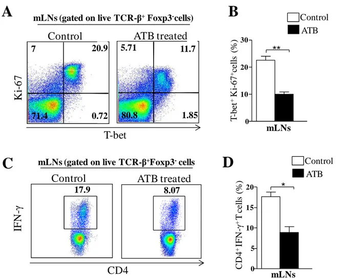
63 Fig 1. Immune responses against T. gondii are down-regulated in the mLNs of ATB
mice. Mice were treated with a cocktail of antibiotics (ATB) for 4 weeks and then infected with 10 cysts of T. gondii strain ME49 C1. Control mice were also infected. At day 9 of infection, mesenteric lymph nodes (mLNs) cells were harvested and single-cell suspensions were either immediately stained or cultured with polyclonal stimuli. After 4 hours, stimulated cells were stained for flow cytometry assessment. A: representative plot of T-bet and Ki-67 co-expression in live CD4+T cells. B: Bar graph showing quantification of A. C: representative plot of IFN-γexpression in live
CD4+T cells. D: Bar graph showing quantification of C. The data are representative of 3 different experiments performed with 4 mice each * P< 0.05. ** P < 0.01.
A
5.71 11.7 1.85 80.8 7 20.9 0.72 71.4K
i-6
7
ATB treated
Control
T-bet
mLNs (gated on live TCR-β+Foxp3-cells)
T -b et + K i-6 7 + ce ll s (% )
B
0 10 20 30 mLNs**
Control ATB 17.9 8.07IF
N
-γ
ATB treated
Control
CD4
mLNs (gated on live TCR-β+Foxp3-cells
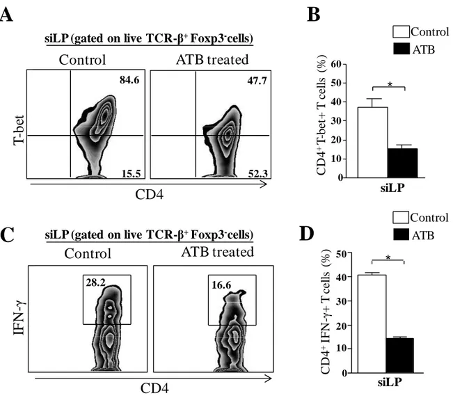
64 Fig 2. Reduced inflammatory immune responses in the small intestine of ATB treated mice infected with T. gondii. Mice were treated with a cocktail of antibiotics for 4 weeks and then infected with 10 cysts of T. gondii strain ME49 C1. Control mice were also infected. At day 9 of infection, small intestine lamina propria (siLP) cells were harvested and single-cell suspensions were either immediately stained or cultured with polyclonal stimuli. After 4 hours, stimulated cells were stained for flow cytometry assessment. A: representative plot of T-bet expression in live CD4+ T cells. B: Bar graph showing quantification of A. C: representative plot of IFN-γ expression in live
CD4+ T cells. D: Bar graph showing quantification of C * P< 0.05. The data are representative of 3 different experiments performed with 4 mice each
A
B
T
-b
et
ATB treated
Control
IF
N
-γ
CD4
16.6 28.2 84.6 15.5 47.7 52.3 0 10 20 30 40 50*
siLP C D 4 +IF N -γ + T c el ls (% ) Control ATB 0 10 20 30 40 50 60 siLP C D 4 +T -b et + T c el ls (% )*
Control ATBATB treated
Control
CD4
siLP (gated on live TCR-β+Foxp3-cells)
siLP (gated on live TCR-β+Foxp3-cells)
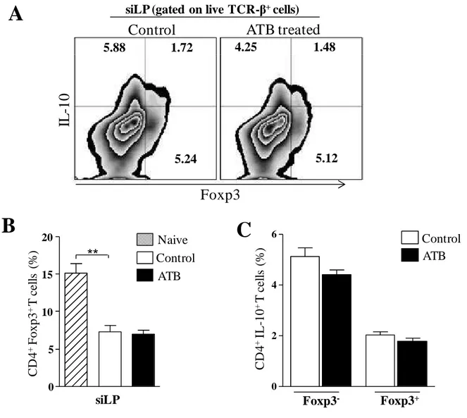
65 Regulatory responses induced by T. gondii oral infection are not affected by
antibiotic treatment
66 Fig 3. Regulatory responses are not affected by depletion of gut microbiota. Mice
were treated with a cocktail of antibiotics for 4 weeks and then infected with 10 cysts of T. gondii strain ME49 C1. Control mice were also infected. At day 9 of infection, small intestine lamina propria cells (siLP) were harvested and single-cell suspensions were either immediately stained or cultured with polyclonal stimuli. After 4 hours, stimulated cells were stained for flow cytometry assessment.. A: representative plot of Foxp3 and IL-10 co-expression in live CD4+ T cells. B: Bar graph showing frequency of Treg cells. C: frequency of IL-10 expression in effector (Foxp3-) CD4+ T cells and Treg cells. The data are representative of 3 different experiments with 4 mice each ** P< 0.01.
5.88 1.72 4.25 1.48
5.24 5.12
Control ATB
siLP Foxp3
-C D 4 + F o x p 3 + T c el ls (% ) C D 4 + IL -1 0 + T c el ls (% )
IL
-1
0
ATB treated
Control
Foxp3
A
0 5 10 15 20 Control ATB NaivesiLP (gated on live TCR-β+cells)
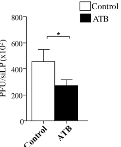
67 Reduced parasite load in the gut of antibiotic treated mice
68 Fig 4. Lower parasite burden at day 9 of infection in ATB treated mice infected
with T. gondii. Mice were treated with a cocktail of antibiotics for 4 weeks and then infected with 10 cysts of T. gondii strain ME49 C1. Control mice were also infected. At day 9 of infection, small intestine lamina propria (siLP) cells were harvested and single-cell suspensions (104 to 106 cells) were added to fibroblast monolayer cultures and titrated by plaque formation. The results of these titrations are reported in Plaque Forming Units (PFUs). The data are representative of 3 different experiments with 4 mice each. *. P< 0.05.
0 200 400 600 800
P
F
U
/s
iL
P
(x10
2 )
Control ATB
69 Less severe immunopathology in germfree mice infected with T. gondii
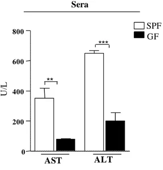
70 ***
**
0 200 400 600 800
U
/L
SPF GF
AST ALT
Sera
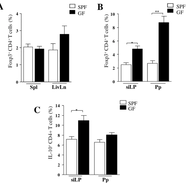
71 Reduced immune responses in germfree mice infected with T. gondii
We next evaluated the adaptive response against the parasite. Expression of T-bet in effector CD4+ T cells was found to be similar between SPF and GF mice (fig. 6 A). Production of IFN-γ by effector CD4+ T cells at day 9 of infection was slightly but no significantly reduced in GF mice (fig. 6 B). In agreement with previous reports associating intestinal Th-17 immune response to a restricted group of bacteria, the segmented filamentous bacteria (SFB) (Ivanov et al., 2008), we could not detect IL-17 production by CD4+ T cells in the lamina propria of the small intestine of GF mice when compared to SPF mice (fig. 6 C).
72 Fig 6. Reduced inflammatory immune responses in the small intestine of germfree mice infected with T. gondii. GF and SPF mice were infected with 10 cysts of T.
gondii strain ME49 C1. At day 9 of infection, small intestine lamina propria
(siLP) cells and Peyer’s patches (Pp) cells were harvested and single-cell suspensions were cultured with polyclonal stimuli. After 4 hours, stimulated cells
were stained for flow cytometry assessment. A: expression of IFN-γin live CD4+
T cells. B: expression of IL-17 A in live CD4+T cells. *** P< 0.001. The data are
73 Fig 7. Germfree mice infected with T. gondii have increased regulatory responses
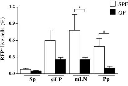
74 Reduced parasite load in the gut of germfree mice
75 0.0
0.3 0.6 0.9 1.2
R
F
P
+ li
v
e
ce
ll
s
(%
)
Sp siLP mLN Pp
*
* SPF
GF
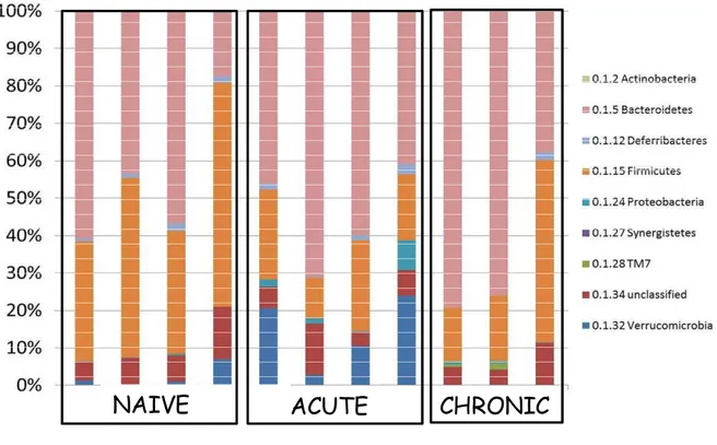
76 Changes in composition of the microbiota during T. gondii-induced ileitis
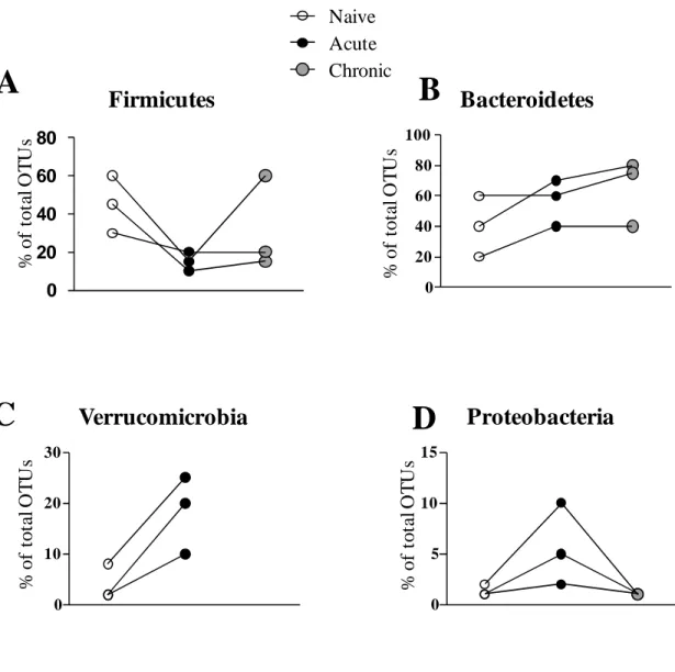
Previous work showed that T. gondii-induced ileitis is accompanied by decreasing species diversity and increasing bacterial load within the gut microbiota. This phenomenon has been shown to aggravate the pathogenesis of the disease (Heimesaat et al., 2006). To further explore these changes we used 454 pyrosequencing of 16S rRNA genes from fecal samples to survey the bacterial diversity of the gut microbiota during T. gondii infection. We sampled fecal pellets from naïve mice, mice at day 9 of infection (peak of acute phase) and at day 45 of infection (chronic phase). Figure 9 represent the taxonomic classification of OTUs found in all samples at the phylum level. Overall, Firmicutes and Bacteroidetes predominated in the gut of naïve mice. Firmicutes, that include Lactobacillus species for example, were reduced during the acute phase of infection in comparison with naïve mice (Fig. 10 A). Bacteroidetes, on the other hand, were increased in samples from acute phase of infection (Fig. 10 B). Verrucomicrobia, a recent describe phylum of bacteria, were found mostly during the acute phase but not in naïve mice or chronic mice (Fig. 10 C). Proteobacteria which includes gamma-proteobacteria considered pathogenic bacteria or pathobionts such as Escherichia coli were found only during the acute phase of infection confirming recent reports that state that the accumulating bacteria that aggravates intestinal inflammation are mainly Enterobacteria such as E. coli and Enterococcus species (Fig. 10 D). Interestingly, the composition of the microbiota after
77 naïve mice indicating that the changes caused by parasitic infection might persist beyond the resolution of the infection (Fig. 9).
NAIVE
ACUTE
CHRONIC
Fig 9. Composition of the gut microbiota at different time-points during T.
gondii infection. Mice were infected with 25 cysts of T. gondii strain ME49
78
Firmicutes
Naive Acute ChronicBacteroidetes
0 20 40 60 80 100Verrucomicrobia
0 10 20 30Proteobacteria
0 5 10 15 % o f to ta l O T U s % o f to ta l O T U s % o f to ta l O T U s % o f to ta l O T U s79 Temporal intestinal immune responses against T. gondii
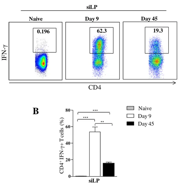
80 Fig 11. Temporal immune responses during T. gondii infection. Mice were
infected with T. gondii strain ME49 C1. At day 9 of infection, small intestine lamina propria (siLP) cells were harvested and single-cell suspensions were cultured with polyclonal stimuli. After 4 hours, cells were stained for flow cytometry assessment. A: representative plot of IFN-γexpression in live CD4+ T cells B: bar graph showing quantification of A. The data are representative of 2 different experiments with 3 mice each ** P< 0.01; *** P< 0.001.
Day 9 Day 45
Naive
0.196 62.3 19.3
siLP
IF
N
-γ
CD4
0 20 40 60 80 siLP C D 4 + IF N -γ + T c el ls (%) Day 9
81 T. gondii-induced ileitis induces systemic translocation of commensal bacteria
T. gondii-induced ileitis promotes a deregulation of intestinal mucosal
82
A
B
Fig 12. Systemic translocation of bacteria in T. gondii oral infection. Mice were infected with 10 cysts of T. gondii strain ME49 C1. At day 9 of infection, different tissues were collected. A: mesenteric lymph nodes (mLns), spleens and livers were homogeneized in saline and serial dilutions were plated in Tryptic Soy agar plates. Colonies were counted after 48 hours of culture. The result was reported as the number of colony forming units (CFU) per organ. B: Small intestine of infected mice were fixed in 10% formalin and 5µm slides
were prepared and stained with anti-LPS core antigen. The data are representative of 2 different experiments with 4 mice each. ND: not detected..
mLN 0 1 2 3 4 L o g C F U /t is su e Spleen 0 1 2 3 4 L o g C F U /t is su e Liver 0 1 2 3 4 L o g C F U /t is su e Naive Day 9
ND ND ND
20 X 40 X
83 Antibody responses against commensal bacteria are triggered during the acute
mucosal T. gondii infection
Given that we can readily observe systemic translocation in response to parasitic infection we decided to investigate the adaptive immune response to commensal bacteria and the consequences of such responses. In order to study whether translocation of commensal bacteria during acute T. gondii infection can activate adaptive immune responses against commensal bacterial antigens, C57BL/6 mice were infected orally with the type II T. gondii strain ME 49 and fresh fecal pellets were collected at day 9 of infection. The fecal pellets were homogeneized and serial dilutions were plated in a rich media and incubated in aerobic conditions. Single colonies obtained from plates of higher dilutions were expanded in broth and plated in several differential media. Two gram-negative bacteria and one gram-positive were isolated and identified by chemical and morphological methods. The three bacteria that accumulated and were found in higher quantities during the acute phase of the infection were identified as E. coli, Enterococcus faecalis and P. mirabilis. These three strains were used as whole bacterial lysate to evaluate specific B cell responses.
84 infection indicating that the response is long-lasting (fig 13 A). The total IgG levels in response to E. coli antigens were also higher in infected mice in both time points analyzed (fig 13 B) as well as IgM levels (fig 13 C). Interestingly, we did not find detectable levels of E. coli-specific IgA at the peak of infection (day 9). The IgA titers were restored after 45 days of infection to levels comparable to those observed in naïve mice. We did not find significant levels of any of the antibodies in germfree mice sera.
We also examined B cell responses against E. faecalis and P. mirabilis. The Pan Ig titers against E. faecalis are increased at days 9 and 45 after infection in comparison to naïve levels although they are lower than the titers observed for E. coli responses (fig. 14 A). The Pan Ig titers after 45 days of infection are significant higher than the titers found at day 9. There was also an increased in E. faecalis-specific IgG2c and IgG1 levels in response to T. gondii infection (fig. 14 B and C). We also found increased antibody responses against P. mirabilis after T. gondii infection (fig. 15 A and B). Our data shows that there is an induction of B cell response against commensal bacteria during acute phase of T. gondii oral infection.