Indomethacin can downregulate the levels of
inflam-matory mediators in the hippocampus of rats
sub-mitted to pilocarpine-induced status epilepticus
Michele Juliane Vieira,I,IISandra Regina Perosa,IGustavo Adolfo Argan˜araz,III Jose´ Antoˆnio Silva Jr,IV Esper Abra˜o Cavalheiro,IMaria da Grac¸a Naffah-MazzacorattiI,V
IUniversidade Federal de Sa˜o Paulo, Departamento de Neurologia/Neurocirurgia, Sa˜o Paulo/SP, Brazil.IISociedade Beneficente Israelita Brasileira, Hospital
Albert Einstein, Neurofisiologia Clı´nica, Sa˜o Paulo/SP, Brazil.IIIUniversidade de Brası´lia, Faculdade de Cieˆncias da Sau´de, Laborato´rio de Patologia
Molecular, Brasilia/DF, Brazil.IVUniversidade Nove de Julho, Departamento de Educac¸a˜o Fı´sica, Programa de Po´s-Graduac¸a˜o em Cieˆncias da Reabilitac¸a˜o,
Sa˜o Paulo/SP, Brazil.VUniversidade Federal de Sa˜o Paulo, Departamento de Bioquı´mica, Sa˜o Paulo/SP, Brazil.
OBJECTIVE:Refractory status epilepticus is one of the most life-threatening neurological emergencies and is characterized by high morbidity and mortality. Additionally, the use of anti-inflammatory drugs during this period is very controversial. Thus, this study has been designed to analyze the effect of a low dose of indomethacin (a COX inhibitor) on the expression of inflammatory molecules.
METHOD: The hippocampus of rats submitted to pilocarpine-induced long-lasting status epilepticus was analyzed to determine the expression of inflammatory molecules with RT-PCR and immunohistochemistry. RESULTS:Compared with controls, reduced levels of the kinin B2 receptors IL1band TNFawere found in the hippocampus of rats submitted to long-lasting status epilepticus and treated with indomethacin.
CONCLUSIONS:These data show that low doses of indomethacin could be employed to minimize inflammation during long-lasting status epilepticus.
KEYWORDS: Indomethacin; Inflammatory Mediators; Epilepsy.
Vieira MJ, Perosa SR, Argan˜araz GA, Silva Jr JA, Cavalheiro EA, Naffah-Mazzacoratti MG. Indomethacin can downregulate the levels of inflammatory mediators in the hippocampus of rats submitted to pilocarpine-induced status epilepticus. Clinics. 2014;69(9):621-626.
Received for publication onNovember 26, 2013;First review completed onJanuary 13, 2014;Accepted for publication onApril 10, 2014 E-mail: naffahmazz.nexp@epm.br
Tel.: 55 11 5576-4846
& INTRODUCTION
Epilepsy has traditionally been considered to be a neuronal disease and several recent studies have suggested that astrocytes, microglia, blood-derived leukocytes and blood-brain barrier (BBB) breakdown are involved in the pathogenesis of this disease (1,2). Experimental and clinical evidence has demonstrated the increased synthesis of specific inflammatory mediators and the upregulation of their receptors in the epileptic brain, indicating that some proinflammatory pathways are activated in seizure foci. Patients with refractory temporal lobe epilepsy (TLE) frequently present with hippocampal sclerosis (HS), which has been related to high levels of IL1band nitric oxide (NO) in the hippocampal formation (3). Lymphocyte infiltrates were also present in the hippocampus of these patients,
indicating that the immune system participates in this disease.
Several molecules are involved in these proinflammatory pathways, including molecules involved in disturbance to the BBB, molecules in the cyclooxygenase (COX2) signaling pathway, related prostaglandins, classical cytokines and their downstream targets and toll-like receptors (1,4).
Refractory status epilepticus (SE) is one of the most life-threatening neurological emergencies and it is characterized by high morbidity and mortality (5). This severe condition sometimes worsens the prognosis and, at times, long-lasting seizures lead to refractory TLE in adulthood (6). Therefore, several approaches have been used to diminish brain sequelae due to SE. Treatment involves intravenous anesthetics and antiepileptic drugs, including topiramate, which has been associated with a decrease in tissue excitability, blocking glutamate a -amino-3-hydroxy-5-methyl-4-isoxazolepropionic acid (AMPA) receptors (7).
Previous studies from our group have shown increased levels of prostaglandin F2a(PGF2a)in the hippocampus of rats submitted to pilocarpine-induced long-lasting SE (8). We have also demonstrated that the administration of high doses of indomethacin (a COX inhibitor) increases rat death due to tonic seizures, blocking changes in the PG concentration. Copyrightß2014CLINICS– This is an Open Access article distributed under
the terms of the Creative Commons Attribution Non-Commercial License (http:// creativecommons.org/licenses/by-nc/3.0/) which permits unrestricted non-commercial use, distribution, and reproduction in any medium, provided the original work is properly cited.
No potential conflict of interest was reported.
These data reveal the dual effect of anti-inflammatory drugs in the treatment of epilepsy. In accordance with our previous results, Jeong et al. (9) reported that acetylsalicylic acid treatment also leads to increased death among epileptic animals. In contrast, several authors have shown that COX2 inhibition can control P-glycoprotein (PgP) expression, improving the penetration of antiepileptic drugs into the brain and restoring the pharmacosensitivity to these drugs (10,11). Additionally, COX2 deficiency reduces excitotoxic damage to the epileptic brain (12). According to Levin et al. (4), COX2 knockout mice presented with earlier mortality after pilocarpine-induced SE. Dore´ et al. (13) also reported that antagonists of the PGE2 receptor provide a neuropro-tective effect, decreasing cell damage and reducing stroke severity. These findings make this issue very controversial. Thus, this study has been designed to analyze the effect of a low dose of indomethacin on inflammatory molecule expression in the hippocampus of rats submitted to pilocarpine-induced, long-lasting SE.
& MATERIALS AND METHODS Wistar rat treatment
The animal experiments were performed with institu-tional ethics approval and all efforts were made to minimize animal suffering. Moreover, the animals were given assistance with eating and hydration during the initial recovery period after SE to improve their condition and endurance. Wistar adult male rats, weighing 250 g, were housed in groups of three to four per cage and kept at a controlled room temperature, humidity and light–dark cycle (12512 h). Chow pellets and tap water were available ad
libitum. The rats received a single dose of pilocarpine (350 mg/kg, intraperitoneal [i.p.]). To prevent peripheral cholinergic effects, scopolamine methyl nitrate was sub-cutaneously injected at a dose of 1 mg/kg 30 min before pilocarpine administration. Animals that progressed to SE were divided into two groups: pilo (group B) and pilo+indomethacin (group C) (0.5 mg/kg i.p.). The
indo-methacin was injected at different time intervals after SE (30 min, 1 h, 2 h and 4 h). The animals were sacrificed 5 h after SE. Saline-treated animals (group A) were sacrificed 5 h after saline and scopolamine methyl nitrate injections and were used as control groups.
These groups were used to quantify the mRNA levels of kinin B1 and B2 receptors, TNFaand IL-1bwith a real-time PCR assay (n = 4 per group). The spatial and temporal localization of both kinin receptors and cytokines were analyzed using immunohistochemistry (n = 3 per group).
Quantitative real-time TaqmanTMPCR
For the biochemical evaluation of kinin B1 and B2 receptors, TNFaand IL-1b, brains were collected from the following groups: saline-treated rats (control group; group A); animals that received scopolamine methyl nitrate plus pilocarpine injection (350 mg/kg i.p.) and were sacrificed 5 h after SE onset (pilocarpine-treated group; group B); and animals that received scopolamine methyl nitrate plus pilocarpine injection (350 mg/kg i.p.) plus indomethacin (0.5 mg/kg i.p.) (indomethacin-treated group; group C). Rats from the aforementioned groups (n = 4 for each group) were sacrificed and their brains were isolated and dissected. Rat hippocampi were then frozen in liquid nitrogen and stored at -80
˚
C. Thawed tissue was homogenized in 1 ml ofTRIzol reagent (Gibco BRL, Gaithersburg, MD) and total RNA was isolated according to the manufacturer’s instruc-tions. Samples were amplified in 20ml reactions using the TaqManTMAmplification System with an ABI PRISMH7000 Sequence Detection System (Applied Biosystems, Foster City, CA, USA). Real-time PCR was performed with 900 ng of cDNA for kinin B1 and B2 receptors, IL1b and TNFa, whereas 100 ng of cDNA was used for glyceraldehyde 3-phosphate dehydrogenase (GAPDH), which served as an internal standard. Oligonucleotide primer and fluorogenic probe sets for TaqmanTMReal-Time PCR were designed for kinin receptors and GAPDH using the Assays-by-Design Service (Applied Biosystems) to meet all TaqmanTMdesign guidelines. Probes were synthesized with the reporter dye 6-carboxyfluorescein (6-FAM) covalently linked to the 59 end and the quencher dye 6-carboxy-tetramethyl-rhoda-mine (TAMRA) linked to the 39 end of the probe. Each reaction was carried out with 10ml of Master Mix (Applied Biosystems), 1ml of a mix containing two primers (18ml each) and a probe 5ml specific to mRNA of Kinin B1 receptor (forward primer: 59 -CCAGGGTTCGTCATCACT-ATCTG-39, reverse primer: 59 -GCAAAAGGAAGAAGGAC-AAGACTAA-39), the kinin B2 receptor (forward primer: 59 -CCCTTCCTCTGGGTCCTCTT-39, reverse primer: 59 -CAG-AACACGCTGAGGACAAAGA-3), IL-1b (forward primer: 59-CACCTCTCAAGCAGAGCACAG-39, reverse primer: 59 -GGGTTCCATG GTGAAGTCAAC-39), TNFa (forward primer: 59- AAATGGGCTCCCTCTATCAGTTC-39, reverse primer: 59-TCTGCTTGGTGGTTTGCTACGAC- 39), or GAPDH (forward primer: 59 -GGGCAGCCCAGAACATC-AT-39, reverse primer: 59 -CCGTTCAGCTCTGGGATGAC-39). The cycling conditions were as follows: 50
˚
C for 2 min and 95˚
C for 10 min, followed by 50 cycles of 95˚
C for 15 s (melting step) and 60˚
C for 1 min (annealing/extension step).Data analysis
Increases in the level of reporter dye fluorescence during the 50 cycles of amplification were monitored using Sequence Detector Software (SDS version 1.6; Applied Biosystems). The PCR amplification cycle in which a given fluorescence threshold is reached is the first parameter used for analyzing mRNA expression and is referred to as Ct. The higher the initial copy level, the lower the Ct value. A normalized value is obtained by subtracting the Ct of GAPDH from the Ct of kinin B1, B2, IL-1b or TNFa, resulting in the DCt. It is uncommon to use DCt as a measure of relative expression due to its logarithmic nature; thus, the 2-DCt parameter is used to express the relative expression level (14). The mRNA expression levels of the kinin B1 and B2 receptors and cytokines are presented as n-fold differences relative to the levels expressed during different experimental model phases and were compared using one-way ANOVA followed by Tukey’s post-test. The data are presented as the means ¡ S.E.M.; p,0.05 was considered statistically significant.
Immunohistochemistry
perfusion and post-fixed in paraformaldehyde (4%). The brains were sectioned at 40mm, collected and washed with PBS (pH 7.4). All sections were treated with 3% H2O2in PBS to quench the endogenous peroxidase and were then incubated for 2 h in PBS containing 0.3% Triton X-100 and bovine serum albumin. Subsequently, the sections were incubated overnight at 4
˚
C with the following primary antibodies: anti-B2 receptor (BD Transduction) diluted 15100, anti-IL1b (IBL) diluted 15100, anti-TNFa (IBL)diluted 15100 and anti-B1 (Santa Cruz) diluted 1550 in 2%
albumin. After three washes in PBS, the sections were incubated with the secondary biotinylated antibody for 2 h. The complex was then incubated with the ABC Kit (Vector) and visualized using 3,39-diaminobenzidine (1 mg/ml in 1% H2O2). The sections were subsequently dehydrated, coverslipped and analyzed by light microscopy using bright-field illumination.
& RESULTS
Immunoreactivity of the kinin B1 and B2 receptors, IL-1band TNFain the hippocampus
Immunohistochemical analysis revealed the distribution of the kinin B1 receptor in all hippocampal formations of all studied groups (Figure 1, panel A–C). Regions such as CA1, CA2, CA3 and the hilus as well as the granular cells from the dentate gyrus were stained. However, the staining intensity of kinin B1 was very different among the groups studied. Increased immunoreactivity was observed in the CA1 region of rats in groups B and C (Figure 2, panels A-C). On the other hand, the CA3 region showed
less immunoreactivity for the kinin B1 receptor when groups B and C were compared with group A (Figure 3, panels A-C).
Immunoreactivity for the kinin B2 receptor was also visualized in all hippocampal formations of all studied groups (Figure 1, panels A’-C’). Increased staining was noted in the CA1 and CA3 regions of rats from group B compared with rats from group A (Figures 2 and 3, panels A’-B’).
Immunohistochemical analysis of IL-1bshowed increased staining in the CA1 region of rats from group B compared with those of group A (Figures 2 and 3, A’’-C’’).
TNFa staining was decreased in the CA3 of rats from groups B and C compared with rats from group A (Figure 3, panels A’’’-C’’’). Interestingly, the CA1 and CA2 regions were not labeled by TNFa antibody. Note that all hippocampal regions of rats treated with indomethacin had decreased levels of cytokines.
Real-time PCR analysis of the kinin B1 and B2 receptors, IL-1band TNFa
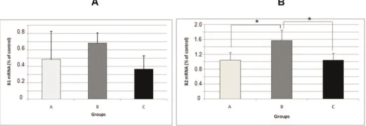
The mRNA levels of the kinin receptors, IL1band TNFa in Wistar rats were analyzed using a semi-quantitative RNA assay. There were no alterations in kinin B1 mRNA in any of the groups that were studied (Figure 4 A). The kinin B2 receptor mRNA level was increased in group B (1.565¡0.2841) compared with group A (1.042¡ 0.2057) (p,0.05) (Figure 4 B). However, when comparing the animals in group B (1.565¡0.2841) with those in group C (1.038¡0.1857), we observed a decrease in the expression of this receptor (p,0.05) (Figure 4 B). There was an increase in
the IL-1bmRNA in group B (0.4450¡0.1622) compared with group A (0.2150¡0.09950) (p,0.05) (Figure 5 A). On the other hand, the level of IL-1b mRNA was decreased in group C (0.1850¡0.06608) compared with group B (0.4450¡0.1622) (p,0.05) (Figure 5 A). Quantitative real-time PCR also revealed a significant increase in the mRNA
expression of TNFa in the animals in group B (0.6275¡0.2364) compared with those in group A (0.2700¡0.09661) (p,0.05) (Figure 5 B). When the animals in group B (0.6275¡0.2364) were compared with the animals in group C (0.3325¡0.1284), no differences in TNFaexpression levels were observed.
Figure 2 -Photomicrographs of the CA1 region of hippocampi processed for immunohistochemistry showing expression of the kinin B1 receptor (A, B and C), the kinin B2 receptor (A’, B’ and C’) and IL1b(A’’, B’’ and C’’). Groups: A – saline-treated group; B – pilocarpine-treated group; and C – indomethacin-pilocarpine-treated group. Scale bars: 0.02 mm.
& DISCUSSION
In this study, we found that rats submitted to long-lasting SE exhibited increased expression of IL1b and TNFa, as previously reported by other authors. These cytokines are related to pro-convulsing action (15), increased excitotoxi-city, inhibition of glutamate uptake by astrocytes and decreased GABA release (16,17). We also found that the administration of indomethacin (a non-selective COX inhibitor) reduced IL1band TNFasynthesis and expression, providing neuroprotection against inflammatory processes. With respect to the kinin B1 and B2 receptors, only the B2 receptor was upregulated by SE and indomethacin admin-istration returned this receptor to its normal levels. These data show that the inflammation-induced kinin B1 receptor is not modulated by this drug. Because the kinin B2 receptor has been considered to play a protective role in the nervous system (18), its downregulation by indomethacin seems to be a maladaptive process.
In a lithium plus pilocarpine model of epilepsy, Voutsinos-Porche et al. (19) found increased expression of IL1b, NFkb and COX2 in structures that are prone to develop neuronal damage and in activated astrocytes. COX2 inhibitors such as parecoxib have neuroprotective effects, but they have not been effective in modifying epileptogen-esis in a pilocarpine-model of epilepsy (20). Other authors who used the selective COX2 inhibitor SC58236 have shown that this inhibition does not modify either the epileptogenic process or the frequency of chronic seizures (21). In contrast, Okada et al. (22) used indomethacin and observed a reduction in the seizure duration in mice that were genetically susceptible to seizures.
Zibell et al. (23) demonstrated that seizure activity induces the expression of the BBB efflux transporter P-glycoprotein (Pgp), limiting the penetration of antiepileptic drugs into the brain. This induction was associated with the activation of NMDA glutamate receptors as well as COX2 and arachidonic acid production. In this way, the highly
Figure 4 -mRNA expression levels of the kinin B1 receptor (A) and kinin B2 receptor (B) in the hippocampus of rats in the pilocarpine model of epilepsy as evaluated with a real-time PCR assay. All data have been normalized to the levels of GAPDH. The data are presented as the mean¡S.E.M. (n = 5 per group).
selective COX2 inhibitor NS-398, an indomethacin-related molecule, blocked the overexpression of Pgp in pilocarpine-induced seizures.
Taken together, these data show that the inhibition of COX during the development of neurodegenerative pro-cesses can be dose- and time-dependent, which was reported previously by other authors (23,24). As reported by our group (8), the administration of high doses of indomethacin prior to pilocarpine injection increases the mortality rate due to tonic seizures in rats, indicating a dual effect involving COX inhibition in this epileptic model. Additionally, numerous specific cell types, including neurons of various subcategories and different glial cells, express and respond to inflammatory mediators in dis-tinctly different ways. Therefore, a given molecule might be beneficial for a particular population of cells but harmful for other populations (25). Several authors have debated the efficacy of anti-inflammatory drugs in epilepsy treatment, highlighting the antiepileptic action of some types of PGs, such as PGD2 and PGE2, but noting that PGs may contribute to the feedback inhibition of neurotransmitter release, possibly reducing excitation (26). In this sense, the blockage of or decrease in COX activity in the early stages of SE, which reduces the levels of inflammatory mediators, seems to be insufficient for blocking epileptogenesis, but it could be effective in neuronal protection or for the control of otherwise refractory seizures.
Therefore, our data show that the administration of indomethacin decreases the levels of inflammatory media-tors when used in the early phases of SE onset in an epilepsy model induced by pilocarpine. However, caution should be exercised when using anti-inflammatory drugs in epilepsy because several pathways are activated during this process.
& ACKNOWLEDGMENTS
This work was supported by Fundac¸a˜o de Amparo a Pesquisa do Estado de Sa˜o Paulo (FAPESP), Conselho Nacional de Desenvolvimento Cientı´fico e Tecnolo´gico (CNPQ), Institutos Nacionais de Cieˆncia e Tecnologia (INCT) and Programa de Apoio a Nu´cleos de Exceleˆncia (PRONEX).
& AUTHOR CONTRIBUTIONS
Vieira MJ and Perosa SR developed the experimental model of epilepsy and the immunohistochemistry protocol; additionally, they assisted with manuscript writing. Silva Jr JA performed real-time PCR. Cavalheiro EA, Argan˜araz GA and Naffah-Mazzacoratti MG outlined the main ideas and wrote the manuscript.
& REFERENCES
1. Friedman A, Dingledine R. Molecular cascades that mediate the influence of inflammation on epilepsy. Epilepsia. 2011;52 Suppl 3:33-9, http://dx.doi.org/10.1111/j.1528-1167.2011.03034.x.
2. Devinsky O, Schein A, Najjar S. Epilepsy associated with systemic autoimmune disorders. Epilepsy Curr. 2013;13(2):62-8, http://dx.doi. org/10.5698/1535-7597-13.2.62.
3. Varella PP, Santiago JF, Carrete H Jr., Yacubian EM, Centeno RS, Caboclo LO, et al. Relationship between fluid-attenuated inversion-recovery (FLAIR) signal intensity and inflammatory mediator’s levels in the hippocampus of patients with temporal lobe epilepsy and mesial temporal sclerosis. Arq Neuropsiquiatr. 2011;69(1):91-9, http://dx.doi. org/10.1590/S0004-282X2011000100018.
4. Levin JR, Serrano G, Dingledine R. Reduction in delayed mortality and subtle improvement in retrograde memory performance in pilocarpine-treated mice with conditional neuronal deletion of cyclooxygenase-2 gene. Epilepsia. 2012;53(8):1411-20, http://dx.doi.org/10.1111/j.1528-1167.2012.03584.x.
5. Sutter R1, Marsch S, Fuhr P, Ru¨egg S. Mortality and recovery from refractory status epilepticus in the intensive care unit: a 7-year observational study. Epilepsia. 2013;54(3):502-11.
6. Cendes F, Ragazzo PC, da Costa V, Martins LF. Corpus callosotomy in treatment of medically resistant epilepsy: preliminary results in a pediatric population. Epilepsia. 1993;34(5):910-7, http://dx.doi.org/10. 1111/j.1528-1157.1993.tb02111.x.
7. Hottinger A, Sutter R, Marsch S, Ru¨egg S. Topiramate as an adjunctive treatment in patients with refractory status epilepticus: an observational cohort study. CNS Drugs. 2012;26(9):761-72, http://dx.doi.org/10.2165/ 11633090-000000000-00000.
8. Naffah-Mazzacoratti MG, Bellı´ssimo MI, Cavalheiro EA. Profile of prostaglandin levels in the rat hippocampus in pilocarpine model of epilepsy. Neurochem Int. 1995;27(6):461-6, http://dx.doi.org/10.1016/ 0197-0186(95)80003-4.
9. Jeong KH, Kim JY, Choi YS, Lee MY, Kim SY. Influence of aspirin on pilocarpine-induced epilepsy in mice. Korean J Physiol Pharmacol. 2013;17(1):15-21.
10. Zibell G, Unkru¨er B, Pekcec A, Hartz AM, Bauer B, Miller DS, et al. Prevention of seizure-induced up-regulation of endothelial P-glycoprotein by COX-2 inhibition. Neuropharmacology. 2009;56(5):849-55, http://dx. doi.org/10.1016/j.neuropharm.2009.01.009.
11. Schlichtiger J, Pekcec A, Bartmann H, Winter P, Fuest C, Soerensen J, Potschka H. et al. Celecoxib treatment restores pharmacosensitivity in a rat model of pharmacoresistant epilepsy. Br J Pharmacol. 2011;60(5):1062-71.
12. Takemiya T, Maehara M, Matsumura K, Yasuda S, Sugiura H, Yamagata K. Prostaglandin E2 produced by late induced COX-2 stimulates hippocampal neuron loss after seizure in the CA3 region. Neurosci Res. 2006;56(1):103-10, http://dx.doi.org/10.1016/j.neures.2006.06.003. 13. Dore´ S. GPCR antagonists as an alternative to COX-2 inhibitors: a case
for the PGE2 EP1 receptor. Trends Pharmacol Sci. 2006;27(9):458-60, http://dx.doi.org/10.1016/j.tips.2006.07.001.
14. Livak KJ, Schmittgen TD. Analysis of relative gene expression data using real-time quantitative PCR and the 2(-Delta Delta C(T)) Method. Methods. 2001;25(4):402-8, http://dx.doi.org/10.1006/meth.2001.1262. 15. Vezzani A, Conti M, De Luigi A, Ravizza T, Moneta D, Marchesi F, et al.
Interleukin-1beta immunoreactivity and microglia are enhanced in the rat hippocampus by focal kainate application: functional evidence for enhancement of electrographic seizures. J Neurosci. 1999;19(12):5054-65. 16. Ye ZC, Sontheimer H. Cytokine modulation of glial glutamate uptake: a possible involvement of nitric oxide. Neuroreport. 1996;7(13):2181-5, http://dx.doi.org/10.1097/00001756-199609020-00025.
17. Zeise ML, Espinoza J, Morales P, Nalli A. Interleukin-1beta does not increase synaptic inhibition in hippocampal CA3 pyramidal and dentate gyrus granule cells of the rat in vitro. Brain Res. 1997;768(1-2):341-4. 18. Argan˜araz GA, Regina Perosa S, Cristina Lencioni E, Bader M, Abra˜o
Cavalheiro E, da Grac¸a Naffah-Mazzacoratti M, et al. Role of kinin B1 and B2 receptors in the development of pilocarpine model of epilepsy. Brain Res. 2004;1013(1):30-9.
19. Voutsinos-Porche B, Koning E, Kaplan H, Ferrandon A, Guenounou M, Nehlig A, et al. Temporal patterns of the cerebral inflammatory response in the rat lithium-pilocarpine model of temporal lobe epilepsy. Neurobiol Dis. 2004;17(3):385-402, http://dx.doi.org/10.1016/j.nbd. 2004.07.023.
20. Polascheck N, Bankstahl M, Lo¨scher W. The COX-2 inhibitor parecoxib is neuroprotective but not antiepileptogenic in the pilocarpine model of temporal lobe epilepsy. Exp Neurol. 2010;224(1):219-33, http://dx.doi. org/10.1016/j.expneurol.2010.03.014.
21. Holtman L, van Vliet EA, van Schaik R, Queiroz CM, Aronica E, Gorter JA. Effects of SC58236, a selective COX-2 inhibitor, on epileptogenesis and spontaneous seizures in a rat model for temporal lobe epilepsy. Epilepsy Res. 2009;84(1):56-66, http://dx.doi.org/10.1016/j.eplepsyres. 2008.12.006.
22. Okada K, Yamashita U, Tsuji S. Cyclooxygenase system contributes to the maintenance of post convulsive period of epileptic phenomena in the genetically epileptic El mice. J UOEH. 2006;28(3):265-75.
23. Baran H, Vass K, Lassmann H, Hornykiewicz O. The cyclooxygenase and lipoxygenase inhibitor BW755C protects rats against kainic acid-induced seizures and neurotoxicity. Brain Res. 1994;646(2):201-6. 24. Serrano GE, Lelutiu N, Rojas A, Cochi S, Shaw R, Makinson CD, et al.
Ablation of cyclooxygenase-2 in forebrain neurons is neuroprotective and dampens brain inflammation after status epilepticus. J Neurosci. 2011;31(42):14850-60, http://dx.doi.org/10.1523/JNEUROSCI.3922-11. 2011.
25. Dedeurwaerdere S, Callaghan PD, Pham T, Rahardjo GL, Amhaoul H, Berghofer P, et al. PET imaging of brain inflammation during early epileptogenesis in a rat model of temporal lobe epilepsy. EJNMMI Res. 2012;2(1):60, http://dx.doi.org/10.1186/2191-219X-2-60.